Abstract
Mitogen activation of mRNA decay pathways likely involves specific endoribonucleases, such as G3BP, a phosphorylation-dependent endoribonuclease that associates with RasGAP in dividing but not quiescent cells. G3BP exclusively cleaves between cytosine and adenine (CA) after a specific interaction with RNA through the carboxyl-terminal RRM-type RNA binding motif. Accordingly, G3BP is tightly associated with a subset of poly(A)+ mRNAs containing its high-affinity binding sequence, such as the c-myc mRNA in mouse embryonic fibroblasts. Interestingly, c-myc mRNA decay is delayed in RasGAP-deficient fibroblasts, which contain a defective isoform of G3BP that is not phosphorylated at serine 149. A G3BP mutant in which this serine is changed to alanine remains exclusively cytoplasmic, whereas a glutamate for serine substitution that mimics the charge of a phosphorylated serine is translocated to the nucleus. Thus, a growth factor-induced change in mRNA decay may be modulated by the nuclear localization of a site-specific endoribonuclease such as G3BP.
The stability of an mRNA influences gene expression by affecting the steady-state level of the mRNA as well as the rate at which the mRNA disappears following transcriptional repression and accumulates following transcriptional induction (23, 36, 37). This level of regulation is particularly important for proteins that are active for a brief period, such as growth factors, transcription factors, and proteins that control cell cycle progression. Indeed, many proto-oncogenes, cytokines, and lymphokines are rapidly and transiently activated by extracellular stimuli, and the rapid disappearance of these messages is due not only to a shutoff of transcription but also to their short half-lives (38). The stability of these mRNAs depends, at least in part, upon specific cis-acting elements found either in the coding region or more frequently in the 3′ untranslated region (UTR). The 3′ UTR destabilizing elements can be quite variable in sequence and length, but some are characterized by AU-rich regions (ARE) containing one or more AUUUA pentamers (2, 12). In some cases, the latter sequence is sufficient to destabilize a normally long-lived mRNA, such as β-globin mRNA (41). Moreover, ARE-directed mRNA degradation is influenced by many exogenous factors, including phorbol esters, calcium ionophores, cytokines, and transcription inhibitors, consistent with the possibility that AREs play a critical role in the regulation of gene expression during cell growth and differentiation (2, 7, 29, 36).
While many ARE-specific RNA binding proteins have been described previously (16, 19, 27, 31, 32, 51), the molecular mechanism by which these proteins target mRNA for rapid degradation is not known. It is also not clear whether AREs are the actual target for ribonucleases and/or whether ARE-specific RNA binding proteins are involved in deadenylation that is observed prior to the decay of many vertebrate mRNAs containing these elements (10, 21, 42, 47). In Saccharomyces cerevisiae, two alternative pathways for mRNA degradation can be distinguished: a deadenylation-dependent pathway, in which degradation occurs after the loss of the poly(A) tail, and a deadenylation-independent pathway, in which degradation is stimulated by premature termination of translation (11). The finding that several genes encoding RNA turnover components are highly conserved (1, 30, 44) and recent characterization of the vertebrate poly(A)-specific RNase, PARN (15), suggest that the two pathways analyzed in yeast also exist in mammalian cells. Nevertheless, direct evidence is lacking, and the relative contributions of these pathways to the degradation of specific mammalian mRNAs are unknown. In addition, vertebrate cells make extensive use of endonucleases to catalyze mRNA decay (39) that does not seem to require deadenylation. However, the endoribonucleases responsible for mRNA turnover in mammalian cells remain largely unidentified (39). Currently, G3BP, a protein that associates with Ras GTPase-activating protein p120 (RasGAP) (33), has been demonstrated to harbor an intrinsic endonuclease activity that cleaves the ARE of mouse c-myc mRNA in vitro (18). G3BP, with a predicted molecular mass of 52 kDa, contains a carboxyl (C)-terminal RNA binding domain (33), the RRM-type domain, an amino (N)-terminal domain (1 to 120) homologous to nuclear transporter factor 2 (NTF2), and a central domain rich in acidic residues (140 to 240). G3BP provides a unique paradigm of enzyme regulation because it is the only endoribonuclease known to require site-specific phosphorylation for its catalytic activity (18). Another interesting feature of G3BP is that both its phosphorylation and its association with RasGAP in the particulate fraction of cells are affected by extracellular stimuli, consistent with the possibility that G3BP plays a role in modulating mRNA stability via external signals.
Here we provide evidence that G3BP binds specific sequences via its C-terminal RRM domain and behaves as a highly active, single-strand-specific endoribonuclease that exclusively cleaves between CA dinucleotides. The c-myc mRNA, which contains a high-affinity G3BP binding site in its 3′ UTR, decays more rapidly in control fibroblasts than in fibroblasts deficient in p120 RasGAP. Importantly, these RasGAP−/− fibroblasts contain a G3BP isoform lacking phosphorylation at Ser-149. By site-directed mutagenesis, we demonstrate that phosphorylation at this site may regulate G3BP subcellular localization. Thus, G3BP fulfills the criteria of an endoribonuclease that can couple signal transduction to mRNA decay and can potentially give rise to a functional differentiation between transcriptional and posttranscriptional controls.
MATERIALS AND METHODS
Oligonucleotides.
The sequences of the synthetic oligonucleotides (Genosys-Sigma) used in this study as templates or primers for PCR are the following (in the form of name, sequence given 5′ to 3′):
S1, CCCGACACCCGCGGATCCATGGGCACTATTTATATCAAC; S2, CGCGGATCCTAATACGACTCACTATAGGGGCCACCAACGACA; Sub 0, CACCAACGACAGTTGATATAAAT;
Sub R, CACCAACGACA(N)12GTTGATATAAAT; Sub U, CACCAACGACA(T)12GTTGATATAAAT; Sub A, CACCAACGACA(A)12GTTGATATAAAT; Sub C, CACCAACGACA(C)12GTTGATATAAAT; Sub G, CACCAACGACA(G)12GTTGATATAAAT; Sub CS, CACCAACGACAACCCATACGCAGGTTGATATAAAT; Sub 2CS, CACCAACGACAACCCATACGCAGACCCATACGCAGGTTGATATAAAT; Sub ACS, CACCAACGACATGGGTATGCGTCGTTGATATAAAT; c-myc S, CTCAACGACAGCAGCTCGCC; c-myc A, CGTGGCACCTCTTGAGGACCAGTG; GAPDH S1, CAGTCCATGCCATCACTGCC; GAPDH A1, GCCTGCTTCACCACCTTCTTG; GAPDH S2, ACAGTCCATGCCATCACTGCC; GAPDH A2, GCCTGCTTCACCACCTTCTTG.
Purification of recombinant G3BP, Northwestern analysis, and systematic evolution of ligands by exponential enrichment (SELEX) experiments.
To mutate the N- and C-terminal regions of human G3BP generating G3BPΔN and G3BPΔC, PCR was used to amplify segments of the human G3BP cDNA from position 897 to 1398 and 216 to 1050, respectively, taking the first nucleotide of the initiating methionine as position 1. The amplified fragments were cloned into the transfer vector pVL1393 (Invitrogen), and recombinant proteins were produced and purified from baculovirus-infected Sf9 cells as described previously (33).
Proteins were resolved on sodium dodecyl sulfate (SDS)–10% polyacrylamide gels and electrophoretically transferred to nitrocellulose membranes in 10 mM 3-(cyclohexylamino)-1-propanesulfonic acid, pH 11.0, containing 10% methanol, for 2 h. Nitrocellulose filters were washed three times in phosphate-buffered saline (PBS) and incubated for at least 1 h with several changes of binding buffer (10 mM Tris-HCl [pH 7.5], 50 mM NaCl, 1 mM EDTA, 0.02% bovine serum albumin, 0.02% polyvinylpyrrolidone, 0.02% Ficoll). For optimal binding, the filters were not stored at this stage but rather were used immediately. They were incubated for 1 h in binding buffer containing 5 × 10⁻⁵ nmol/ml of 32P-labeled probes and 10 μg of tRNA/ml as carrier. After binding, filters were washed five times with buffer (10 mM Tris-HCl [pH 7.5], 50 mM NaCl, 1 mM EDTA), dried at room temperature, and visualized by autoradiography.
For in vitro genetic selection of G3BP RNA ligands, a nucleic acid library possessing 5′ and 3′ fixed regions surrounding a 12-nucleotide (nt) randomized region was generated as described (13), using the random oligonucleotide pool (Sub R). Binding of the randomized RNA pool to immobilized recombinant G3BP was carried out by Northwestern analyses as described above. The protein-bound RNA was extracted from the protein/filter as described previously (13). The RNA was reverse transcribed using the 3′ primer S1, and PCR amplification was carried out using both S1 and the 5′ S2 primers. The resulting PCR product was transcribed with T7 RNA polymerase (13). The RNA was gel purified and used in a subsequent round of G3BP binding. This process was continued for five cycles. The PCR product generated during the fifth round was restricted with BamHI and ligated into pUC 19, and the resulting plasmids were used to transform competent DH5 bacteria. Clonal inserts were sequenced using standard methods.
In vitro transcription and RNase assays.
The c-myc 3′ UTR RNA fragment was prepared by in vitro transcription of the pkSGMW1 plasmid (18) linearized at BglII, with T3 RNA polymerase (Promega). Synthetic DNA templates, containing homopolymers (Sub A, Sub U, Sub C and Sub G), the best-guess high-affinity G3BP consensus site present in both orientations (Sub CS and Sub ACS) or duplicated (Sub 2CS), as well as constant regions for primer annealing (Sub 0), were amplified by PCR using S1 and S2 primers and transcribed by T7 polymerase (Promega). Thus, all these substrates, designed for the SELEX experiment, harbor the same two constant regions. Full-length transcripts were labeled with [γ-32P]ATP and T4 polynucleotide kinase (Gibco BRL) and then purified on denaturing polyacrylamide gels. RNase assays were performed with 10 fmol (10,000 cpm) of radiolabeled RNA. The RNase reaction buffer contained 50 mM Tris HCl (pH 6), 150 mM NaCl, and 10% glycerol. Purified recombinant G3BP from baculovirus (0.2 to 8 pmol) and 10 μg of yeast tRNA were mixed with 10 μl of reaction mixture prior to addition of the labeled probe. The samples were incubated for 10 min at 25°C and extracted with phenol-chloroform, and following ethanol precipitation the cleavage products were resolved on either 8 or 12% polyacrylamide–8 M urea gels.
Labeled transcripts were also digested by sequencing grade RNase T1, RNase U2, RNase B. cereus, and RNase Phy M (Pharmacia Biotech) according to the manufacturer's protocols.
Cell culture, immunoprecipitation, and immunoblotting.
Wild-type (wt) and RasGAP−/− mouse fibroblasts were obtained as described previously (46) and were maintained in Dulbecco's modified Eagle's medium supplemented with 10% fetal calf serum and antibiotics (50 U of penicillin/ml and 50 μg of streptomycin/ml) at 37°C in 5% CO2–95% air. They were rendered quiescent by serum starvation for 24 h. Total extracts were prepared as described previously (33). Protein concentrations were determined by the Bio-Rad protein assay using bovine serum albumin as a standard.
G3BP immunoprecipitations were performed with the anti-G3BP monoclonal antibody (1F1) as described previously (18). Proteins were resolved on SDS–10% polyacrylamide gels and electrophoretically transferred to nitrocellulose membranes in 3-(cyclohexylamino)-1-propanesulfonic acid, as described above. Nitrocellulose membranes were incubated overnight at 4°C in blocking solution (5% dried milk in PBS) and then incubated with the following dilutions of primary antibody in blocking solution for 1 h at room temperature: 1/3,000 for anti-G3BP (200 ng/ml); 1/500 for anti-p120GAP (GP200), and 1/300 for anti-glyceraldehyde-3-phosphate dehydrogenase (GAPDH) in a blocking solution supplemented with 0.05% Tween 20 (18). Nitrocellulose filters were washed three times in PBS, and the bound antibodies were detected using an appropriate anti-immunoglobulin G-horseradish peroxidase conjugate followed by enhanced chemiluminescence (ECL; Amersham) according to the manufacturer's protocol.
32P Labeling, phosphoamino acid and phosphopeptide mapping.
For metabolic labeling, 500 μCi of inorganic [32P]phosphate (Amersham) was used per 60-mm-diameter dish of proliferating fibroblasts. Labeling was carried out in 6 ml of phosphate-free Dulbecco's modified Eagle's medium, in the presence of 10% phosphate-free fetal calf serum, for 4 or 8 hours at 37°C in a CO2 incubator. Cells were lysed as described previously (18), and lysates were precleared for 1 h with 50 μl of protein G-Sepharose beads (Pharmacia). The lysates were clarified by centrifugation at 12,000 × g for 30 s at 4°C. An aliquot of the lysates was used to determine the protein concentration by the Bio-Rad protein assay using bovine serum albumin as a standard. Equal amounts of proteins were immunoprecipitated with an anti-G3BP antibody. Immune complexes were collected on protein G-Sepharose beads, washed three times with buffer consisting of 20 mM HEPES (pH 7.5), 150 mM NaCl, 0.1% Triton X-100, 1 mM EGTA, 10 mM pyrophosphate, 1 mM MgCl2, 1 mM Na3VO4, 10 mM Na4P2O7, 100 mM NaF, 1 μg of leupeptin per ml, 1 μg of trypsin inhibitor per ml, 1 μg of pepstatin A per ml, 2 μg of aprotinin per ml, 1 0 μg of benzamidine per ml, 1 mM phenylmethylsulfonyl fluoride, 1 μg of antipain, and 1 μg of chymostatin per ml, and then washed three times in PBS buffer, solubilized in 2× Laemmli sample buffer, boiled for 5 min, analyzed by SDS-polyacrylamide gel electrophoresis on a 10% gel, and stained with Coomassie blue. After drying of the gel and autoradiography, the G3BP band was excised and in-gel digested by sequencing grade-modified trypsin (from Promega) as reported previously (49). The resulting peptides were either purified by successive reverse-phase high-pressure liquid chromatography columns using different phases, pHs, and organic modifiers as described previously (43). Purifications were followed by UV recording at 220 nm and radioactivity measurements using a Procise model 492 peptide/protein sequencer from Perkin-Elmer. Radioactive peptides were also covalently attached to Sequelon polyvinylidene difluoride aryl membranes (Perspective) according to the manufacturer's instructions. Membranes were loaded onto the Procise 492 sequencer, and sequencing was carried out using the anilino thiazolinone (ATZ) program without modification except that solvent S3 was 80% methanol (17). Collected ATZ fractions were counted in a liquid scintillation counter. Phosphopeptide analysis was performed as reported previously (6) using pH 1.9 buffer in the first dimension and phosphochromatography buffer in the second dimension.
Poly(A) tail determination, RT-PCR, and Northern blot analysis.
G3BP-containing complexes were purified by immunoprecipitation as described above, using 2 ml of total extracts from RasGAP+/− and RasGAP−/− cells, in a quiescent phase or after 1 or 8 h of serum stimulation. RNA species recovered from total extracts, the immune complexes, and the precipitation supernatants were treated by proteinase K (200 mg/ml in 100 mM Tris-HCl [pH 7.6], 150 mM NaCl, 12.5 mM EDTA, 1% SDS), extracted with phenol-chloroform, and precipitated with ethanol. Purified RNAs were subsequently treated with 2 units of RQ1 DNase (Promega) for 20 min at 37°C and extracted with phenol-chloroform. Half of the samples was treated with 5 units of RNase T1 (Pharmacia) in 15 μl of 20 mM Tris-HCl (pH 6.8) at 37°C for 2 h. Thirty microliters of RNase A (45 μg/ml) in buffer (20 mM Tris [pH 8], 0.5 M NaCl and 1 mM MgCl2) was added to the reaction mixtures, and the incubation was continued for 2 h at 37°C. The completely hydrolyzed RNA fragments were extracted, 5′-end labeled with [γ-32P]ATP and T4 polynucleotide kinase (Gibco BRL), analyzed by electrophoresis on 10% denaturing polyacrylamide gels, and revealed by autoradiography.
The other half of the samples was subjected to reverse transcription with 400 units of M-MLV reverse transcriptase (GIBCO-BRL) and 400 ng of poly(dT)15 primer in a final volume of 50 μl. PCR amplifications were performed with 0.5 or 2 μl of the reverse transcription (RT) reactions, 40 pmol of each primer (myc S and myc A for mouse c-myc mRNA, GAPDH S1 and GAPDH A1 for mouse GAPDH mRNA), 100 μM deoxynucleoside triphosphate, 1 mM MgCl2, and 2.5 U of AGSGold Taq DNA polymerase (Hybaid-AGS) in a final volume of 50 μl. After 25 cycles of PCR (30 s at 60°C for GAPDH or 55°C for c-myc, 30 s at 72°C, 30 s at 95°C), 2 μl of each PCR was analyzed on agarose gels and stained with ethidium bromide.
For quantitative RT-PCR analysis, total RNAs were extracted from cells harvested at different time points using Tri-Reagent (Sigma) according to the supplier's instructions. RQ1 DNase-treated RNA samples (5 μg) were reverse transcribed as described above. After first-strand synthesis, the cDNA was quantified by a ready-to-use reaction mix for PCR, containing Syber Green I dye for real-time detection and quantification of the PCR product (Roche Molecular Biochemicals). Fluorescence was detected with the LightCycler system (Roche Molecular Biochemicals). c-myc amplification primers were c-myc S for the forward primer and c-myc A for the reverse primer. Amplification primers for GAPDH were GAPDH S2 for the forward primer and GAPDH A2 for the reverse primer. Primer pairs were tested to ensure a robust amplification signal of expected size with no additional bands. Melting curves were generated to determine the temperature that maximized fluorescence from Syber green 1 binding to amplicon and minimized fluorescence due to primer dimers. PCR amplifications were performed for GAPDH (3 min at 95°C, 5 s at 70°C, and 15 s at 72°C) and for c-myc (3 min at 95°C, 10 s at 65°C, and 15 s at 72°C). The amount of c-myc message in each RNA sample was quantified and normalized to GAPDH content. Relative amounts of c-myc cDNA were expressed as a percentage of the maximal value.
Generation of GFP-G3BP fusion proteins and cell transfection.
Mutations were introduced in the G3BP coding sequence using specific primers surrounding the two phosphorylation sites Ser-149 and Ser-232 by PCR. Full-length cDNAs corresponding to G3BP phosphorylation mutants were first subcloned by PCR in pBlueBacHis2-B vector (Invitrogen) between the BglII and EcoRI sites. Then, humanized green fluorescent protein (GFP) (pEGFP-C1, GenBank accession no. U55762; CLONTECH) was fused in frame to the NH2 terminus of these cDNAs by inserting the BglII-EcoRI fragment. All open reading frames and in-frame fusions were entirely sequenced to verify their integrity. Sequences of the oligonucleotides used for all PCR amplifications are available upon request.
RasGAP+/− and RasGAP−/− mouse embryo fibroblasts (MEFs) were grown on gelatin-coated coverslips and transfected with 1 μg of plasmid DNA encoding N-terminal GFP-G3BP, -G3BP(S149A), -G3BP(S149E), -G3BP(S232E), -G3BP(S232A), -G3BP(S149E S232A), -G3BP(S149E S232E), -G3BP(S149A, S232E), or -G3BP(S149A, S232A) fusion constructs using the FuGENE 6 kit (Roche Diagnostics) according to the manufacture's protocols. Twenty-four hours after transfection, cells were washed twice in PBS and fixed in 4% formaldehyde–PBS for 10 min at room temperature. After two washes with PBS, cells were treated with 70% ethanol–PBS overnight and washed twice with PBS. Coverslips were mounted in 50% glycerol containing 1 μg of 4′,6′-diamidino-2-phenylindole/ml. Immunofluorescence was performed using confocal scanning microscopy (Leica).
To detect endogenous G3BP, quiescent or proliferating RasGAP+/− and RasGAP−/− cells were fixed and permeabilized as described above. They were then incubated with a 1/100 dilution of monoclonal antibody against G3BP in blocking solution for 2 h at room temperature. Cells were washed two times with PBS, and the bound antibodies were detected using anti-mouse immunoglobulin G-fluorescein isothiocyanate conjugate (Sigma) according to the manufacturer's protocols. The nuclei of cells were stained with 1 μg of 4′,6′-diamidino-2-phenylindole/ml.
RESULTS
G3BP exclusively cleaves the 3′ UTR of c-myc at CA sites.
To further characterize the RNase activity of G3BP, we used purified recombinant protein overexpressed in insect cells using the baculovirus system (18). This protein proved to be phosphorylated as in mammalian cells, and it thereby carries a modification essential for G3BP RNase activity (18). Purified, recombinant G3BP failed to degrade poly-rU, poly-rG, poly-rC, or poly-rA homopolymers (data not shown), suggesting that a relatively specific sequence and/or structure was required for its RNase activity. Using an unlabeled 147-nt RNA probe containing the sequence spanning positions 2094 to 2195 of mouse c-myc mRNA (c-myc 3′ UTR) as a substrate, we found that fragments generated by recombinant G3BP have 5′-OH termini that could be phosphorylated using radioactively labeled ATP and T4 polynucleotide kinase without previous treatment with alkaline phosphatase (data not shown). Thus, recombinant G3BP generates 5′ OH at the cleavage site and, like other enzymes that activate 2′-OH of ribose, its activity is metal independent (data not shown). The latter finding is significant because it indicates that cleavage was not the result of G3BP-mediated catalytic RNA structure, which would require MgCl2 coordination at the cleavage site. This was confirmed using the c-myc substrate in which phosphorothioate linkages were incorporated. The phosphorothioates would to interfere with cleavage if incorporated at critical positions for binding and/or catalysis by putative RNA structures induced by G3BP. However, G3BP cleaved all modified substrates with the same efficiency as the unmodified substrate (data not shown), demonstrating that G3BP acts as a site-specific endoribonuclease.
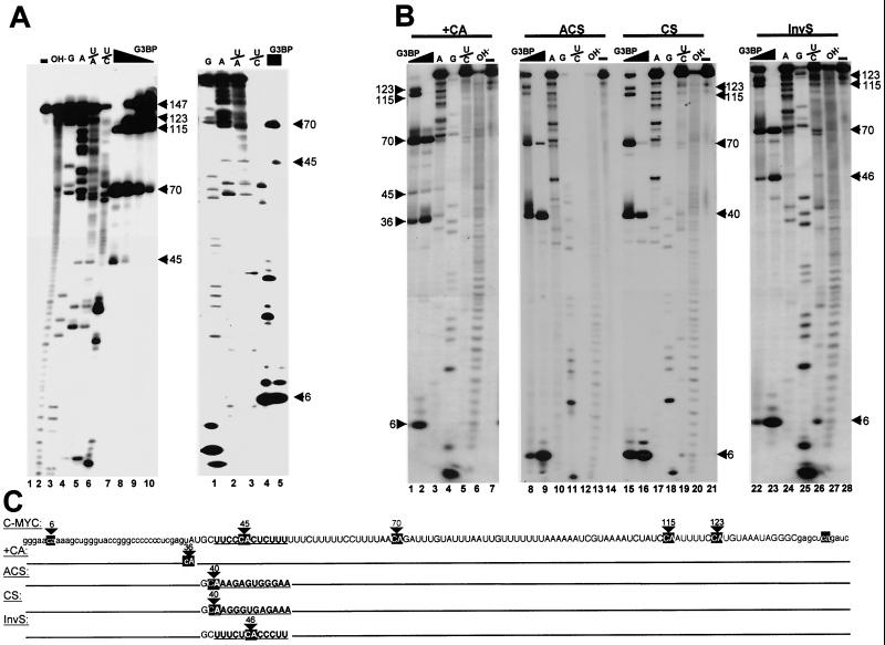
To elucidate the origin of G3BP degradation products, RNase assays were performed using a c-myc substrate labeled only at its 5′ end (Fig. 1A). The control reactions show the cleavage products generated by the following: (i) single-strand-specific endonucleases RNase T1 and RNase U2, which specifically cleave 3′-adjacent to G and A residues, respectively, (ii) RNase Phy M, which hydrolyses phosphodiester bonds 3′-adjacent to A and U residues (U/A), (iii) RNase B. cereus, which cleaves 3′ to pyrimidines (U/C) (left panel, lanes 3 to 6, and right panel, lanes 1 to 4). These controls demonstrated that G3BP cleaves the c-myc substrate exclusively at cytosines that are followed by adenines (left panel, lanes 7 to 10, and right panel, lane 5). However, cleavage was not equivalent at all CA phosphodiester bonds. A characteristic initial cleavage product was detected as a doublet of 123 and 115 nt at low protein concentration (left panel, lane 10). Higher concentrations of enzyme (left panel, lanes 7 to 9, and right panel, lane 5) resulted in the appearance of shorter fragments of 70, 45, and 6 nt, implying that most 3′ CA cleavage sites were preferred over 5′-terminal sites. Under this standard assay, 8 pmol of G3BP was sufficient to produce a complete digestion of 10 μg of the full-length substrate into these three fragments in 5 min at 20°C. This condition is hereafter referred to as 1 U of enzyme.
FIG. 1.
G3BP specifically cleaves wt and c-myc 3′ UTR mutants at CA sites. (A) 5′ 32P-labeled c-myc 3′ UTR transcripts (left panel, lane 1) were digested, using the RNA sequencing kit, with alkaline hydrolysis (lane 2), RNase T1 (cleaves 3′ to G residues) (lane 3), RNase U2 (cleaves 3′ to A residues) (lane 4), RNase Phy M (hydrolyses phosphodiester bonds 3′-adjacent to A and U residues [U/A]) (lane 5), RNase B. cereus (cleaves 3′ to pyrimidines U/C) (lane 6), or purified recombinant G3BP at concentrations of 8 pmol (lane 7), 4 pmol (lane 8), 2.4 pmol (lane 9), or 0.8 pmol (lane 10). Equivalent samples in lanes 3 to 7 were subjected to a short run electrophoresis (right panel, lanes 1 to 5, respectively). (B) 5′ 32P-labeled +CA (lanes 1 to 7), ACS (lanes 8 to 14), CS (lanes 15 to 21) and InvS (lanes 22 to 28) c-myc 3′ UTR mutants were either left unreacted (lanes 7, 14, 21, and 28) or digested with 2.4 pmol (lanes 1, 8, 15 and 22) or 8 pmol (lanes 2, 9, 16, and 23) of recombinant G3BP, RNase U2 (lanes 3, 10, 17, and 24), RNase T1 (lane 4, 11, 18, and 25), RNase B. cereus (lane 5, 12, 19, and 26), or alkaline hydrolysis (6, 13, 20, and 27). (C) Sequence of the c-myc 3′ UTR. Sequences from position 40 to 52 relative to the 5′ end which were mutated in ACS, CS, and InvS transcripts are noted in bold characters and underlined. The cleavage sites at CA dinucleotides are indicated by arrows. Nucleotides shown in lowercase characters are present in the transcription vector.
To confirm the preferential cleavage at the 3′ end of c-myc, we created a new CA site 36 nt from the 5′ end by a U-to-C conversion. As expected, higher concentrations of G3BP (1 U) were required to achieve efficient cleavage of this new site than with most downstream sites, which were cut extensively at lower concentrations (0.3 U) (Fig. 1B, compare lanes 1 and 2). Notably, the CA site 45 nt from the 5′ end constitutes an exception to this rule, as it was protected from cutting at any concentration of G3BP (lanes 1 and 2). Interestingly, the latter site was also protected in the wt c-myc 3′ UTR (Fig. 1A, lanes 7 to 10). Since this site is embedded in a sequence that could serve as a high-affinity G3BP binding site (see below), it is possible that this CA dinucleotide is blocked by direct binding of G3BP molecules or, alternatively, is engaged in an inaccessible structure. To distinguish between these two possibilities, we analyzed the effects of mutations that change sequences surrounding this cleavage site. We first replaced the sequence from position 40 to 52 relative to the 5′ end with its complementary sequence in both orientations (Fig. 1C, ACS and CS substrates). This destroys the authentic CA site but creates a new CA site five nucleotides upstream (Fig. 1B, lanes 8 to 21). These mutations were predicted to affect both secondary structure formation and/or G3BP binding, which could limit the accessibility of the cleavage site. The mutant substrates were cleaved with wt efficiency at all CA sites, including the new CA site at position 40, in both substrates (lanes 8 and 9 and lanes 15 to 16), suggesting that the cleavage site must be sufficiently exposed in a single-stranded conformation. To test whether strong binding of G3BP could also render the cleavage site inaccessible, sequences from position 40 to 52 relative to the 5′ end were replaced by the same sequence in the opposite orientation (Fig. 1C, InvS substrate), maintaining a CA site at position 46 but affecting the binding of G3BP (see below). This mutation restored cleavage to the new CA site (lanes 22 and 23), implying that strong binding of the endoribonuclease to a cleavage site may reduce its cleavage efficiency. Therefore, the specificity of G3BP is not determined solely by the site of cleavage but may also be modulated at the level of its binding to specific RNA sequences.
RNA binding is a prerequisite for cleavage at CA sites.
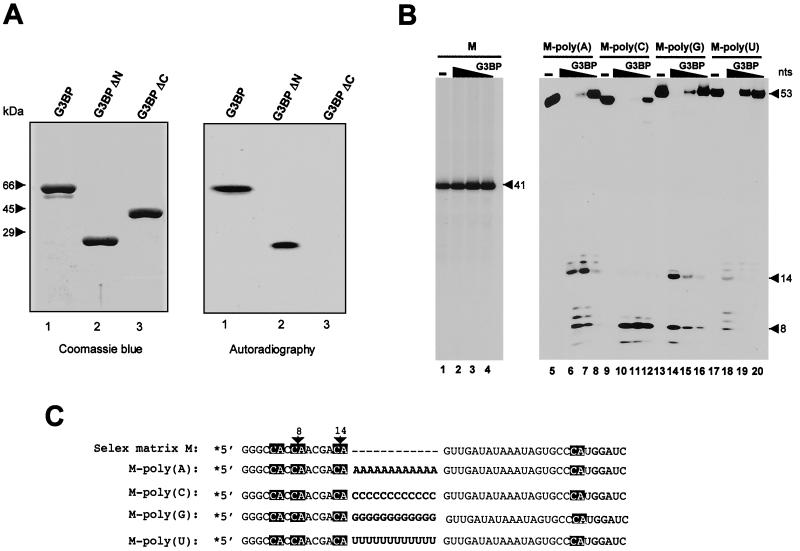
The determination of specific binding sequences for G3BP in solution proved to be elusive due to its high specific activity. We therefore immobilized the purified enzyme by Western blotting on nitrocellulose filters and tested its ability to bind radioactive RNA. As shown in Fig. 2A, the c-myc RNA probe readily bound to immobilized G3BP (right panel, lane 1), and no cleavage product was detected when the bound probe was eluted and analyzed by electrophoresis (data not shown), indicating that it is possible to uncouple the abilities of G3BP to bind and to degrade RNA. G3BP-RNA interaction likely involves the RRM and RGG boxes at the C terminus of the protein, since a truncated version of G3BP in which the C-terminal domain was selectively removed (G3BPΔC) left panel lane 3) failed to bind the c-myc RNA (right panel, lane 3). In contrast, N-terminal truncation of G3BP (G3BPΔN) left panel, lane 2), which removes as much as 299 amino acid residues, did not impair its RNA binding activity (right panel, lane 2). However, neither G3BPΔC nor G3BPΔN could degrade c-myc RNA under standard RNase assays, implying that the native structure of G3BP is required for its RNase activity (data not shown).
FIG. 2.
G3BP RNA binding activity and targeted cleavage by homopolymers. (A) Coomassie blue staining (left panel) of recombinant wt G3BP (lane 1), NH2-terminally truncated G3BP (lane 2), and COOH-terminally truncated G3BP (lane 3) purified after overexpression using a baculovirus system (see Materials and Methods). Right panel, autoradiography of Northwestern analysis of the same proteins, using 32P-labeled c-myc 3′ UTR as a probe. (B) 5′-end-labeled transcripts harboring constant regions either alone (M, lanes 1 to 4), or separated with 12 A residues [M-poly(A), lanes 5 to 8], 12 C residues [M-poly(C), lanes 9 to 12], 12 G residues [M-poly(G), lanes 13 to 16], or 12 U residues [M-poly(U), lanes 17 to 20] were digested with 8 pmol (lanes 2, 6, 10, 14, and 18), 2.4 pmol (lanes 3, 7, 11, 15, and 19), or 0.8 pmol (lanes 4, 8, 12, 16, and 20) of purified recombinant G3BP. (C) Sequences of the various transcripts. G3BP cleavage sites at CA dinucleotides are indicated by arrows. Homopolymers between constant regions are in boldface, and shading refers to empty region.
Further experiments established that the binding of G3BP to target RNA is a prerequisite for cleavage at CA sites. As shown in Fig. 2B, a 41-nt synthetic RNA that did not bind to immobilized G3BP (data not shown) was refractory to cleavage at all concentrations of G3BP (lanes 2 to 4), even though it contains four putative CA cleavage sites located 5, 8, 14, or 34 nt from the 5′ end (Fig. 2C, M substrate). This substrate, designed for SELEX experiments (see below), harbors two constant regions for primer annealing, which can be separated by randomized nucleotides. In order to test whether binding of G3BP was essential for cleavage, an initial experiment was carried out with substrates in which homopolymers of A, U, C, or G, ranging in size between 12 and 14 nt, were inserted between the constant regions [Fig. 2C, M-poly(A), M-poly(C), M-poly(G), and M-poly(U) substrates]. The abilities of these substrates to bind G3BP were confirmed by Northwestern analyses (data not shown).
The finding that all four substrates were cut by G3BP shows that these homopolymers were able to confer cleavage at CA sites contained in the constant regions (Fig. 2B, M-poly(A) [lanes 5 to 8], M-poly(C) [lanes 9 to 12], M-poly(G) [lanes 13 to 16], and M-poly(U) [lanes 17 to 20]). However, these substrates differed in their sensitivities to various concentrations of G3BP. Substrates containing poly(C) demonstrated strong sensitivity (lanes 10 to 12), since 100% of the full-length substrate was cleaved with 0.3 U of G3BP (lane 11) and 50% of initial cleavage products were detected with only 0.1 U of the enzyme (lane 12). Substrates harboring homopolymers of poly(A) or poly (G) (lanes 5 to 8 and 13 to 16, respectively) were less sensitive, since 1 U of G3BP was required to achieve complete cleavage (lanes 6 and 14) and only background cleavage was detected with 0.1 U of the enzyme (lanes 8 and 16). The substrate with poly(U) showed the weakest sensitivity, since more than 0.3 U of G3BP was necessary before cleavage products were observed (lanes 18 to 20). Furthermore, the substrate containing poly(C) was more frequently cut at the CA site 8 nt from the 5′ end (lanes 10 to 12), while the other substrates were cut equally well at sites located 8 and 14 nt from the 5′ end. Due to a heterogeneity at the 5′ end of M-poly(A) and M-poly(U) T7 transcripts, additional bands with sizes greater than 8 and 14 nt were detected (lanes 6 to 8 and lanes 18 to 20). The reason for differences in sensitivities of the various substrates to G3BP cleavage is currently unknown. It could be attributed to secondary and/or tertiary structure that the various homopolymers might form with the cleavage sites. The possibility that the effects of homopolymers were due to spacing between the 5′ and 3′ constant regions of the M substrate, rather than direct binding of G3BP, can be discounted because the M-ACS substrate (see below), which created exactly the same spacing between these two regions, was refractory to cleavage due to an absence of G3BP binding. Taken together, these results indicate that extended regions like homopolymers could serve as potential binding sites for G3BP and thereby confer cleavage to proximal CA sites.
Substrate specificity of G3BP.
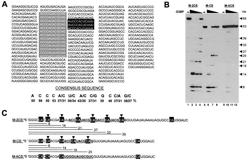
In order to study the RNA-binding specificity of G3BP, we performed an iterative in vitro genetic selection (SELEX) from a pool of random sequences (45). Full-length G3BP protein immobilized on nitrocellulose filters was used as a selection matrix. In vitro selection was carried out using a large molar excess of a pool of 55-nt synthetic RNA molecules containing a randomized region of 12 nt flanked by constant regions for primer annealing as described above. After five cycles of selection-amplification, cDNA fragments corresponding to selected RNAs were cloned and the internal sequences of 105 independent clones were determined. Sequence alignment, using the ClustalW program, allowed us to design a best-guess high-affinity G3BP consensus site: ACCCA(U/C)(A/C)(C/G)G(C/A)AG (Fig. 3A). Interestingly, six independent clones perfectly matched the consensus sequence (ACCCAUACGCAG), and seven differed by only one or two nucleotides. As mentioned above, sequences that are present in the 3′ UTR of c-myc (UCCCACUCUUU) were also found among G3BP-selected RNAs, indicating that G3BP high-affinity binding sites could occur within known mRNA sequences. This possibility was further confirmed by performing a BLAST search in the genomic database with selected G3BP-consensus sequences, and mRNAs containing these sequences are shown in Table 1.
FIG. 3.
In vitro selection and amplification of high-affinity RNA target sequences for G3BP. (A) The sequences of individual clones after five cycles are shown. A consensus sequence was derived from the score of each nucleotide in the randomized sequence. Sequences that match the deduced consensus or deviate by 1 or 2 nt are boxed in grey. Sequences that are present in the c-myc 3′ UTR are boxed in black. (B) 5′-end-labeled transcripts containing constant regions separated by two copies (M-2CS, lanes 1 to 4), or one copy (M-CS, lanes 5 to 8) of the consensus sequence or its complementary sequence (M-ACS, lanes 9 to 12) were digested with 8 pmol (lanes 2, 6, and 10), 2.4 pmol (lanes 3, 7, and 11), or 0.8 pmol (lanes 4, 8, and 12) of purified recombinant G3BP. (C) Sequences of the various transcripts. The consensus sequence and its complementary sequence are noted in bold characters and underlined. G3BP cleavage sites at CA dinucleotides are indicated by arrows.
TABLE 1.
cDNAs that contain G3BP SELEX consensus sequencea
| Accession no. | species | Protein | Position in mRNA |
|---|---|---|---|
| AF001691 | Homo sapiens | Cornified envelope precursor | Coding sequence |
| AB018254 | Homo sapiens | KIAA0711 | 3′ UTR |
| U22526 | Homo sapiens | 2,3-oxidosqualene-Ianosterol cyclase | 3′ UTR |
| NM-001261 | Homo sapiens | Cyclin-dependent kinase 9 | Coding sequence |
| NM-002340 | Homo sapiens | Lanosterol synthase | 3′ UTR |
| NM-002350 | Homo sapiens | v-yes-1 Yamaguchi sarcoma virus-related oncogene homolog (LYN) | 5′ UTR |
| NM-001874 | Homo sapiens | Carboxypeptidase M (CPM) | 5′ UTR |
| NM-003678 | Homo sapiens | NF2/meningioma region of 22q12 (PK1.3) | Coding sequence |
| NM-005415 | Homo sapiens | Solute carrier family 20 (phosphate transporter), member 1 | 5′ UTR |
| M16038 | Homo sapiens | Lyn tyrosine kinase | 5′ UTR |
| AF102059 | Homo sapiens | Gibbon ape leukemia virus receptor 1 | 5′ UTR |
| AB042027 | Mus musculus | Goblin mRNA for Golgi-associated band 4.1-like protein | Coding sequence |
| NM-016850 | Mus musculus | Interferon regulatory factor 7 (Irf7) | Coding sequence |
| L33415 | Mus musculus | Integral membrane protein (Nramp2) | Coding sequence |
| AB036882 | Mus musculus | Midnolin | 5′ UTR |
| X60672 | Mus musculus | Radixin | 3′ UTR |
| U28775 | Mus musculus | Odorant receptor gene | Coding sequence |
| NM-012610 | Rattus norvegicus | Nerve growth factor receptor | Coding sequence |
A perfect match to the G3BP consensus sequence (ACCCAUACGCAG) was present in 488 vertebrate-expressed sequence tags and in 159 independent clones of vertebrate genomic sequences using BLAST, on the NCBI server (S. F. Altschul et al., 1997; http://www.ncbi.nlm.nih.gov). mRNAs from human, mouse, and rat containing the SELEX consensus sequence are shown. Sequence accession numbers are for GenBank.
To assess the specificity of binding and cleavage of these sequences by G3BP, radiolabeled RNA probes containing either one (Fig. 3C, M-CS substrate) or two (Fig. 3C, M-2CS substrate) tandemly repeated consensus ACCCAUACGCAG sequences were tested by RNase assays. Note that M-2CS has two additional Us at the beginning of the first consensus sequence. As shown in Fig. 3B, M-2CS was a better substrate than M-CS, since 0.3 U of G3BP was sufficient to cleave most of the input labeled M-2CS RNA while 50% of input M-CS RNA remained undegraded (compare lanes 3 and 7). As expected, cleavage occurred exclusively at CA sites (Fig. 3C). Notably, the first CA site within the consensus sequence of the M-CS substrate was less frequently cleaved than other CA sites in this RNA (cleavage at this site gave rise to the 19-nt fragment), implying that the binding of G3BP to its cognate sequence may prevent cleavage at this site. Interestingly, no preferential cleavage was observed with the M-2CS substrate, which contained two possible G3BP binding sites. All the CA sites were cleaved at the same frequency, showing that the intermittent binding of G3BP to either one of the consensus sequences may be responsible for the efficient cleavage of the M-2CS substrate. Again, binding of G3BP to its substrate was a prerequisite for cutting, as demonstrated by the finding that replacement of the consensus sequence by its antisense sequence in the M-CS RNA (M-ACS substrate) rendered this substrate refractory to cleavage (lanes 10 to 12). Even at higher concentrations of G3BP (1 U), only background cleavage was detected (lane 10). Taken together, the results indicate that site-specific cleavage at CA dinucleotides depends on a specific interaction between G3BP and its substrate.
G3BP forms a stable complex with poly(A)+ mRNAs in both RasGAP+/− and RasGAP−/− fibroblasts.
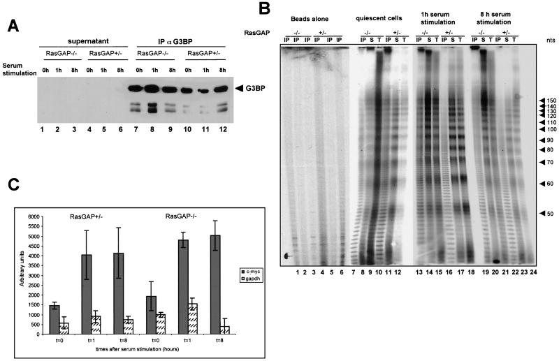
The results shown above demonstrate that G3BP interacts and cleaves the 3′ UTR of c-myc, in vitro, and as such might initiate mRNA turnover. Since c-myc mRNA is also decayed by rapid deadenylation (9, 10), we wished to determine whether, in vivo, G3BP associates with RNAs containing poly(A) tails. To address this issue, RNAs interacting with G3BP were recovered by immunoprecipitation from total extracts prepared from quiescent or dividing MEFs using G3BP-specific antibodies. Given the previously demonstrated association between G3BP and p120RasGAP during cell proliferation (18, 33), we used RasGAP+/ −and RasGAP−/− MEFs isolated from RasGAP mutant embryos at day 9.5 of development (46). The RasGAP+/− cells have the same genetic background as RasGAP−/− cells and therefore served as controls for cells expressing RasGAP. Western blot analysis showed that G3BP was efficiently depleted from both RasGAP+/− and RasGAP−/− extracts following immunoprecipitation (Fig. 4A, lanes 7 to 12). G3BP was not detected in the supernatants (lanes 1 to 6). Coimmunoprecipitated RNAs from these cells were extracted and treated with RNase T1 and RNase A, and the resulting resistant poly(A) tails were labeled at their 5′ end with [γ-32P]ATP and T4 polynucleotide kinase. Labeled poly(A) oligomers were then fractionated on a 12% polyacrylamide–urea denaturing gel and detected by autoradiography. Control experiments with total RNA contained in the extracts showed a characteristic pattern of poly(A) oligomers differing in length by roughly 10 to 12 nt (Fig. 4B, lanes 9, 12, 15, 18, 21, and 24). The same ladder was also detected with RNAs coimmunoprecipitated with G3BP (lanes 7, 10, 13, 16, 19, and 22). In contrast, no poly(A) oligomers were detected with the corresponding amount of immunoprecipitates of protein G without antibody that were previously incubated with the same extracts (lanes 1 to 6), indicating that cellular RNAs associated with G3BP show heterogeneous lengths of their poly(A) tracts. Interestingly, comparison of the poly(A) oligomers in adjacent lanes of the same gel showed no obvious difference between repeat lengths for total RNAs from quiescent or serum-stimulated RasGAP−/− cells (lanes 9, 15, and 21) and RasGAP+/− cells (lanes 12, 18, and 24).
FIG. 4.
Cellular RNAs associated with G3BP. (A) G3BP was affinity purified by an anti-G3BP antibody immobilized on protein G-Sepharose, from total extracts of quiescent RasGAP−/− (lane 7) and RasGAP+/− (lane 10) cells or following serum stimulation for 1 h (lanes 8 and 11, respectively) or 8 h (lanes 9 and 12, respectively). Immunoprecipitates and corresponding supernatants (lanes 1 to 6) were analyzed by immunoblotting using anti-G3BP antibodies. (B) The poly(A) tails of RNAs from immunoprecipitates (IP) (lanes 7, 10, 13, 16, 19, and 22), supernatant (S) (lanes 8, 11, 14, 17, 20, and 23), and total extracts (T) (lanes 9, 12, 15, 18, 21, and 24) of quiescent RasGAP−/− (lanes 7 to 9) and RasGAP+/− (lanes 10 to 12) cells or those which were serum stimulated for 1 h (lanes 13 to 15 and lanes 16 to 18, respectively) or 8 h (lanes 19 to 21 and 22 to 24, respectively) were determined as described in Materials and Methods. (C) RNAs associated with affinity-purified G3BP from RasGAP−/− and RasGAP+/− quiescent cells or those cells which were serum stimulated for 1 and 8 h were extracted. The c-myc (grey bars) and GAPDH (white dotted bars) mRNAs from immunoprecipitates were detected by RT-PCR using specific primers. The amplified products were analyzed on an agarose gel, visualized by ethidium bromide, and quantified by fluorography. Background RT-PCR amplification from immunoprecipitates with beads alone in the absence of anti-G3BP antibodies was subtracted from the plotted values. Error bars resulting from two independently performed experiments, each measured in triplicate, are shown.
To determine whether G3BP-containing complexes also contained c-myc mRNA, RNA recovered in these complexes were analyzed by RT-PCR using oligo(dT) for priming the first-strand DNA synthesis and specific primers to either c-myc or GAPDH mRNAs. The results in Fig. 4C clearly show that c-myc, but not GAPDH, was among the mRNAs efficiently selected by G3BP from serum-stimulated RasGAP+/− and RasGAP−/− cells. However, both c-myc and GAPDH mRNAs were detected in the immunoprecipitation supernatants (data not shown), indicating that only a fraction of c-myc mRNA was associated with G3BP.
Decay of c-myc mRNA in RasGAP+/− and RasGAP−/− fibroblasts.
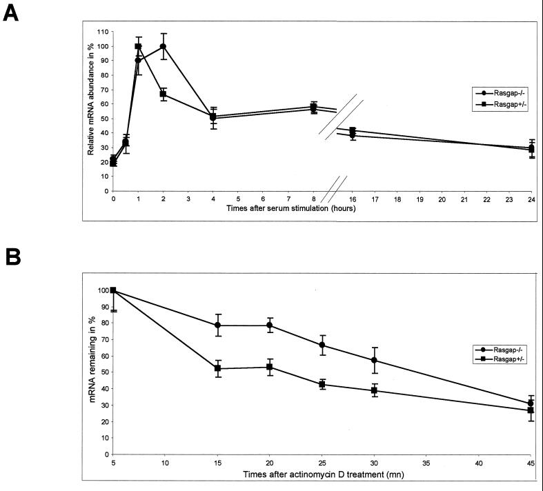
To analyze c-myc mRNA levels in the various cultures, cells were starved by serum deprivation for 24 h, RNAs were isolated after different times of serum stimulation, and the mRNA levels were determined by quantitative real-time RT-PCR amplification using the LightCycler system (Roche Molecular Biochemicals). By real-time PCR analysis, the PCR product was measured as it accumulated, allowing accurate quantification of mRNA levels without the ambiguities associated with traditional RT-PCR. The data, averaged from three independent experiments and normalized to an internal GAPDH control, are depicted in graph form in Fig. 5A. The maximum signal in each case was considered 100%. c-myc mRNA levels in RasGAP+/− cells were maximal after 1 h of serum stimulation and decayed between 1 and 16 h (Fig. 5A). Interestingly, the levels of c-myc mRNA measured after 2 h of serum stimulation of RasGAP−/− cells did not decrease and were two fold higher than in RasGAP+/− cells. This demonstrates that RasGAP depletion affects the c-myc mRNA levels during the initial phase of serum stimulation.
FIG. 5.
(A) c-myc mRNA expression in RasGAP+/ −and RasGAP−/− cells. After 24 h of serum starvation, expression was stimulated by serum addition for the indicated number of hours. RNA isolated from cells harvested at each time point was reverse transcribed and assayed for c-myc and GAPDH cDNAs using real-time quantitative PCR. All values were scaled to the level obtained from GAPDH, considered as a constitutively expressed mRNA. One hundred percent RNA is arbitrarily assigned to the time point which gave the highest signal. (B) Comparison of c-myc mRNA decay in RasGAP+/− and RasGAP−/− cells. Quiescent cells were stimulated with serum for 1 h and treated with 5 μg of actinomycin D/ml. At various times thereafter, cells were harvested and total RNA was prepared. The level of c-myc mRNA was quantified as in panel A and plotted. Error bars resulting from three independent experiments, each measured in duplicate, are shown.
To determine if c-myc mRNA is destabilized more rapidly during this early stimulation period in RasGAP+/− cells compared to RasGAP−/− cells, quiescent cells were stimulated with serum for 1 h and then exposed to actinomycin D to inhibit transcription. RNAs were isolated at various times after inhibition of transcription, and the levels of c-myc mRNA were measured and normalized to GAPDH mRNA levels using RT followed by real-time PCR analysis. Each time point was repeated three times, and the quantified data are presented graphically in Fig. 5B. Given that all mRNA survival curves, plotted as a function of actinomycin D chase time, display a short lag period of 5 min before the onset of decay, the maximum signal obtained at 5 min of actinomycin D chase was considered 100%. As previously observed with other cell lines, the c-myc mRNA appeared to be extremely unstable in RasGAP+/− cells, with an apparent half-life of 15 min, whereas in RasGAP−/− cells the c-myc mRNA decay was delayed, with an apparent half-life of 35 min. This finding agrees with steady-state mRNA measurements and is consistent with the hypothesis that RasGAP modulates the level of c-myc mRNA degradation, presumably via G3BP, during the initial period following growth factor stimulation. The observation that c-myc mRNA in both RasGAP+/− and RasGAP−/− cells decreased to the same steady-state levels after 4 h of serum stimulation also indicated that the absence of RasGAP did not prevent massive degradation of c-myc mRNA.
RasGAP−/− cells harbor a phosphorylation-deficient isoform of G3BP.
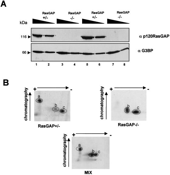
Since the stability of c-myc mRNA was different between RasGAP+/− and RasGAP−/− cells, we next determined whether the absence of p120 RasGAP had any effect on G3BP expression and/or phosphorylation. Western blotting (Fig. 6A) confirmed the absence of expression of RasGAP in cells that were homozygous for the RasGAP mutation (lanes 3, 4, 7, and 8), whereas the protein was expressed in heterozygous cells (lanes 1, 2, 5, and 6). Notably, all cells were found to contain similar levels of G3BP (lanes 1 to 8), implying that the deletion of the RasGAP gene did not affect G3BP expression. The possibility that the G3BP gene was mutated in RasGAP−/− cells was also excluded, as sequencing of the entire cDNA with specific primers did not reveal any mutation (data not shown).
FIG. 6.
(A) Proteins contained in total extracts derived from two cultures of exponentially growing RasGAP+/− (lanes 1, 2, 5, and 6) and RasGAP−/− (lanes 3, 4, 7, and 8) cells were analyzed by immunoblot using anti-G3BP and anti-p120 GAP antibodies. Amounts of loaded proteins were 15 μg (lanes 2, 4, 6, and 8) and 30 μg (lanes 1, 3, 5, and 7). (B) Phosphotryptic peptide mapping of immunopurified 32P-labeled G3BP from dividing RasGAP+/− (left panel) and RasGAP−/− (right panel) cells. The bottom panel represents the analysis of the mixture of the samples in both panels.
Previously, we showed that phosphorylation levels of G3BP were modified during proliferation and that phosphorylation was a critical parameter for G3BP RNase activity in vitro. We therefore compared the phosphorylation status of G3BP in RasGAP+/− and RasGAP−/− cells. Proteins were metabolically labeled with [32P]orthophosphate, immunopurified with anti-G3BP antibody, and subjected to phosphotryptic peptide mapping. In agreement with previous work of members of our group (18), three labeled phosphopeptides characterized the tryptic pattern of G3BP from RasGAP+/− cells (Fig. 6B). Two of these peptides were identical to those forming the trypsin digestion pattern of labeled immunopurified G3BP from RasGAP−/− cells (Fig. 6B), but one phosphopeptide was absent. An additional difference between the two patterns was the level of phosphopeptide b phosphorylation (Fig. 6B). This phosphopeptide was more highly phosphorylated in RasGAP−/− cells than in RasGAP+/− cells.
To determine the precise phosphorylation sites, radioactive phosphopeptides from RasGAP+/− cells were purified by reverse-phase high-pressure liquid chromatography (see Materials and Methods) and subjected to amino acid sequencing. This analysis established that peptides spanning residues 133 to 159 and 230 to 247 contained the radioactive residues identified as Ser-149 and Ser-232, respectively. Using the baculovirus system, expression of recombinant proteins, which were mutated in either serine, allowed us to confirm that phosphopeptides a and c corresponded to phosphorylation at serines 149 and 232, respectively (Tourrière et al., unpublished results). However, due to contamination with other unlabeled peptides, it was not possible to unambiguously map the site of phosphorylation of phosphopeptide b. Altogether, these data demonstrate that RasGAP−/− cells harbor a G3BP isoform in which Ser-149 is not phosphorylated.
Glutamate substitution at position Ser-149 induces translocation of G3BP from the cytoplasm to the nucleus.
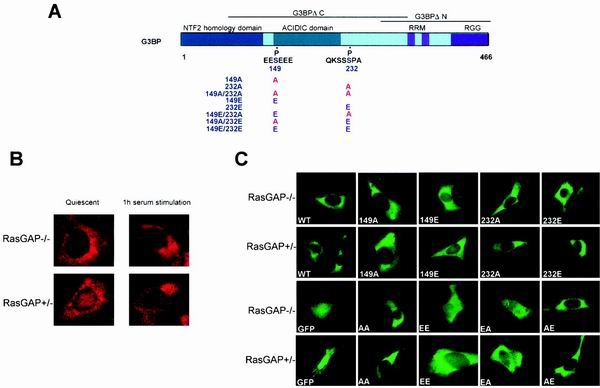
Ser-149 is located at the C-terminal end of the NTF2 homology domain, a specific domain that could behave as a nuclear transport carrier (see Discussion) mediating the cellular localization of G3BP. To test whether phosphorylation of Ser-149 affects G3BP cellular localization, we first performed immunolocalization studies using monoclonal anti-G3BP antibodies. Given that G3BP was hyperphosphorylated on serine residues in resting cells compared to proliferating cells (18), we assessed whether the presence or absence of serum led to changes in the cellular distribution of G3BP and whether the absence of RasGAP would have any effect on this distribution. In contrast to the exclusively cytoplasmic localization of G3BP in proliferating RasGAP+/− cells (Fig. 7B, lower right panel), a fraction of G3BP localized to the nucleus of RasGAP+/− resting cells (Fig. 7B, lower left panel). However, no changes in G3BP distribution were observed between serum-stimulated and serum-starved RasGAP−/− cells (Fig. 7B, upper left and right panels), suggesting that the absence of phosphorylation at Ser-149 is detrimental for G3BP nuclear translocation.
FIG. 7.
(A) Schematic representation of G3BP structural domains: NTF2 homology domain (in blue), acidic domain (in green), and RRM domain (in pink). The positions of the major phosphorylation sites are indicated. (B) Indirect immunofluorescent staining of quiescent and serum stimulated RasGAP−/− (top panels) or RasGAP+/− (bottom panels) cells with anti-G3BP antibodies. (C) Cellular localization of GFP fusion proteins in RasGAP+/− and RasGAP−/− cells. Direct fluorescence of GFP (GFP), GFP-G3BP (WT), GFP-S149A (149A), GFP-S149E (149E), GFP-S232A (232A), GFP-S232E (232E), and GFP-double mutants (EA, EE, AE, and AA) were performed 20 h after transfection. Expression of fusion proteins was confirmed by immunoblot analysis using an anti-GFP antibody (data not shown).
To test this hypothesis more directly, the GFP was fused in frame to the amino terminus of wt G3BP or any of several phosphorylation mutants (Fig. 7A). Ser-149 and Ser-232 were mutated to either alanine (S149A and S232A) in order to destroy the phosphorylation site or to glutamate (S149E and S232E) to mimic the charge of the phosphorylated serine residue. Both single- and double-mutant constructs were generated, and the resultant fusion proteins were transiently expressed in either RasGAP+/− or RasGAP−/− proliferating cells. These fusion proteins were localized using direct GFP fluorescence (Fig. 7C), and the expression of full-length proteins was confirmed by Western blotting using anti-GFP antibodies (data not shown). In contrast to the ubiquitously expressed GFP (GFP panels), GFP-G3BP corresponding to wt G3BP fused to GFP was clearly present throughout cytoplasm (WT panels), similar to the endogenous cellular G3BP in proliferating cells (Fig. 7B, right panels). However, no difference in the distribution of GFP-G3BP was observed between RasGAP+/− and RasGAP−/− cells, confirming that the absence of phosphorylation of Ser-149 did not affect the cytoplasmic localization of G3BP. Accordingly, GFP-G3BP(S149A) had the same cellular distribution as GFP-G3BP (149A panels). In sharp contrast, GFP-G3BP(S149E) displayed a rather broader distribution in both the cytoplasm and the nucleus (149E panels). The effect of glutamate substitution was specific for Ser-149, because the same mutation of Ser-232 (GFP-G3BP(S232E)) did not change G3BP distribution compared to GFP-G3BP or GFP-G3BP(S232A) (panels 232A and 232E). Furthermore, all double mutants that harbored the S149E mutation translocated to the nucleus (panels EE and EA), indicating that the phosphorylation of Ser-149 is important for G3BP nuclear entry. These findings are in complete agreement with the nuclear localization of G3BP in resting RasGAP+/− cells, which accumulate a hyperphosphorylated isoform of G3BP on serine residues (18). Unlike bona fide phosphorylation, the charge of glutamate substitution at Ser-149 is not sensitive to dephosphorylation, and thus the cellular distribution of S149E mutants was affected neither by serum stimulation nor by the presence or absence of RasGAP.
DISCUSSION
Despite numerous reports implicating hormonal and other extracellular stimuli in the activation of vertebrate mRNA decay by endonucleolytic cleavage, the enzymes responsible for these cleavages are still largely unidentified (40). G3BP, initially discovered through its direct interaction with the SH3 domain of RasGAP (33), was shown to harbor a phosphorylation-dependent endoribonuclease activity (18). The enzyme is only active on RNAs that contain its target sequences, and it exclusively cleaves between CA dinucleotides. Our data suggest that the recognition of specific sequences by G3BP serves as an entry point for the enzyme, which can then act in a processive manner to cleave multiple CA sites within the same molecule. Targeting G3BP to the nucleus via phosphorylation of Ser-149 could be the mechanism by which G3BP integrates into assembling mRNP complexes in the nucleus and degrades its target mRNAs. In support of this hypothesis, the absence of such phosphorylation in RasGAP-deficient cells correlates with a delay in degradation of c-myc mRNA, a cellular mRNA whose 3′ UTR contains a high-affinity G3BP binding site. Thus, G3BP has unique characteristics required for selective mRNA degradation: it can be targeted to specific sequences through its RRM domain and accumulates in the nucleus following RasGAP signaling-dependent phosphorylation of Ser-149.
The specificity of G3BP is different from that of a 65-kDa human site-specific single-strand endoribonuclease that functionally resembles Escherichia coli RNase E (48). While both G3BP and the 65-kDa protein have similar eletrophoretic mobilities on an SDS-polyacrylamide gel and cleave the 3′ UTR of c-myc mRNA in vitro, the 65-kDa protein-mediated cleavage occurs at several positions that resemble RNase E cleavage sites with no preference for a single sequence motif (48), thus demonstrating a substrate specificity different from that of G3BP. Furthermore, the 65-kDa protein cross-reacted with RNase E antibodies, indicating that it shares a conserved epitope with RNase E. In contrast, G3BP has no obvious sequence homology to RNase E, except that both proteins have domains that mediate interaction with RNA. Indeed, both G3BP and RNase E were demonstrated to bind RNA by Northwestern blots (14).
G3BP RNase activity and c-myc mRNA decay.
Many mRNAs in mammalian cells decay via a sequential pathway involving rapid conversion of polyadenylated molecules to a poly(A)-deficient state, followed by rapid degradation of the poly(A)-deficient molecules (10, 21, 42, 47). A central focus of these cell-cycle-regulated mRNA turnover pathways could be the initiation of degradation by an endonucleolytic cleavage, usually in the 3′ UTR of target transcripts. The ability of G3BP to cleave at several CA sites in the c-myc 3′ UTR, which is known to mediate rapid mRNA decay (22), suggests that G3BP might be a component of an mRNA degradation system that controls normal cell growth and differentiation. Both in vitro and in vivo data showed that c-myc decays by a sequential pathway involving deadenylation followed by degradation of the mRNA body, generating easily observed 3′-terminal decay intermediates (8). Strikingly, the major 3′-terminal decay product detected in these studies was generated by cleavage at a CA site in the 3′ UTR of c-myc mRNA, and the enzymatic activity responsible for this cleavage was, like G3BP, associated with polysomes (8). Also, G3BP preferentially cleaved 3′-end CA sites rather than 5′-end sites in the mouse c-myc 3′ UTR (Fig. 1).
However, the 3′ UTR is not the sole determinant of the half life of c-myc mRNA, and G3BP is unlikely to be the only endoribonuclease involved in c-myc mRNA decay. The coding region of c-myc mRNA contains a 180- to 320-nt purine-rich segment (CRD) that interacts with a protein that shields it from endonucleolytic cleavage (5, 34). The endonuclease that attacks this region is also tightly bound to polysomes but, unlike G3BP, is magnesium dependent (28). Given that the CRD is an mRNA instability element independent of other c-myc regions, it is possible that c-myc mRNA is degraded through a variety of mechanisms to ensure proper steady-state levels. The existence of alternative pathways for degradation of c-myc mRNA would be to provide redundancy in the event of inactivation of one pathway, because of mutation, for example. While there is still no direct proof of G3BP acting as an endoribonuclease on c-myc mRNA in vivo, mutation of the RasGAP gene, which affects both the phosphorylation status and localization of G3BP, leads to a twofold increase in the c-myc mRNA half-life. Furthermore, immunoprecipitation experiments conclusively demonstrate (Fig. 4C) that a fraction of c-myc mRNA is tightly associated with G3BP. Since G3BP requires binding for cleavage (in vitro data), our findings would argue that c-myc mRNA is one of its physiologically meaningful substrates. Other activities may be operative in RasGAP−/− cells besides G3BP-dependent mRNA degradation which prevent a larger increase in the c-myc mRNA half life. Alternatively, absence of phosphorylation at Ser-149 in RasGAP−/− cells may be less inhibitory to G3BP activity in vivo than in vitro.
RNA binding specificity of G3BP and mRNA-binding proteins that influence mRNA stability.
The protein-RNA interaction observed between G3BP and its target RNA sequence is of considerable importance in regulating mRNA decay rates. The RRM-type RNA binding domain in G3BP may compete with the binding of several mRNA binding proteins that shield these messages from endonucleolytic attacks (36, 37). The poly(A) binding protein is one of these mRNAs, protecting proteins that bind to poly(A) tails and prevent mRNA degradation (3, 4). While the mechanism by which poly(A) binding protein exerts its effect on mRNA stability is not fully understood, it may be via the recruitment of proteins that interfere with mRNA degradation. Therefore, it is significant that mRNAs associated with G3BP have poly(A) tails (Fig. 4).
The observation that phosphorylation significantly influences G3BP cellular localization (see below), together with the finding that phosphorylation of G3BP is affected by RasGAP gene depletion, strongly suggest that this modification influences the role of G3BP in mediating mRNA decay. Indeed, G3BP could translocate into the nucleus following phosphorylation of Ser-149, increasing its local concentration and thus competing with the binding of proteins which could inhibit mRNA degradation. While direct evidence for such a competition model is lacking, it is significant that G3BPΔN, which has the ability to bind the same RNA sequences as wt G3BP (Fig. 2), inhibits the in vitro cleavage by wt G3BP (Tourrière et al., unpublished results). Thus, G3BP phosphorylated at Ser-149 may initially bind its target mRNA in the nucleus and compete with RNA binding proteins that provide ongoing protection from the degradation in the cytoplasm. Absence of Ser-149 phosphorylation in RasGAP-deficient cells would then be expected to be detrimental for RNA degradation control and could give a rationale for the delay in c-myc mRNA decay.
G3BP localization and relationship to RasGAP.
By using the covalent cross-linking reagent 1-ethyl-3(dimethylamine)-propyl carbodiimide, we previously showed that in vivo, there is a direct interaction of RasGAP and G3BP in dividing but not quiescent cells (18). We have also found that the complex between RasGAP and G3BP is localized exclusively in the subcellular particulate fraction of dividing cells, whereas free G3BP and RasGAP are primarily cytosolic. This, together with the finding that the level of G3BP phosphorylation changes during the transition from resting to dividing cells, suggests that RasGAP could play a role in changing the phosphorylation status of G3BP. Consistent with this idea, G3BP from RasGAP−/− cells is not phosphorylated at Ser-149, a modification which seems to modulate G3BP subcellular distribution. The mechanism by which RasGAP maintains the phosphorylation of Ser-149 is currently unclear. The interaction with the SH3 domain of RasGAP could either prevent, by steric hindrance, the action of a phosphatase that constitutively dephosphorylates this residue, or it could recruit G3BP to a specific kinase. We favor the latter possibility because phosphorylation of Ser-149 is maintained in quiescent cells even though G3BP is not associated with RasGAP in this state (18). However, we cannot exclude other indirect effects caused by the absence of RasGAP in the RasGAP−/− cells, such as aberrant Ras regulation, reduced p190 tyrosine phosphorylation (46), and the presence or absence of factor(s) that associate with G3BP.
G3BP is a stable protein whose expression does not vary during the cell cycle (18) or following RasGAP gene deletion, (Fig. 6A) but, as demonstrated here, it is posttranslationally modified by phosphorylation. This modification concerns serine residues that appear to carry the information required to modulate, at least in vitro, the RNase activity of G3BP (18) and its localization in the cell. We have mapped two phosphorylation sites and shown by site-directed mutagenesis that the highly conserved Ser-149 residue plays a critical, but possibly not exclusive, role in the localization of G3BP. Recent studies indicate that nuclear localization signal (NLS) function can be precisely regulated, with phosphorylation being the main mechanism controlling NLS-dependent nuclear import of a number of proteins (24, 25). In particular, the rate of nuclear import of the prototypical NLS-containing simian virus large tumor antigen (T-ag) is modulated by a CKII site 13 amino acids N-terminal of the NLS (26). Although no putative NLS can be found in the G3BP sequence, it is striking that the Ser-149 is present within a CK II consensus site. This phosphorylation site is 20 amino acids C-terminal to the NTF2 homology domain, a putative domain that is expected to play a key role in nuclear transport (20, 50). Indeed, NTF2 is a nuclear transport carrier that mediates the uptake of cytoplasmic RanGDP into the nucleus and is therefore essential for maintaining an appropriate concentration of Ran across the nuclear envelope (35). The NTF2-like domain of G3BP could similarly bind factors at the nuclear pore, which would eventually facilitate its nuclear import. The phosphorylation of Ser-149 could either change the conformation of G3BP to render the NTF2-like domain accessible or prevent the binding of a cytoplasmic factor(s) that mask this domain. Biochemical studies to determine which step of nuclear transport is affected by phosphorylation of Ser-149 are under way.
ACKNOWLEDGMENTS
H.T., I.G., and K.C. contributed equally to this work.
We are grateful to Edouard Bertrand, George Lutfalla, and Gilles Uzé for technical assistance and helpful discussions. We thank N. Taylor, B. Hipskind, and J. Soret for pertinent comments on the manuscript.
This work was supported by Association pour la Recherche sur le Cancer (ARC) and the CNRS. Special thanks go to Fabienne Parker for purified recombinant proteins. H.T. was supported by graduate fellowships from the Ministère de l' Education Nationale, de la Recherche et de la Technologie (MENRT) and ARC. K.C. and I.G. were supported by fellowships from Rhône Poulenc Rorer.
REFERENCES
- 1.Bashkirov V I, Scherthan H, Solinger J A, Buerstedde J M, Heyer W D. A mouse cytoplasmic exoribonuclease (mXRN1p) with preference for G4 tetraplex substrates. J Cell Biol. 1997;136:761–773. doi: 10.1083/jcb.136.4.761. [DOI] [PMC free article] [PubMed] [Google Scholar]
- 2.Belasco J, Brawerman G. Control of messenger RNA stability. San Diego, Calif: Academic Press; 1993. [Google Scholar]
- 3.Bernstein P, Peltz S W, Ross J. The poly(A)-poly(A)-binding protein complex is a major determinant of mRNA stability in vitro. Mol Cell Biol. 1989;9:659–670. doi: 10.1128/mcb.9.2.659. [DOI] [PMC free article] [PubMed] [Google Scholar]
- 4.Bernstein P, Ross J. Poly(A), poly(A) binding protein and the regulation of mRNA stability. Trends Biochem Sci. 1989;14:373–377. doi: 10.1016/0968-0004(89)90011-x. [DOI] [PubMed] [Google Scholar]
- 5.Bernstein P L, Herrick D J, Prokipcak R D, Ross J. Control of c-myc mRNA half-life in vitro by a protein capable of binding to a coding region stability determinant. Genes Dev. 1992;6:642–654. doi: 10.1101/gad.6.4.642. [DOI] [PubMed] [Google Scholar]
- 6.Boyle W J, van der Geer P, Hunter T. Phosphopeptide mapping and phosphoamino acid analysis by two-dimensional separation on thin-layer cellulose plates. Methods Enzymol. 1991;201:110–149. doi: 10.1016/0076-6879(91)01013-r. [DOI] [PubMed] [Google Scholar]
- 7.Brewer G. Regulation of c-myc mRNA decay in vitro by a phorbol ester-inducible, ribosome-associated component in differentiating megakaryoblasts. J Biol Chem. 2000;275:33336–33345. doi: 10.1074/jbc.M006145200. [DOI] [PubMed] [Google Scholar]
- 8.Brewer G. Characterization of c-myc 3′ to 5′ mRNA decay activities in an in vitro system. J Biol Chem. 1998;273:34770–34774. doi: 10.1074/jbc.273.52.34770. [DOI] [PubMed] [Google Scholar]
- 9.Brewer G. Evidence for a 3′-5′ decay pathway for c-myc mRNA in mammalian cells. J Biol Chem. 1999;274:16174–16179. doi: 10.1074/jbc.274.23.16174. [DOI] [PubMed] [Google Scholar]
- 10.Brewer G, Ross J. Poly(A) shortening and degradation of the 3′ A+U-rich sequences of human c-myc mRNA in a cell-free system. Mol Cell Biol. 1988;8:1697–1708. doi: 10.1128/mcb.8.4.1697. [DOI] [PMC free article] [PubMed] [Google Scholar]
- 11.Caponigro G, Parker R. Mechanisms and control of mRNA turnover in Saccharomyces cerevisiae. Microbiol Rev. 1996;60:233–249. doi: 10.1128/mr.60.1.233-249.1996. [DOI] [PMC free article] [PubMed] [Google Scholar]
- 12.Chen C Y, Shyu A B. AU-rich elements: characterization and importance in mRNA degradation. Trends Biochem Sci. 1995;20:465–470. doi: 10.1016/s0968-0004(00)89102-1. [DOI] [PubMed] [Google Scholar]
- 13.Chen H, Brown D, Gold L. Novel methods of generating specific oligonucleotide inhibitors of viral polymerases. Methods Enzymol. 1996;275:503–520. doi: 10.1016/s0076-6879(96)75028-0. [DOI] [PubMed] [Google Scholar]
- 14.Claverie-Martin F, Wang M, Cohen S N. ARD-1 cDNA from human cells encodes a site-specific single-strand endoribonuclease that functionally resembles Escherichia coli RNase E. J Biol Chem. 1997;272:13823–13828. doi: 10.1074/jbc.272.21.13823. [DOI] [PubMed] [Google Scholar]
- 15.Dehlin E, Wormington M, Korner C G, Wahle E. Cap-dependent deadenylation of mRNA. EMBO J. 2000;19:1079–1086. doi: 10.1093/emboj/19.5.1079. [DOI] [PMC free article] [PubMed] [Google Scholar]
- 16.DeMaria C T, Brewer G. AUF1 binding affinity to A+U-rich elements correlates with rapid mRNA degradation. J Biol Chem. 1996;271:12179–12184. doi: 10.1074/jbc.271.21.12179. [DOI] [PubMed] [Google Scholar]
- 17.Fesquet D, Labbe J C, Derancourt J, Capony J P, Galas S, Girard F, Lorca T, Shuttleworth J, Doree M, Cavadore J C. The MO15 gene encodes the catalytic subunit of a protein kinase that activates cdc2 and other cyclin-dependent kinases (CDKs) through phosphorylation of Thr161 and its homologues. EMBO J. 1993;12:3111–3121. doi: 10.1002/j.1460-2075.1993.tb05980.x. [DOI] [PMC free article] [PubMed] [Google Scholar]
- 18.Gallouzi I E, Parker F, Chebli K, Maurier F, Labourier E, Barlat I, Capony J P, Tocque B, Tazi J. A novel phosphorylation-dependent RNase activity of GAP-SH3 binding protein: a potential link between signal transduction and RNA stability. Mol Cell Biol. 1998;18:3956–3965. doi: 10.1128/mcb.18.7.3956. [DOI] [PMC free article] [PubMed] [Google Scholar]
- 19.Gao F B, Carson C C, Levine T, Keene J D. Selection of a subset of mRNAs from combinatorial 3′ untranslated region libraries using neuronal RNA-binding protein Hel-N1. Proc Natl Acad Sci USA. 1994;91:11207–11211. doi: 10.1073/pnas.91.23.11207. [DOI] [PMC free article] [PubMed] [Google Scholar]
- 20.Gorlich D, Kutay U. Transport between the cell nucleus and the cytoplasm. Annu Rev Cell Dev Biol. 1999;15:607–660. doi: 10.1146/annurev.cellbio.15.1.607. [DOI] [PubMed] [Google Scholar]
- 21.Greenberg M E, Shyu A B, Belasco J G. Deadenylylation: a mechanism controlling c-fos mRNA decay. Enzyme. 1990;44:181–192. doi: 10.1159/000468756. [DOI] [PubMed] [Google Scholar]
- 22.Herrick D J, Ross J. The half-life of c-myc mRNA in growing and serum-stimulated cells: influence of the coding and 3′ untranslated regions and role of ribosome translocation. Mol Cell Biol. 1994;14:2119–2128. doi: 10.1128/mcb.14.3.2119. [DOI] [PMC free article] [PubMed] [Google Scholar]
- 23.Jacobson A, Peltz S W. Interrelationships of the pathways of mRNA decay and translation in eukaryotic cells. Annu Rev Biochem. 1996;65:693–739. doi: 10.1146/annurev.bi.65.070196.003401. [DOI] [PubMed] [Google Scholar]
- 24.Jans D A. The regulation of protein transport to the nucleus by phosphorylation. Biochem J. 1995;311:705–716. doi: 10.1042/bj3110705. [DOI] [PMC free article] [PubMed] [Google Scholar]
- 25.Jans D A, Hubner S. Regulation of protein transport to the nucleus: central role of phosphorylation. Physiol Rev. 1996;76:651–685. doi: 10.1152/physrev.1996.76.3.651. [DOI] [PubMed] [Google Scholar]
- 26.Jans D A, Jans P. Negative charge at the casein kinase II site flanking the nuclear localization signal of the SV40 large T-antigen is mechanistically important for enhanced nuclear import. Oncogene. 1994;9:2961–2968. [PubMed] [Google Scholar]
- 27.Katz D A, Theodorakis N G, Cleveland D W, Lindsten T, Thompson C B. AU-A, an RNA-binding activity distinct from hnRNP A1, is selective for AUUUA repeats and shuttles between the nucleus and the cytoplasm. Nucleic Acids Res. 1994;22:238–246. doi: 10.1093/nar/22.2.238. [DOI] [PMC free article] [PubMed] [Google Scholar]
- 28.Lee C H, Leeds P, Ross J. Purification and characterization of a polysome-associated endoribonuclease that degrades c-myc mRNA in vitro. J Biol Chem. 1998;273:25261–25271. doi: 10.1074/jbc.273.39.25261. [DOI] [PubMed] [Google Scholar]
- 29.Lindstein T, June C H, Ledbetter J A, Stella G, Thompson C B. Regulation of lymphokine messenger RNA stability by a surface-mediated T cell activation pathway. Science. 1989;244:339–343. doi: 10.1126/science.2540528. [DOI] [PubMed] [Google Scholar]
- 30.Mitchell P, Petfalski E, Shevchenko A, Mann M, Tollervey D. The exosome: a conserved eukaryotic RNA processing complex containing multiple 3′→5′ exoribonucleases. Cell. 1997;91:457–466. doi: 10.1016/s0092-8674(00)80432-8. [DOI] [PubMed] [Google Scholar]
- 31.Myer V E, Fan X C, Steitz J A. Identification of HuR as a protein implicated in AUUUA-mediated mRNA decay. EMBO J. 1997;16:2130–2139. doi: 10.1093/emboj/16.8.2130. [DOI] [PMC free article] [PubMed] [Google Scholar]
- 32.Nakagawa J, Waldner H, Meyer-Monard S, Hofsteenge J, Jeno P, Moroni C. AUH, a gene encoding an AU-specific RNA binding protein with intrinsic enoyl-CoA hydratase activity. Proc Natl Acad Sci USA. 1995;92:2051–2055. doi: 10.1073/pnas.92.6.2051. [DOI] [PMC free article] [PubMed] [Google Scholar]
- 33.Parker F, Maurier F, Delumeau I, Duchesne M, Faucher D, Debussche L, Dugue A, Schweighoffer F, Tocque B. A Ras-GTPase-activating protein SH3-domain-binding protein. Mol Cell Biol. 1996;16:2561–2569. doi: 10.1128/mcb.16.6.2561. [DOI] [PMC free article] [PubMed] [Google Scholar]
- 34.Prokipcak R D, Herrick D J, Ross J. Purification and properties of a protein that binds to the C-terminal coding region of human c-myc mRNA. J Biol Chem. 1994;269:9261–9269. [PubMed] [Google Scholar]
- 35.Ribbeck K, Lipowsky G, Kent H M, Stewart M, Gorlich D. NTF2 mediates nuclear import of Ran. EMBO J. 1998;17:6587–6598. doi: 10.1093/emboj/17.22.6587. [DOI] [PMC free article] [PubMed] [Google Scholar]
- 36.Ross J. mRNA stability in mammalian cells. Microbiol Rev. 1995;59:423–450. doi: 10.1128/mr.59.3.423-450.1995. [DOI] [PMC free article] [PubMed] [Google Scholar]
- 37.Ross J. Control of messenger RNA stability in higher eukaryotes. Trends Genet. 1996;12:171–175. doi: 10.1016/0168-9525(96)10016-0. [DOI] [PubMed] [Google Scholar]
- 38.Schiavi S C, Belasco J G, Greenberg M E. Regulation of proto-oncogene mRNA stability. Biochim Biophys Acta. 1992;1114:95–106. doi: 10.1016/0304-419x(92)90009-n. [DOI] [PubMed] [Google Scholar]
- 39.Schoenberg D R, Chernokalskaya E. Ribonucleases involved in eukaryotic mRNA turnover. In: Harford J, Morris D R, editors. mRNA metabolism and post-transcriptional gene regulation. New York, N.Y: Wiley; 1997. pp. 217–240. [Google Scholar]
- 40.Schoenberg D R, Cunningham K S. Characterization of mRNA endonucleases. Methods. 1999;17:60–73. doi: 10.1006/meth.1998.0708. [DOI] [PubMed] [Google Scholar]
- 41.Shaw G, Kamen R. A conserved AU sequence from the 3′ untranslated region of GM-CSF mRNA mediates selective mRNA degradation. Cell. 1986;46:659–667. doi: 10.1016/0092-8674(86)90341-7. [DOI] [PubMed] [Google Scholar]
- 42.Shyu A B, Belasco J G, Greenberg M E. Two distinct destabilizing elements in the c-fos message trigger deadenylation as a first step in rapid mRNA decay. Genes Dev. 1991;5:221–231. doi: 10.1101/gad.5.2.221. [DOI] [PubMed] [Google Scholar]
- 43.Strausfeld U, Fernandez A, Capony J P, Girard F, Lautredou N, Derancourt J, Labbe J C, Lamb N J. Activation of p34cdc2 protein kinase by microinjection of human cdc25C into mammalian cells. Requirement for prior phosphorylation of cdc25C by p34cdc2 on sites phosphorylated at mitosis. J Biol Chem. 1994;269:5989–6000. [PubMed] [Google Scholar]
- 44.Tharun S, Parker R. Analysis of mutations in the yeast mRNA decapping enzyme. Genetics. 1999;151:1273–1285. doi: 10.1093/genetics/151.4.1273. [DOI] [PMC free article] [PubMed] [Google Scholar]
- 45.Tuerk C. Using the SELEX combinatorial chemistry process to find high affinity nucleic acid ligands to target molecules. Methods Mol Biol. 1997;67:219–230. doi: 10.1385/0-89603-483-6:219. [DOI] [PubMed] [Google Scholar]
- 46.van der Geer, Henkemeyer P M, Jacks T, Pawson T. Aberrant Ras regulation and reduced p190 tyrosine phosphorylation in cells lacking p120-Gap. Mol Cell Biol. 1997;17:1840–1847. doi: 10.1128/mcb.17.4.1840. [DOI] [PMC free article] [PubMed] [Google Scholar]
- 47.Voeltz G K, Steitz J A. AUUUA sequences direct mRNA deadenylation uncoupled from decay during Xenopus early development. Mol Cell Biol. 1998;18:7537–7545. doi: 10.1128/mcb.18.12.7537. [DOI] [PMC free article] [PubMed] [Google Scholar]
- 48.Wennborg A, Sohlberg B, Angerer D, Klein G, von Gabain A. A human RNase E-like activity that cleaves RNA sequences involved in mRNA stability control. Proc Natl Acad Sci USA. 1995;92:7322–7326. doi: 10.1073/pnas.92.16.7322. [DOI] [PMC free article] [PubMed] [Google Scholar]
- 49.Wilm M, Shevchenko A, Houthaeve T, Breit S, Schweigerer L, Fotsis T, Mann M. Femtomole sequencing of proteins from polyacrylamide gels by nano-electrospray mass spectrometry. Nature. 1996;379:466–469. doi: 10.1038/379466a0. [DOI] [PubMed] [Google Scholar]
- 50.Yoneda Y, Hieda M, Nagoshi E, Miyamoto Y. Nucleocytoplasmic protein transport and recycling of Ran. Cell Struct Funct. 1999;24:425–433. doi: 10.1247/csf.24.425. [DOI] [PubMed] [Google Scholar]
- 51.Zhang W, Wagner B J, Ehrenman K, Schaefer A W, DeMaria C T, Crater D, DeHaven K, Long L, Brewer G. Purification, characterization, and cDNA cloning of an AU-rich element RNA-binding protein, AUF1. Mol Cell Biol. 1993;13:7652–7665. doi: 10.1128/mcb.13.12.7652. [DOI] [PMC free article] [PubMed] [Google Scholar]


