ABSTRACT
Concomitant mitral and aortic valve stenosis in a patient with mitral annular calcification and porcelain aorta poses a unique problem to the surgical team. Transcatheter aortic and mitral valve replacements in native valves offer a viable option for such selected group of patients. We present the case of a 54-year-old male who presented with severe aortic stenosis (AS) and severe mitral stenosis (MS) but was deemed high risk for surgery owing to intense calcification of the aorta and mitral annular calcification, and successfully underwent transcatheter double native valve replacement.
Keywords: Native valve, porcelain aorta, TAVR, TDVR, TMVR, transcatheter
INTRODUCTION
Transcatheter heart valve interventions have revolutionized the treatment for valvular heart diseases and provide an alternative to surgical valve replacement in patients with frailty or multiple comorbidities. While transfemoral transcatheter aortic valve replacement (TAVR) has established itself as a definitive procedure,[1] transcatheter mitral valve replacement (TMVR) is slowly emerging as an off-label therapy for treatment of patients with severe mitral valve disease at high risk for conventional mitral valve surgery.[2] Simultaneous transcatheter valvular replacements at both aortic and mitral positions have been reported, but are scarce, and definitive role is still to be pronounced. We describe here the technique and peri-operative management of transcatheter double native valve replacement (TDVR) (aortic and mitral) using total percutaneous transfemoral access.
CASE PRESENTATION
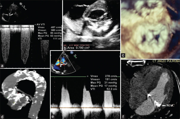
A 54-year-old gentleman, a known case of hypertension and diabetes mellitus type 2, presented with progressive effort intolerance and bilateral pitting pedal edema. His echocardiogram showed severe calcific mitral stenosis (mitral valve area 0.79 cm2, peak and mean gradients of 31 and 17 mmHg respectively) with severe mitral annular calcification (MAC); severe calcific aortic stenosis (aortic valve area 0.25 cm2, peak and mean gradients of 95 and 63 mmHg respectively), and mild LV systolic dysfunction with an EF of 45%. He was considered for surgical double valve replacement, but was deemed a high-risk candidate for surgical intervention, owing to presence of porcelain aorta.
On the basis of ECG gated CT angiography, the multidisciplinary heart team at our institute recommended concomitant transcatheter mitral valve replacement (TMVR) through a transfemoral transseptal (TS) approach and transcatheter aortic valve replacement (TAVR) through a transfemoral approach [Figure 1].
Figure 1.
Pre-procedure images (a): Gradient across the Aortic valve; (b): Mitral Valve area in 2D Echo; (c): 3D Echo view of mitral valve; (d): Severe and diffused calcification in aorta; (e): Gradient across the Mitral Valve; (f): Mitral Annular Calcification in Axial view CT Angiogram (Arrow)
Procedure
The procedure was done under conscious sedation and transthoracic echocardiography (TTE) guidance for TAVR followed by general anesthesia with 3D transesophageal echocardiography (TEE) for TMVR. On the morning of the procedure, a 6Fr sheath and a triple lumen catheter was cannulated in the right IJV and left radial artery access was obtained under local anesthesia. Baseline measurements were obtained for bi-spectral index (BIS) and cerebral oximetry (INVOS™). Infusion of Dexmedetomidine was started and titrated to a BIS of around 70-80.
After preparation and drape, access was obtained in bilateral common femoral artery and common femoral vein. A 5Fr balloon tip temporary pacing catheter was placed at RV apex through left common femoral vein. A 5Fr angled pigtail catheter was advanced and positioned in Non Coronary Cusp (NCC) sinus. Coronary and femoral angiograms were performed. Two 6Fr Perclose ProGlide (Abbott Vascular) suture-mediated closure systems were placed at 90° to each other in left common femoral artery. Heparin was given at a dose of 1 mg/kg and ACT kept around 300. The access site was serially dilated. Then 14Fr sheath was finally placed in left common femoral artery over a stiff wire. The aortic valve was crossed using 5Fr AR2 catheter, exchanged to pigtail catheter and hemodynamic measurements were recorded. Following balloon pre-dilatation of aortic valve, the patient went into a complete heart block and became dependent on ventricular pacing. Pacing was initiated at 60 beats/minute. Then a 29 mm Medtronic Core Valve™ Evolut™ PRO was delivered from the left groin and was adequately positioned across the aortic valve (deep placement to prevent LVOT obstruction) as per the fluoroscopic and echo guidance. Aortic valve was inflated under rapid ventricular pacing (RVP) at 180 beats/minute. After post deployment aortic valve dilatation, hemodynamics showed no significant gradients. Hemodynamics of the patient remained normal with pacing and inotropic requirement was minimal. Arterial pressures were closely monitored during and immediately after rapid ventricular pacing, and were titrated with infusion of norepinephrine and boluses of phenylephrine to keep systolic pressure above 60 mmHg.
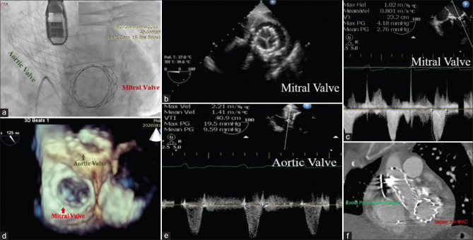
Once the hemodynamic status of the patient was stable for some time after TAVR, general anesthesia was given for TMVR. The patient was induced using Fentanyl 2 mcg/kg, Etomidate 0.3 mg/kg, and Vecuronium 0.1 mg/kg, intubated with an ETT 8.5 mm ID and put on mechanical ventilator. A 3D TEE probe was inserted to assist during TMVR. ACT was reassessed and the TMVR procedure was performed through right femoral vein. The atrial septal puncture was done by conventional method at infero-posterior septum under 3D trans-esophageal echo and fluoroscopic guidance. An 8.5Fr Agilis EPI™ Steerable Introducer was advanced into LA. Balloon atrial septostomy was done followed by balloon pre-dilatation of mitral valve. A 29 mm Edwards Sapien 3 Heart Valve was advanced through right femoral vein and valve alignment was done at RA-IVC junction. Then valve was advanced through inter-atrial septum and positioned across the mitral valve under fluoroscopic and 3D TEE guidance. The valve was finally deployed within mitral annulus by slowly inflating the balloon under rapid ventricular pacing. The delivery system was then withdrawn. The mean gradients across the mitral and aortic valves post-procedure were 2.7 mmHg and 9.5 mmHg respectively.
The Sheath was removed and Perclose ProGlide was deployed in left common femoral artery. Pelvic angiogram showed good hemostasis. The right common femoral artery closed by Angio-Seal VIP vascular closure device, right common femoral vein was closed by figure of 8 suture and left common femoral vein sheath was removed.
A dual chamber permanent pacemaker (Evity 8 DR-T, Biotronic) was implanted simultaneously as the patient had low chances of reverting to sinus rhythm (extensive intracardiac calcification and pre-procedure RBBB and LAHB).
The heparin was neutralized with protamine and patient reversed with neostigmine-glycopyrrolate combination and extubated in cath lab.
The post-procedure stay of the patient was uneventful and the patient was discharged home on 5th post-procedure day [Figure 2].
Figure 2.
Post-Procedure images. (a): Mitral and Aortic Valve prostheses (indicated by Arrows) in Fluoroscopy; (b): Mitral prosthesis in 2D TEE (c): Gradient across Mitral Valve post-procedure; (d): 3D TEE view of Mitral valve; (e): Gradient across Aortic valve post-procedure; (f): Aortic and Mitral prostheses in CT scan
DISCUSSION
Surgical valve replacement may not always be a feasible option because co-morbidities, advanced age, high surgical risk, frailty, or calcification may preclude surgery in many patients.[3,4] Transcatheter valve therapy has evolved constantly. Over the past few years, TAVR has quickly adapted to various patient populations in a way that it is now indicated irrespective of the patients’ predicted surgical risk.[5]
While TAVR has become more and more main-stream, transcatheter mitral valve replacement hasn’t had quite the initial success, mainly due to complex mitral valve anatomy, difference in etiology of diseases and proximity to LVOT predisposing to outflow obstruction.[6] With pre-procedural simulation of valve insertion within the native mitral orifice, we can assess the aorto-mitral and the LVOT-mitral valve angle and identify patients at an increased risk of obstruction of the outflow tract by calculating the neo-LVOT area.[7] Neo-LVOT area of 250 mm2 or more is considered to identify a lower risk of LVOT obstruction.[8]
Simultaneous transcatheter double valve replacement is rare, but can be performed in inoperable high surgical risk patients. Some centers have reported experience with concomitant transapical mitral and aortic valve implantation in patients who have failed prosthetic mitral and aortic valves (TDVIVR).[9] There is also a recent report of successful transapical implantation of mitral and aortic valve in native valves with severe AS and MS (TDVR).[10] Bashir et al., had reported first case of successful transfemoral aortic and transseptal mitral valve replacement in native aortic and mitral valves.[11] Fanari et al., also reported successful simultaneous TAVR and transseptal TMVR using SAPIEN S3 for severe AS and severe native MS with associated severe MAC.[12]
In transcatheter double valve replacements, D’Onofrio[13] has suggested that aortic valve deployment should be done first as it reduces the afterload and improves the hemodynamics for the mitral procedure; reduces the chances of hemodynamic deterioration in case of mitral prosthesis failure causing severe mitral regurgitation; and reduces the chances of deployed valve malposition.
Transcatheter double valve replacement is not a routine procedure. Nevertheless, a patient with severe calcific aortic stenosis, severe mitral stenosis with severe MAC and calcification in whole of ascending and arch of aorta presented as a perfect indication for combined transcatheter procedure. Anesthesia technique for our procedure was a combination of conscious sedation with local anesthesia and general anesthesia. The TAVR was done under conscious sedation with transthoracic echocardiography (TTE) and fluoroscopy guidance as this was a routine technique for TAVR at our institute. Also, we wanted to ensure success of TAVR procedure before proceeding for TMVR under general anesthesia allowing usage of 3D Transesophageal echocardiography (TEE).
CONCLUSION
After careful evaluation by experienced heart teams, combined native TAVR/TMVR via transfemoral access can be successfully performed. The use of a fully percutaneous transfemoral approach avoids complications related to thoracotomy or transapical access and also reduces hospital stay. This therapeutic option may be considered as valuable alternative to surgical DVR in extremely high-risk patients, provided suitable anatomy for TDVR.
Declaration of patient consent
The authors certify that they have obtained all appropriate patient consent forms. In the form the patient (s) has/have given his/her/their consent for his/her/their images and other clinical information to be reported in the journal. The patients understand that their names and initials will not be published and due efforts will be made to conceal their identity, but anonymity cannot be guaranteed.
Financial support and sponsorship
Nil.
Conflicts of interest
There are no conflicts of interest.
REFERENCES
- 1.Mahmaljy H, Tawney A, Young M. Transcatheter Aortic Valve Replacement. [Updated 2022 May 5]. In: StatPearls [Internet] Treasure Island (FL): StatPearls Publishing; 2022. [Last accessed on 2022 Aug 25]. Available from: https://www.ncbi.nlm.nih.gov/books/NBK431075/ [PubMed] [Google Scholar]
- 2.Guerrero M, Salinger M, Levisay J, Feldman T. Transcatheter Mitral Valve Replacement Therapies - American College of Cardiology. [online] American College of Cardiology. 2022. [Last accessed on 2022 Aug 25]. Available at: <https://www.acc.org/latest-in-cardiology/articles/2017/04/28/09/32/transcatheter-mitral-valve-replacement-therapies> .
- 3.Iung B, Cachier A, Baron G, Messika-Zeitoun D, Delahaye F, Tornos P, et al. Decision-making in elderly patients with severe aortic stenosis: Why are so many denied surgery? Eur Heart J. 2005;26:2714–20. doi: 10.1093/eurheartj/ehi471. [DOI] [PubMed] [Google Scholar]
- 4.Nataf P, Pavie A, Jault F, Bors V, Cabrol C, Gandjbakhch I. Intraatrial insertion of a mitral prosthesis in a destroyed or calcified mitral annulus. Ann Thorac Surg. 1994;58:163–7. doi: 10.1016/0003-4975(94)91092-8. [DOI] [PubMed] [Google Scholar]
- 5.FDA Expands TAVR Indication to Low-Risk Patients-American College of Cardiology [Internet]. American College of Cardiology. 2020. [Cited on 2020 Nov 7]. Available from: https://www.acc.org/latest-in-cardiology/articles/2019/08/16/13/49/fda-expands-tavr-indication-to-lowrisk-patients#:~:text=The%20U.S.%20Food%20and%20Drug,stenosis%20at%20low%20surgical%20risk .
- 6.Von Ballmoos M, Kalra A, Reardon M. Complexities of transcatheter mitral valve replacement (TMVR) and why it is not transcatheter aortic valve replacement (TAVR) ASVIDE. 2018;5:880–880. doi: 10.21037/acs.2018.10.06. [DOI] [PMC free article] [PubMed] [Google Scholar]
- 7.Blanke P, Naoum C, Dvir D, Bapat V, Ong K, Muller D, et al. Predicting LVOT obstruction in transcatheter mitral valve implantation. JACC Cardiovasc Imaging. 2017;10:482–5. doi: 10.1016/j.jcmg.2016.01.005. [DOI] [PubMed] [Google Scholar]
- 8.Guerrero M, Salinger M, Pursnani A, Pearson P, Lampert M, Levisay J, et al. Transseptal transcatheter mitral valve-in-valve: A step by step guide from preprocedural planning to postprocedural care. Catheter Cardiovasc Interv. 2017;92:E185–96. doi: 10.1002/ccd.27128. [DOI] [PubMed] [Google Scholar]
- 9.Savoj J, Iftikhar S, Burstein S, Hu P. Transcatheter double valve-in-valve replacement of aortic and mitral bioprosthetic valves. Cardiol Res. 2019;10:193–8. doi: 10.14740/cr863. [DOI] [PMC free article] [PubMed] [Google Scholar]
- 10.Elkharbotly A, Delago A, El-Hajjar M. Simultaneous transapical transcatheter aortic valve replacement and transcatheter mitral valve replacement for native valvular stenosis. Catheter Cardiovasc Interv. 2015;87:1347–51. doi: 10.1002/ccd.26078. [DOI] [PubMed] [Google Scholar]
- 11.Bashir M, Sigurdsson G, Horwitz P, Zahr F. Simultaneous transfemoral aortic and transseptal mitral valve replacement utilising SAPIEN 3 valves in native aortic and mitral valves. EuroIntervention. 2017;12:1649–52. doi: 10.4244/EIJ-D-16-00953. [DOI] [PubMed] [Google Scholar]
- 12.Fanari Z, Mahmaljy H, Nandish S, Goswami N. Simultaneous transcatheter transfemoral aortic and transeptal mitral valve replacement using Edward SAPIEN S3. Catheter Cardiovasc Interv. 2017;92:988–92. doi: 10.1002/ccd.27410. [DOI] [PubMed] [Google Scholar]
- 13.D’Onofrio A, Zucchetta F, Gerosa G. Simultaneous transapical aortic and mitral valve-in-valve implantation for double prostheses dysfunction: Case report and technical insights. Catheter Cardiovasc Interv. 2014;84:509–12. doi: 10.1002/ccd.25498. [DOI] [PubMed] [Google Scholar]


