PLOS ONE logoLink to PLOS ONE
. 2023 Mar 9;18(3):e0282911. doi: 10.1371/journal.pone.0282911

3D-printed eye model: Simulation of intraocular pressure

Hidenaga Kobashi 1,2,*, Masaaki Kobayashi 3
Editor: Aparna Rao4
PMCID: PMC9997944  PMID: 36893149

Abstract

Purpose

To develop artificial eye models using 3D printing and to evaluate the correlation between different corneal thicknesses and intraocular pressures (IOPs).

Methods

We designed 7 artificial eye models using a computer-aided design system and fabricated them using 3D printing. Corneal curvature and axial length were based on the Gullstrand eye model. Hydrogels were injected into the vitreous cavity, and seven different corneal thicknesses (200 to 800 μm) were prepared. In this proposed design, we also produced different corneal stiffnesses. A Tono-Pen AVIA tonometer was used by the same examiner to perform five consecutive IOP measurements in each eye model.

Results

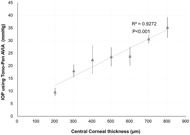
Different eye models were ideally created using 3D printing. IOP measurements were successfully performed in each eye model. The corneal thickness was significantly correlated with IOP (R2 = 0.927; 𝑃<0.001).

Conclusion

The 3D-printed eye model is useful for evaluating IOP measurements. This technique might be a promising alternative to the conventional porcine eye model.

Introduction

Three-dimensional (3D) printing technology, also known as additive manufacturing technology, is a technology that manufactures building blocks layer by layer from combinations of materials by applying three-dimensional data. The primary process involves delivery of a series of thin three-dimensional computer-aided design (CAD) or computed tomography cross-sections by the software to the printer for rapid imaging from bottom to top [1]. Three-dimensional printing is an all-inclusive term for a variety of methods that use digital data to produce 3D objects made of various materials (both synthetic and organic) [2, 3]. This technology can rapidly produce customized devices and prostheses at low costs via simple development and design processes and is capable of a single item at a time, each unique in shape and design. Such products may be as diverse as instruments that aid in early detection of common ocular conditions, diagnostic and therapeutic devices built specifically for individual patients, 3D-printed contact lenses and intraocular implants, and models that assist in surgery planning, improve patient and medical staff education. The role of 3D printing in the field of ophthalmology and eyecare is evolving, particularly after the introduction of high-resolution pico- to micrometer-scale 3D printers. Several applications, such as orbital implants, ocular prostheses, intraocular devices, ophthalmic models, surgical instruments, and surgical simulation devices, have been developed [4, 5]. To the best of our knowledge, to date, there have been no studies on 3D-printed eyeballs simulating different intraocular pressures (IOPs). A high IOP can induce glaucoma in models. Generally, the central corneal thickness (CCT) significantly affects IOP readings obtained by tonometry [6]. Previous studies have induced a higher IOP model by changing the height of a drip stand in porcine eyes [7, 8], but this technique is limited to a wet laboratory curriculum. To simplify the simulation of the elevated IOP model, we developed artificial eye models by changing the CCT based on a 3D printing approach. A good understanding of this technology will be beneficial to glaucoma research. The aim of this study was to evaluate the association between different CCTs and the IOP in 3D-printed eyeball models.

Methods

Design of the eyeball model

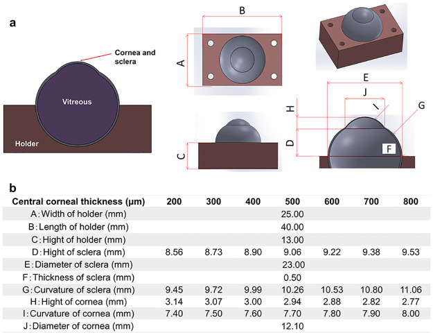
The 3D-printed eyeball model was generated by CAD software and a simulation environment represented by ANSYS Workbench 19.1. In the current study, the eyeball consisted of the cornea, sclera, and vitreous humor to highlight the change in corneal thickness. The optical model was based on Gullstrand’s simplified schematic eye [3]. Fig 1a shows the structure and main components of the eye model, which is available as parameterizable CAD geometry. When we manufactured the eyeball, the “Material Jetting” style was used as an additive manufacturing. This process is as follows:

Fig 1.

Fig 1

a) Schematic diagram of the 3D printed eye model as a computer-aided design construction. b) Characteristics of ocular biometric parameters in the cornea, sclera, and eyeball holder.

  1. Jet liquid ultra violet (UV) curable resin onto the print tray.

  2. Use a roller to level the UV curable resin.

  3. Use a UV lamp to harden the UV curable resin.

  4. Repeat processes 1–3 and build the models.

The material shore hardness is changed by the material jetting pattern. The output of the UV lamp and the rotational speed of the roller (600 rpm) were held constant during printing. This means that the material shore hardness is not changed by the output of the UV lamp and the speed of the roller.

To easily measure the IOP using a Tono-Pen AVIA tonometer (Reichert Inc., Depew, New York, USA), we created a cuboid eye holder. The aqueous and vitreous humor were simulated by the injection of hydrogels into the vitreous cavity. To induce different corneal stiffnesses, seven different CCTs were prepared for every 100-μm increase, and the curvatures of the cornea and sclera were simultaneously varied (Fig 1b). To simply control the change in the IOP, we changed only the CCT in the current study. Some parameters were fixed as the default values. To assess the deviation between the printed specimen and the CAD design, we aimed to measure the actual value of the CCT using an ultrasound pachymeter (SP-100; Tomey, Nagoya, Japan). Three consecutive measurements were performed in each eye model.

In addition to the design of the eye geometry, an adequate description of the material properties is required for the 3D-printing simulation. Table 1 shows the material parameters of the eye model.

Table 1. Material parameters of the eye model.

Cornea (500 μm) Vitreous Holder
Product name Stratasys FLX9740-DM Stratasys Gel Support Stratasys VeroClear
Stiffness of material Rubber-like Gel Hard
Tensile strength 3–4 MPa - 50–65 MPa
Young’s modulus of elasticity Un open (industrial secrets) - 2000–3000 Mpa
Elongation break 190–210% - 10–25%
Tensile tear resistance 6.0–8.0 kg/cm - -
Flexural strength Un open (industrial secrets) - 75–110 Mpa
Flexural module Un open (industrial secrets) - 2200–3200 Mpa

Intraocular pressure measurements

Experiments with different CCTs in the eyeball model demonstrated different elastic responses when measuring pressure readings using the Tono-Pen AVIA tonometer. The range of IOP measurements was 5 to 55 mmHg in the tonometer. All series were taken five times by the same examiner (H.K.) using the tonometer at each corneal thickness level, and the mean value and standard deviation of each parameter were recorded. To determine the repeatability of the IOP measurement, intraclass correlation coefficients (ICCs) were calculated for the 5 repeated measurements at each corneal thickness. The ICC ranges from 0 to 1 and is commonly classified as follows: less than 0.75 indicates poor repeatability, 0.75 to less than 0.90 shows moderate repeatability, and greater than 0.90 represents high repeatability. The experiments were carried out at room temperature (24 ± 1 °C).

Data analysis

Analyses were performed with Statistical Analysis Software (version 9.4; SAS Institute, Cary, NC). Linear regression was employed to analyze the CCT in the model eye and IOP to evaluate the linear regression coefficient (R2). The normality of all data was first checked by the Kolmogorov‒Smirnov test. Because the use of parametric statistics was possible, the Pearson correlation coefficient was used to assess the correlation between the IOP and the CCT in each eye model. The outcome measures are reported as the mean ± standard deviation. A P value of < 0.05 was considered statistically significant.

Results

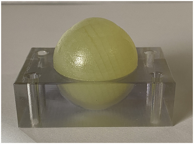
Fig 2 shows a representative 3D-printed eyeball with a central corneal thickness of 500 μm. All eyeballs were successfully created at each corneal thickness. Table 2 presents the mean IOP of 3D-printed model eyeballs at each central corneal thickness. The repeatability of the IOP measurement was high, with an ICC of 0.969 (95% confidence interval: 0.909 to 0.994). The IOPs were significantly correlated with the CCTs (R2 = 0.927, p<0.001) (Fig 3).

Fig 2. Whole eyeball and holder simulated using 3D-printing technology.

Fig 2

Table 2. Intraocular pressure of the 3D-printed model eyeballs for each central corneal thickness.

Central corneal thickness (μm) 200 300 400 500 600 700 800
Mean (mmHg) 9.6 18.0 22.4 23.6 23.8 30.6 35.2
Standard deviation 1.5 2.4 5.5 3.7 3.6 1.7 4.0

Fig 3. Graph showing a significant correlation between central corneal thickness and intraocular pressure in the 3D-printed eyeball models.

Fig 3

Table 3 presents the actual value of the CCT measured using an ultrasound pachymeter.

Table 3. Actual value of the central corneal thickness measured using ultrasound.

Central corneal thickness
CAD design (μm) 200 300 400 500 600 700 800
Ultrasound pachymeter (μm) 201.3 ± 7.6 303.0 ± 6.2 403.7 ± 7.2 504.7 ± 7.4 601.7 ± 12.1 699.7 ± 11.5 803.0 ± 2.0

Discussion

In this study, we successfully developed variable IOP eyeball models using 3D printing technology. In the 3D-printed eyeball model, the IOP increased as the CCT increased, which is similar to the results of a previous study in humans [6]. IOP measurements are dependent on the CCT. The current approach is the first to assess the correlation between IOP and CCT in 3D-printed eyeballs. This technique is quite unique and useful for basic research on ocular tonometers and glaucoma treatment because a special wet laboratory unit is needed. However, the true IOP might be unknown because it is impossible to determine the exact IOP that represents the pressure at the optic disc. Generally, corneal biomechanics play a role in the IOP in the human eye. Thus, we mimicked different IOP models with increasing CCTs using 3D-printed eye models. Almost all previous reports used ex vivo porcine eyeballs by changing the height of saline bottles to evaluate the elevated IOP [7, 8]. Our model might be a promising alternative to the conventional porcine eye model.

To simplify the methodology in this study, we changed the CCT and accordingly modified parameters such as the curvature and height of the sclera and cornea (Fig 1). We determined the diameter and thickness of the sclera at 23.00 mm and 0.50 mm, respectively. As a positive correlation was shown between the CCT and IOP in Fig 3, the impact of other geometric parameters might be smaller than that of corneal thickness.

With regard to the repeatability of the IOP measurements using the Tono-Pen AVIA, we confirmed the good repeatability for 5 consecutive measurements by the same examiner, as the standard deviation was almost within 5 mmHg at each corneal thickness. Our results of the reputability of the Tono-Pen AVIA in 3D-printed eyeballs are similar to those reported in the human eye [9]. However, it is difficult for the Tono-Pen AVIA to determine the correctness of the IOP measurements in 3D-printed eye models because the Tono-Pen AVIA itself has not been calibrated and designed to be used with those models. It would have been more beneficial to compare the IOP measured with other tannometers, such as iCare and noncontact tonometers. With regard to the reliability of the CCT in the CAD model, we evaluated the actual value of the CCT using an ultrasound pachymeter. The difference in the CCT between the CAD model and the ultrasound measurements was clinically acceptable because of the small difference (within 5 μm), as shown in Table 3.

There are three limitations to this study. First, the accurate morphological structure of the eyeball was not realized in the 3D-printed model because no crystalline lens, anterior chamber, or iris was created in this study. However, the primary aim was to develop a 3D-printed eyeball and simulate different IOP models with various corneal thicknesses. Second, precise corneal biomechanics were not achieved in these rubber-like model eyes. Based on the Gullstrand model of the cornea, we simulated stiffness, strength, and resistance values similar to those of an artificial cornea, as shown in Table 1. Further investigation is necessary to develop a 3D-printed cornea for IOP evaluation. Third, our 3D-printed eyeball models did not include iris, aqueous humor, or lens components, all of which affect IOP dynamics in humans, due to technological limitations.

In conclusion, a method for creating an elevated IOP model was demonstrated in a 3D-printed eyeball model. The model was useful for the assessment of the IOP in terms of basic research on glaucoma.

Data Availability

All relevant data are within the manuscript.

Funding Statement

This article is based on results obtained from a project, JPNP0401005, commissioned by the New Energy and Industrial Technology Development Organization (NEDO). The funders had no role in study design, data collection and analysis, decision to publish, or preparation of the manuscript.

References

  • 1.Chen X, Lawrence JM, Wey LT, Schertel L, Jing Q, Vignolini S, et al. 3D-printed hierarchical pillar array electrodes for high-performance semi-artificial photosynthesis. Nat Mater. 2022. Jul;21(7):811–818. Epub 2022 Mar 7. doi: 10.1038/s41563-022-01205-5 . [DOI] [PubMed] [Google Scholar]
  • 2.de SM Ozelim Luan C., and Cavalcante André LB. "Combining microtomography, 3D printing, and numerical simulations to study scale effects on the permeability of porous media." International Journal of Geomechanics 19.2 (2019): 04018194. [Google Scholar]
  • 3.Belot Igor, et al. "A 3D additive manufacturing approach for the validation of a numerical wall-scale model of catalytic particulate filters." Chemical Engineering Journal 405 (2021): 126653. [Google Scholar]
  • 4.Rassow B. Ein Modell für das Gullstrandsche Normalauge [A model of Gullstrand’s "normal eye"]. Ophthalmologica. 1972;164(2):143–8. German. doi: 10.1159/000306725 . [DOI] [PubMed] [Google Scholar]
  • 5.Huang W, Zhang X. 3D Printing: Print the future of ophthalmology. Invest Ophthalmol Vis Sci. 2014. Aug 26;55(8):5380–1. doi: 10.1167/iovs.14-15231 . [DOI] [PubMed] [Google Scholar]
  • 6.Kohlhaas M, Boehm AG, Spoerl E, Pürsten A, Grein HJ, Pillunat LE. Effect of central corneal thickness, corneal curvature, and axial length on applanation tonometry. Arch Ophthalmol. 2006. Apr;124(4):471–6. doi: 10.1001/archopht.124.4.471 . [DOI] [PubMed] [Google Scholar]
  • 7.Tao C, Han Z, Sun Y, Zhou C, Roberts C, Zhou D, et al. Corneal hysteresis with intraocular pressure of a wide range: a test on porcine eyes. J Refract Surg. 2013. Dec;29(12):850–4. doi: 10.3928/1081597x-20131115-05 . [DOI] [PubMed] [Google Scholar]
  • 8.Zhang J, Zhang Y, Li Y, Chen R, Shung KK, Richter G, et al. Correlation of IOP with Corneal Acoustic Impedance in Porcine Eye Model. Biomed Res Int. 2017;2017:2959717. Epub 2017 May 31. doi: 10.1155/2017/2959717 . [DOI] [PMC free article] [PubMed] [Google Scholar]
  • 9.Schweier C, Hanson JV, Funk J, Töteberg-Harms M. Repeatability of intraocular pressure measurements with Icare PRO rebound, Tono-Pen AVIA, and Goldmann tonometers in sitting and reclining positions. BMC Ophthalmol. 2013. Sep 5;13:44. doi: 10.1186/1471-2415-13-44 . [DOI] [PMC free article] [PubMed] [Google Scholar]

Decision Letter 0

Aparna Rao

20 Oct 2022

PONE-D-22-236133D-printed eye model: simulation of intraocular pressurePLOS ONE

Dear Dr Kobashi,

Thank you for submitting your manuscript to PLOS ONE. After careful consideration, we feel that it has merit but does not fully meet PLOS ONE’s publication criteria as it currently stands. Therefore, we invite you to submit a revised version of the manuscript that addresses the points raised during the review process.

Please submit your revised manuscript by Dec 04 2022 11:59PM. If you will need more time than this to complete your revisions, please reply to this message or contact the journal office at plosone@plos.org. When you're ready to submit your revision, log on to https://www.editorialmanager.com/pone/ and select the 'Submissions Needing Revision' folder to locate your manuscript file.

Please include the following items when submitting your revised manuscript:

  • A rebuttal letter that responds to each point raised by the academic editor and reviewer(s). You should upload this letter as a separate file labeled 'Response to Reviewers'.

  • A marked-up copy of your manuscript that highlights changes made to the original version. You should upload this as a separate file labeled 'Revised Manuscript with Track Changes'.

  • An unmarked version of your revised paper without tracked changes. You should upload this as a separate file labeled 'Manuscript'.

If you would like to make changes to your financial disclosure, please include your updated statement in your cover letter. Guidelines for resubmitting your figure files are available below the reviewer comments at the end of this letter.

If applicable, we recommend that you deposit your laboratory protocols in protocols.io to enhance the reproducibility of your results. Protocols.io assigns your protocol its own identifier (DOI) so that it can be cited independently in the future. For instructions see: https://journals.plos.org/plosone/s/submission-guidelines#loc-laboratory-protocols. Additionally, PLOS ONE offers an option for publishing peer-reviewed Lab Protocol articles, which describe protocols hosted on protocols.io. Read more information on sharing protocols at https://plos.org/protocols?utm_medium=editorial-email&utm_source=authorletters&utm_campaign=protocols.

We look forward to receiving your revised manuscript.

Kind regards,

Aparna Rao

Academic Editor

PLOS ONE

Journal Requirements:

When submitting your revision, we need you to address these additional requirements.

1. Please ensure that your manuscript meets PLOS ONE's style requirements, including those for file naming. The PLOS ONE style templates can be found at

https://journals.plos.org/plosone/s/file?id=wjVg/PLOSOne_formatting_sample_main_body.pdf and

https://journals.plos.org/plosone/s/file?id=ba62/PLOSOne_formatting_sample_title_authors_affiliations.pdf

2. Thank you for stating the following financial disclosure:

“This article is based on results obtained from a project, JPNP0401005, commissioned by the New Energy and Industrial Technology Development Organization (NEDO).”

Please state what role the funders took in the study.  If the funders had no role, please state: "The funders had no role in study design, data collection and analysis, decision to publish, or preparation of the manuscript."

If this statement is not correct you must amend it as needed.

Please include this amended Role of Funder statement in your cover letter; we will change the online submission form on your behalf.

3. Thank you for stating the following in the Competing Interests section:

“The author(s) have made the following disclosure(s): H.K.: Employee and equity owner, Toneasy Inc.; Patent, Toneasy Inc. No other disclosures were reported.”

Please confirm that this does not alter your adherence to all PLOS ONE policies on sharing data and materials, by including the following statement: "This does not alter our adherence to  PLOS ONE policies on sharing data and materials.” (as detailed online in our guide for authors http://journals.plos.org/plosone/s/competing-interests).  If there are restrictions on sharing of data and/or materials, please state these. Please note that we cannot proceed with consideration of your article until this information has been declared.

Please include your updated Competing Interests statement in your cover letter; we will change the online submission form on your behalf.

[Note: HTML markup is below. Please do not edit.]

Reviewers' comments:

Reviewer's Responses to Questions

Comments to the Author

1. Is the manuscript technically sound, and do the data support the conclusions?

The manuscript must describe a technically sound piece of scientific research with data that supports the conclusions. Experiments must have been conducted rigorously, with appropriate controls, replication, and sample sizes. The conclusions must be drawn appropriately based on the data presented.

Reviewer #1: No

Reviewer #2: Partly

**********

2. Has the statistical analysis been performed appropriately and rigorously?

Reviewer #1: N/A

Reviewer #2: Yes

**********

3. Have the authors made all data underlying the findings in their manuscript fully available?

The PLOS Data policy requires authors to make all data underlying the findings described in their manuscript fully available without restriction, with rare exception (please refer to the Data Availability Statement in the manuscript PDF file). The data should be provided as part of the manuscript or its supporting information, or deposited to a public repository. For example, in addition to summary statistics, the data points behind means, medians and variance measures should be available. If there are restrictions on publicly sharing data—e.g. participant privacy or use of data from a third party—those must be specified.

Reviewer #1: Yes

Reviewer #2: Yes

**********

4. Is the manuscript presented in an intelligible fashion and written in standard English?

PLOS ONE does not copyedit accepted manuscripts, so the language in submitted articles must be clear, correct, and unambiguous. Any typographical or grammatical errors should be corrected at revision, so please note any specific errors here.

Reviewer #1: Yes

Reviewer #2: No

**********

5. Review Comments to the Author

Please use the space provided to explain your answers to the questions above. You may also include additional comments for the author, including concerns about dual publication, research ethics, or publication ethics. (Please upload your review as an attachment if it exceeds 20,000 characters)

Reviewer #1: This research paper describes the development of a 3D eye model for testing the impact of varying corneal thickness on intraocular pressure. As is already known, corneal thickness has a significant influence on IOP readings obtained by almost any tonometer. Hence the finding that CCT influences IOP readings is not in any way novel. To my understanding, the other rationale for undertaking this study is the dependence on wet labs for studying the impact of various conditions on IOP measurements. However, the authors have not fully described the limitations of current methods and how their method addresses these limitations. This study should have included an in silico simulation assessing the impact of various elements of the setup (e.g., the mechanical holder, material properties geometrical attributes) before proceeding to the printing stage. My other major comments are as follows;

1. Although the main varying parameter is central corneal thickness, other parameters such as curvature of the sclera and cornea also change with each model. This is a significant limitations as not only CCT but also the geometry is likely to affect the IOP readings.

2. There is no description of the additive manufacturing (FDM?, SLA?) method that was used to manufacture the eyeball.

3. There is no mention of elastic modulus of the material that was used to print the cornea and the holder. Furthermore, as the cornea is a viscoelastic material, a rubber-like material (hence a purely elastic material) would not mimic corneal mechanical behavior.

4. The authors should have reported intraclass coefficient of serial measurements instead of describing repeated measurements as having "good repeatability".

5. Although acknowledged as a limitation, failure to factor in lens, iris, aqueous humor is a significant limitation hindering the applicability of this approach in studying IOP dynamics ex vivo.

Reviewer #2: The authors present a new methodology to study the impact of corneal thickness on the intraocular pressure (IOP) measurements. Overall, the paper addresses a quite interesting problem; on the other hand, there are several issues which must be addressed prior to the paper acceptance.

At first, the introduction and general contextualization is quite poor. For example, the authors cite that numerous published studies have considered 3D printing techniques in Health Sciences; however, just a few were cited. Besides, it would be interesting to mention other studies which considered using 3D printing to bring digital models to real life and then perform tests on them. They may consider, for example:

- Combining Microtomography, 3D Printing, and Numerical Simulations to Study Scale Effects on the Permeability of Porous Media by Luan C. de S. M. Ozelim and André L. B. Cavalcante

- A 3D additive manufacturing approach for the validation of a numerical wall-scale model of catalytic particulate filters by Igor Belot, Yixun Sun, David Vidal, Martin Votsmeier, Philippe Causse, François Trochu, François Bertrand.

English level should be enhanced. Sometimes, it becomes a bit confusing to fully understand the sentences.

Regarding the 3D printed model, more information should be provided about the elastic properties of the materials used. For example, which are the Young moduli of the materials? This, together with the corneal thickness, would probably have an impact on the IOP measurements. Besides, it is important to fully compare the mechanical properties of the printing materials to the ones of real human eyes.

The authors should provide more information about the printing procedure. Which were the steps? Also, they should present more details about the printer and its specs (minimum detail, minimum wall thickness etc).

A key aspect which needs to be at least discussed is how the authors assessed the quality of the printed specimens. In short, what are the expected deviations between the printed specimen and the one in the CAD design? Will these deviations impact on the effective corneal thickness? I do not know if the authors have access to a CT scan, but if they do, scanning a few of the printed samples would bring a valuable validation to the thicknesses considered in their analyses.

The authors mention the R-squared and the p-values obtained for the linear fit. Which hypothesis test is the p-value related to?

Since the authors have a complete control over the model, are they capable of measuring the “true” exact IOP?

Since Tono-Pen AVIA has not been calibrated and designed to be used with the 3D printed materials, this would be interesting to actually confirm the measurements are correct. Indicating that the results are reproducible is not sufficient to indicate the correctness of the result itself.

Finally, do the authors have control over the IOP change? How? Are the changes observed only due to the CCT changes? Any additional pressure is later applied to the gels? It is not clear in the paper how the IOP change occurs.

The issues above must be addressed to enhance the quality of the paper and make it publishable in such a high standard journal as PLOS One.

**********

6. PLOS authors have the option to publish the peer review history of their article (what does this mean?). If published, this will include your full peer review and any attached files.

If you choose “no”, your identity will remain anonymous but your review may still be made public.

Do you want your identity to be public for this peer review? For information about this choice, including consent withdrawal, please see our Privacy Policy.

Reviewer #1: Yes: Eray Atalay

Reviewer #2: No

**********

[NOTE: If reviewer comments were submitted as an attachment file, they will be attached to this email and accessible via the submission site. Please log into your account, locate the manuscript record, and check for the action link "View Attachments". If this link does not appear, there are no attachment files.]

While revising your submission, please upload your figure files to the Preflight Analysis and Conversion Engine (PACE) digital diagnostic tool, https://pacev2.apexcovantage.com/. PACE helps ensure that figures meet PLOS requirements. To use PACE, you must first register as a user. Registration is free. Then, login and navigate to the UPLOAD tab, where you will find detailed instructions on how to use the tool. If you encounter any issues or have any questions when using PACE, please email PLOS at figures@plos.org. Please note that Supporting Information files do not need this step.

PLoS One. 2023 Mar 9;18(3):e0282911. doi: 10.1371/journal.pone.0282911.r002

Author response to Decision Letter 0


9 Feb 2023

Dear Academic Editor,

Dr. Aparna Rao,

Thank you for your e-mail regarding our manuscript (PONE-D-22-23613) titled “3D-printed eye model: simulation of intraocular pressure” as well as for the comments from the reviewer. We believe that the paper has been much improved, which was largely a result of the referees’ many thoughtful comments. We would like to respond below to each comment.

To Reviewer #1

This research paper describes the development of a 3D eye model for testing the impact of varying corneal thickness on intraocular pressure. As is already known, corneal thickness has a significant influence on IOP readings obtained by almost any tonometer. Hence the finding that CCT influences IOP readings is not in any way novel. To my understanding, the other rationale for undertaking this study is the dependence on wet labs for studying the impact of various conditions on IOP measurements. However, the authors have not fully described the limitations of current methods and how their method addresses these limitations. This study should have included an in silico simulation assessing the impact of various elements of the setup (e.g., the mechanical holder, material properties geometrical attributes) before proceeding to the printing stage. My other major comments are as follows;

Thank you for your comments on the revision. I have responded the following the comments as below.

1. Although the main varying parameter is central corneal thickness, other parameters such as curvature of the sclera and cornea also change with each model. This is a significant limitations as not only CCT but also the geometry is likely to affect the IOP readings.

To simplify the methodology in this study, we changed the CCT and accordingly modified the parameters such as curvature and height of sclera and cornea (Figure 1). We determined the diameter and thickens of sclera at 23.00 mm and 0.50 mm, respectively. As the positive correlation was shown between CCT and IOP in Figure 3, the impact of other geometric parameters might be smaller than that of corneal thickness.

[Page 8, lines 143-147]: Three sentences have been added in Discussion.

“To simplify the methodology in this study, we changed the CCT and accordingly modified parameters such as the curvature and height of the sclera and cornea (Figure 1). We determined the diameter and thickness of the sclera at 23.00 mm and 0.50 mm, respectively. As a positive correlation was shown between the CCT and IOP in Figure 3, the impact of other geometric parameters might be smaller than that of corneal thickness.”

2. There is no description of the additive manufacturing (FDM?, SLA?) method that was used to manufacture the eyeball.

This method of additive manufacturing is “Material Jetting”.

This process is the following:

1. Jetting some liquid materials which are ultra violet (UV) curable resin on the print tray.

2. Making level UV curable resin by roller.

3. Hardening UV curable resin by UV lamp.

4. Repeat process 1-3 and building models.

Material shore hardness is changed by material jetting pattern. Output of UV lamp and 600 rpm of roller are constant during printing. It means Material shore hardness isn’t changed by output of UV lamp and rpm of roller.

[Page 4, lines 71-80]: Four sentences have been added.

When we manufactured the eyeball, the “Material Jetting” style was used as an additive manufacturing. This process is as follows:

1. Jet liquid ultra violet (UV) curable resin onto the print tray.

2. Use a roller to level the UV curable resin.

3. Use a UV lamp to harden the UV curable resin.

4. Repeat processes 1-3 and build the models.

The material shore hardness is changed by the material jetting pattern. The output of the UV lamp and the rotational speed of the roller (600 rpm) were held constant during printing. This means that the material shore hardness is not changed by the output of the UV lamp and the speed of the roller.

3. There is no mention of elastic modulus of the material that was used to print the cornea and the holder. Furthermore, as the cornea is a viscoelastic material, a rubber-like material (hence a purely elastic material) would not mimic corneal mechanical behavior.

We have added the information of elastic module in printed-corneal and -holder to Table 1. Unfortunately, the young’s modulus of elasticity of the corneal was not disclosed because of the confidential policy. Our strength is that wet lab style using the animal or human eyes is not required, thus our artificial eye balls using 3-D printing is useful for the evaluation of IOP in basic research. Since the difference between the corneal material and rubber-like material is a further limitation, we tried to mimic corneal behavior in terms of elastic modulus.

Table 1. Material parameters of the eye model.

Cornea (500 μm) Vitreous Holder

Product name Stratasys FLX9740-DM Stratasys Gel Support Stratasys VeroClearTM

Stiffness of material Rubber-like Gel Hard

Tensile strength 3-4 MPa - 50-65 MPa

Young’s modulus of elasticity Un open (industrial secrets) - 2000-3000 Mpa

Elongation break 190-210 % - 10-25 %

Tensile tear resistance 6.0-8.0 kg/cm - -

Flexural strength Un open (industrial secrets) - 75-110 Mpa

Flexural module Un open (industrial secrets) - 2200-3200 Mpa

4. The authors should have reported intraclass coefficient of serial measurements instead of describing repeated measurements as having "good repeatability".

Based on the calculation of intraclass correlation coefficients (ICCs), we showed the good repeatability in IOP measurements. The repeatability of IOP was high with 0.969 of an ICC (95% confidence interval: 0.909 to 0.994).

[Page 6, lines 101-105]: Two sentences have been added.

“To determine the repeatability of the IOP measurement, intraclass correlation coefficients (ICCs) were calculated for the 5 repeated measurements at each corneal thickness. The ICC ranges from 0 to 1 and is commonly classified as follows: less than 0.75 indicates poor repeatability, 0.75 to less than 0.90 shows moderate repeatability, and greater than 0.90 represents high repeatability.”

[Page 7, lines 119-121]: One sentence has been added.

“The repeatability of the IOP measurement was high, with an ICC of 0.969 (95 % confidence interval: 0.909 to 0.994).”

5. Although acknowledged as a limitation, failure to factor in lens, iris, aqueous humor is a significant limitation hindering the applicability of this approach in studying IOP dynamics ex vivo.

As you mentioned, our eye models did not have the components of iris, aqueous humor, and lens. These factors affect the IOP dynamics in human. It was hard to realize the precise 3D-printed eyeball model because of the technological limitation.

[Page 10, lines 167-169]: Two sentences have been added.

“Third, our 3D-printed eyeball models did not include iris, aqueous humor, or lens components, all of which affect IOP dynamics in humans, due to technological limitations.”

To Reviewer #2:

The authors present a new methodology to study the impact of corneal thickness on the intraocular pressure (IOP) measurements. Overall, the paper addresses a quite interesting problem; on the other hand, there are several issues which must be addressed prior to the paper acceptance.

Thank you for your positive comments on the revision. We have addressed the reply to your comments to enhance the quality of the paper.

At first, the introduction and general contextualization is quite poor. For example, the authors cite that numerous published studies have considered 3D printing techniques in Health Sciences; however, just a few were cited. Besides, it would be interesting to mention other studies which considered using 3D printing to bring digital models to real life and then perform tests on them. They may consider, for example:

- Combining Microtomography, 3D Printing, and Numerical Simulations to Study Scale Effects on the Permeability of Porous Media by Luan C. de S. M. Ozelim and André L. B. Cavalcante

- A 3D additive manufacturing approach for the validation of a numerical wall-scale model of catalytic particulate filters by Igor Belot, Yixun Sun, David Vidal, Martin Votsmeier, Philippe Causse, François Trochu, François Bertrand.

We have revised and added some sentences in INTRODUCTION. The two references you recommended have been added.

[Page 3, lines 42-49]: Three sentences have been added.

“Three-dimensional printing is an all-inclusive term for a variety of methods that use digital data to produce 3D objects made of various materials (both synthetic and organic) [2,3]. This technology can rapidly produce customized devices and prostheses at low costs via simple development and design processes and is capable of a single item at a time, each unique in shape and design. Such products may be as diverse as instruments that aid in early detection of common ocular conditions, diagnostic and therapeutic devices built specifically for individual patients, 3D-printed contact lenses and intraocular implants, and models that assist in surgery planning, improve patient and medical staff education.”

[Page 11, lines 181-186]: Two references have been added.

“2. Ozelim, Luan C. de SM, and André LB Cavalcante. "Combining microtomography, 3D printing, and numerical simulations to study scale effects on the permeability of porous media." International Journal of Geomechanics 19.2 (2019): 04018194.”

“3. Belot, Igor, et al. "A 3D additive manufacturing approach for the validation of a numerical wall-scale model of catalytic particulate filters." Chemical Engineering Journal 405 (2021): 126653.”

English level should be enhanced. Sometimes, it becomes a bit confusing to fully understand the sentences.

We have revised English in this revision by a native reviewer.

Regarding the 3D printed model, more information should be provided about the elastic properties of the materials used. For example, which are the Young moduli of the materials? This, together with the corneal thickness, would probably have an impact on the IOP measurements. Besides, it is important to fully compare the mechanical properties of the printing materials to the ones of real human eyes.

We have added the information of elastic module in printed-corneal and -holder to Table 1. Unfortunately, the young’s modulus of elasticity of the corneal was not disclosed because of the confidential policy. Our strength is that wet lab style using the animal or human eyes is not required, thus our artificial eye balls using 3-D printing is useful for the evaluation of IOP in basic research. Since the difference between the corneal material and rubber-like material is a further limitation, we tried to mimic corneal behavior in terms of elastic modulus.

Table 1. Material parameters of the eye model.

Cornea (500 μm) Vitreous Holder

Product name Stratasys FLX9740-DM Stratasys Gel Support Stratasys VeroClearTM

Stiffness of material Rubber-like Gel Hard

Tensile strength 3-4 MPa - 50-65 MPa

Young’s modulus of elasticity Un open (industrial secrets) - 2000-3000 Mpa

Elongation break 190-210 % - 10-25 %

Tensile tear resistance 6.0-8.0 kg/cm - -

Flexural strength Un open (industrial secrets) - 75-110 Mpa

Flexural module Un open (industrial secrets) - 2200-3200 Mpa

The authors should provide more information about the printing procedure. Which were the steps? Also, they should present more details about the printer and its specs (minimum detail, minimum wall thickness etc).

This method of additive manufacturing is “Material Jetting”.

This process is the following:

1. Jetting some liquid materials which are ultra violet (UV) curable resin on the print tray.

2. Making level UV curable resin by roller.

3. Hardening UV curable resin by UV lamp.

4. Repeat process 1-3 and building models.

Material shore hardness is changed by material jetting pattern. Output of UV lamp and 600 rpm of roller are constant during printing. It means Material shore hardness isn’t changed by output of UV lamp and rpm of roller. As for its specs (minimum detail, minimum wall thickness etc), Stratasys Japan Co. did not disclose the information because of a confidential policy.

[Page 4, lines 71-80]: Four sentences have been added.

When we manufactured the eyeball, the “Material Jetting” style was used as an additive manufacturing. This process is as follows:

1. Jet liquid ultra violet (UV) curable resin onto the print tray.

2. Use a roller to level the UV curable resin.

3. Use a UV lamp to harden the UV curable resin.

4. Repeat processes 1-3 and build the models.

The material shore hardness is changed by the material jetting pattern. The output of the UV lamp and the rotational speed of the roller (600 rpm) were held constant during printing. This means that the material shore hardness is not changed by the output of the UV lamp and the speed of the roller.

A key aspect which needs to be at least discussed is how the authors assessed the quality of the printed specimens. In short, what are the expected deviations between the printed specimen and the one in the CAD design? Will these deviations impact on the effective corneal thickness? I do not know if the authors have access to a CT scan, but if they do, scanning a few of the printed samples would bring a valuable validation to the thicknesses considered in their analyses.

In this revision, to assess the deviation between the printed specimen and the one in the CAD design, we tried to measure the actual value of CCT using an ultrasound pachymeter (SP-100; Tomey, Nagoya, Japan) (Table 3). Three consecutive measurements were performed in each eye model. The difference in CCT between the CAD and ultrasound style was clinically acceptable because of small difference (within 5 μm).

Table 3. Actual value of the central corneal thickness measured using ultrasound.

Central corneal thickness

CAD design (μm) 200 300 400 500 600 700 800

Ultrasound pachymeter (μm) 201.3 ± 7.6 303.0 ± 6.2 403.7 ± 7.2 504.7 ± 7.4 601.7 ± 12.1 699.7 ± 11.5 803.0 ± 2.0

[Page 5, lines 87-89]: Two sentences have been added.

“To assess the deviation between the printed specimen and the CAD design, we aimed to measure the actual value of the CCT using an ultrasound pachymeter (SP-100; Tomey, Nagoya, Japan). Three consecutive measurements were performed in each eye model.”

[Page 8, line 127]: One sentence has been added.

“Table 3 presents the actual value of the CCT measured using an ultrasound pachymeter.”

[Page 9, lines 156-159]: Two sentences have been added.

“With regard to the reliability of the CCT in the CAD model, we evaluated the actual value of the CCT using an ultrasound pachymeter. The difference in the CCT between the CAD model and the ultrasound measurements was clinically acceptable because of the small difference (within 5 μm), as shown in Table 3.”

The authors mention the R-squared and the p-values obtained for the linear fit. Which hypothesis test is the p-value related to?

The normality of all data was first checked by the Kolmogorov-Smirnov test. Because the use of parametric statistics was possible, the Pearson correlation coefficient was used to assess the correlation of the IOP with CCT in each eye model.

[Page 7, lines 110-113]: Two sentences have been added.

“The normality of all data was first checked by the Kolmogorov‒Smirnov test. Because the use of parametric statistics was possible, the Pearson correlation coefficient was used to assess the correlation between the IOP and the CCT in each eye model.”

Since the authors have a complete control over the model, are they capable of measuring the “true” exact IOP?

Exact answer to the true IOP might be unknow because it is impossible to determine the exact IOP which represents the pressure at optic disc. Generally, corneal biomechanics plays a role in IOP in human eye. Thus, we mimicked the different IOP model with increasing CCT using 3D-printed eye models.

[Page 8, lines 136-140]: Two sentences have been added.

“However, the true IOP might be unknown because it is impossible to determine the exact IOP that represents the pressure at the optic disc. Generally, corneal biomechanics play a role in the IOP in the human eye. Thus, we mimicked different IOP models with increasing CCTs using 3D-printed eye models.”

Since Tono-Pen AVIA has not been calibrated and designed to be used with the 3D printed materials, this would be interesting to actually confirm the measurements are correct. Indicating that the results are reproducible is not sufficient to indicate the correctness of the result itself.

As you mentioned, it is hard for Tono-Pen AVIA to determine the correctness of IOP measurements in 3D-printed eye models because Tono-Pen AVIA itself has not been calibrated and designed to be used with those models. We should have compared the IOP with other tannometers such as iCare and non-contact tonometer.

[Page 9, lines 152-156]: Two sentences have been added.

“However, it is difficult for the Tono-Pen AVIA to determine the correctness of the IOP measurements in 3D-printed eye models because the Tono-Pen AVIA itself has not been calibrated and designed to be used with those models. It would have been more beneficial to compare the IOP measured with other tannometers, such as iCare and noncontact tonometers.”

Finally, do the authors have control over the IOP change? How? Are the changes observed only due to the CCT changes? Any additional pressure is later applied to the gels? It is not clear in the paper how the IOP change occurs.

To simply control the change in the IOP, we changed the only CCT in the current study. The other components of model eyes were identical to each model.

[Page 5, lines 85-86]: One sentence has been added.

“To simply control the change in the IOP, we changed only the CCT in the current study.”

The issues above must be addressed to enhance the quality of the paper and make it publishable in such a high standard journal as PLOS One.

________________________________________

6. PLOS authors have the option to publish the peer review history of their article (what does this mean?). If published, this will include your full peer review and any attached files.

If you choose “no”, your identity will remain anonymous but your review may still be made public.

Do you want your identity to be public for this peer review? For information about this choice, including consent withdrawal, please see our Privacy Policy.

Reviewer #1: Yes: Eray Atalay

Reviewer #2: No

________________________________________

[NOTE: If reviewer comments were submitted as an attachment file, they will be attached to this email and accessible via the submission site. Please log into your account, locate the manuscript record, and check for the action link "View Attachments". If this link does not appear, there are no attachment files.]

While revising your submission, please upload your figure files to the Preflight Analysis and Conversion Engine (PACE) digital diagnostic tool, https://pacev2.apexcovantage.com/. PACE helps ensure that figures meet PLOS requirements. To use PACE, you must first register as a user. Registration is free. Then, login and navigate to the UPLOAD tab, where you will find detailed instructions on how to use the tool. If you encounter any issues or have any questions when using PACE, please email PLOS at figures@plos.org. Please note that Supporting Information files do not need this step.

We believe the manuscript has been prepared and submitted satisfactorily and hope that it will be accepted for publication in Plos One. Thank you for your attention and consideration.

Sincerely yours,

Hidenaga Kobashi, MD, PhD, Department of Ophthalmology, Keio University, School of Medicine, Tokyo, Japan. E-mail address: hidenaga_kobashi@keio.jp

Decision Letter 1

Aparna Rao

27 Feb 2023

3D-printed eye model: simulation of intraocular pressure

PONE-D-22-23613R1

Dear Sir,

We’re pleased to inform you that your manuscript has been judged scientifically suitable for publication and will be formally accepted for publication once it meets all outstanding technical requirements.

Within one week, you’ll receive an e-mail detailing the required amendments. When these have been addressed, you’ll receive a formal acceptance letter and your manuscript will be scheduled for publication.

An invoice for payment will follow shortly after the formal acceptance. To ensure an efficient process, please log into Editorial Manager at http://www.editorialmanager.com/pone/, click the 'Update My Information' link at the top of the page, and double check that your user information is up-to-date. If you have any billing related questions, please contact our Author Billing department directly at authorbilling@plos.org.

If your institution or institutions have a press office, please notify them about your upcoming paper to help maximize its impact. If they’ll be preparing press materials, please inform our press team as soon as possible -- no later than 48 hours after receiving the formal acceptance. Your manuscript will remain under strict press embargo until 2 pm Eastern Time on the date of publication. For more information, please contact onepress@plos.org.

Kind regards,

Aparna Rao

Academic Editor

PLOS ONE

Reviewers' comments:

Reviewer's Responses to Questions

Comments to the Author

1. If the authors have adequately addressed your comments raised in a previous round of review and you feel that this manuscript is now acceptable for publication, you may indicate that here to bypass the “Comments to the Author” section, enter your conflict of interest statement in the “Confidential to Editor” section, and submit your "Accept" recommendation.

Reviewer #2: All comments have been addressed

**********

2. Is the manuscript technically sound, and do the data support the conclusions?

The manuscript must describe a technically sound piece of scientific research with data that supports the conclusions. Experiments must have been conducted rigorously, with appropriate controls, replication, and sample sizes. The conclusions must be drawn appropriately based on the data presented.

Reviewer #2: Yes

**********

3. Has the statistical analysis been performed appropriately and rigorously?

Reviewer #2: Yes

**********

4. Have the authors made all data underlying the findings in their manuscript fully available?

The PLOS Data policy requires authors to make all data underlying the findings described in their manuscript fully available without restriction, with rare exception (please refer to the Data Availability Statement in the manuscript PDF file). The data should be provided as part of the manuscript or its supporting information, or deposited to a public repository. For example, in addition to summary statistics, the data points behind means, medians and variance measures should be available. If there are restrictions on publicly sharing data—e.g. participant privacy or use of data from a third party—those must be specified.

Reviewer #2: Yes

**********

5. Is the manuscript presented in an intelligible fashion and written in standard English?

PLOS ONE does not copyedit accepted manuscripts, so the language in submitted articles must be clear, correct, and unambiguous. Any typographical or grammatical errors should be corrected at revision, so please note any specific errors here.

Reviewer #2: Yes

**********

6. Review Comments to the Author

Please use the space provided to explain your answers to the questions above. You may also include additional comments for the author, including concerns about dual publication, research ethics, or publication ethics. (Please upload your review as an attachment if it exceeds 20,000 characters)

Reviewer #2: All the comments have been addressed in the new version of the manuscript. Therefore, I advise to accept the paper in its current form.

**********

7. PLOS authors have the option to publish the peer review history of their article (what does this mean?). If published, this will include your full peer review and any attached files.

If you choose “no”, your identity will remain anonymous but your review may still be made public.

Do you want your identity to be public for this peer review? For information about this choice, including consent withdrawal, please see our Privacy Policy.

Reviewer #2: No

**********

Acceptance letter

Aparna Rao

1 Mar 2023

PONE-D-22-23613R1

3D-printed eye model: simulation of intraocular pressure

Dear Dr. Kobashi:

I'm pleased to inform you that your manuscript has been deemed suitable for publication in PLOS ONE. Congratulations! Your manuscript is now with our production department.

If your institution or institutions have a press office, please let them know about your upcoming paper now to help maximize its impact. If they'll be preparing press materials, please inform our press team within the next 48 hours. Your manuscript will remain under strict press embargo until 2 pm Eastern Time on the date of publication. For more information please contact onepress@plos.org.

If we can help with anything else, please email us at plosone@plos.org.

Thank you for submitting your work to PLOS ONE and supporting open access.

Kind regards,

PLOS ONE Editorial Office Staff

on behalf of

Dr. Aparna Rao

Academic Editor

PLOS ONE

Associated Data

    This section collects any data citations, data availability statements, or supplementary materials included in this article.

    Data Availability Statement

    All relevant data are within the manuscript.


    Articles from PLOS ONE are provided here courtesy of PLOS

    RESOURCES