A santorinicele is a focal cystic dilatation of the dorsal pancreatic duct termination at the minor papilla 1 . It occurs in patients with pancreas divisum and is a rare cause of recurrent pancreatitis 2 . It is generally diagnosed by magnetic resonance cholangiopancreatography (MRCP) 3 . Endoscopic sphincterotomy of the minor papilla was considered the best treatment for pancreas divisum and santorinicele 4 . We describe a case of recurrent pancreatitis caused by pancreas divisum and santorinicele, diagnosed by endoscopic ultrasound (EUS) and successfully treated with cannulation through the DualKnife incision of the minor papilla after repeated cannulations failed ( Video 1 ).
Video 1 Successful treatment of pancreas divisum with santorinicele diagnosed by endoscopic ultrasonography.
A 54-year-old woman was admitted for three episodes of acute pancreatitis within 3 years. She had no hyperlipidemia or drinking habits. EUS indicated sonographic changes consistent with pancreatitis, with a dilated dorsal pancreatic duct and unclear ventral pancreatic duct, without clear communication between them. An anechoic mass (0.4 × 0.3 cm) was detected in the submucosa of the descending duodenum, communicating with the dorsal pancreatic duct. MRCP confirmed pancreas divisum and santorinicele ( Fig. 1 ).
Fig. 1.
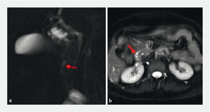
Magnetic resonance cholangiopancreatography showed cystic dilatation at the end of the dorsal pancreatic duct (red arrow).
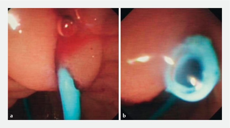
She underwent endoscopic retrograde cholangiopancreatography (ERCP). Duodenoscopy showed a swollen minor papilla and cyst-like change ( Fig. 2 ). The orifice could not be identified, and repeated cannulations failed. Since EUS showed that the cyst originated from the descending duodenum submucosa and the dorsal pancreatic duct was connected to the cyst, the minor papilla was carefully opened with a DualKnife, exposing the suspicious internal orifice ( Fig. 3 ). A 7 F × 8-cm pancreatic stent was inserted ( Fig. 4 ) after cannulation, and pancreatic juice was successfully drained ( Fig. 5 ). During the 4-week follow-up after ERCP, the patient was asymptomatic, and the pancreatic duct stent was removed.
Fig. 2.
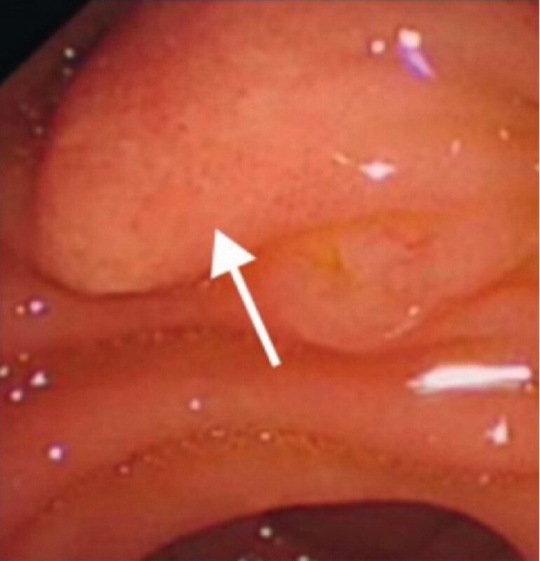
Duodenoscopy showed a swollen minor papilla and cyst-like change (white arrow).
Fig. 3 a.
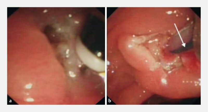
The minor papilla was opened with a DualKnife. b The suspicious internal orifice was successfully found (white arrow).
Fig. 4.
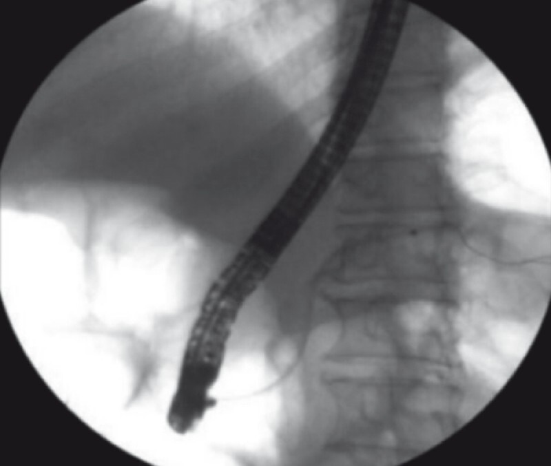
During endoscopic retrograde cholangiopancreatography treatment, a 7 F × 8-cm pancreatic stent was inserted.
Fig. 5.
Cannulation was successful and pancreatic juice was drained.
To our knowledge, this is the first case of pancreas divisum and santorinicele diagnosed by EUS and successfully treated with cannulation through the DualKnife incision of the santorinicele.
Endoscopy_UCTN_Code_CCL_1AZ_2AL
Correction.
Endoscopic ultrasonography successfully diagnosed pancreas divisum and santorinicele He Q, Xie R, Li X al. Endoscopic ultrasonography successfully diagnosed pancreas divisum and santorinicele. Endoscopy 2023 (S1), 55: E527-E528, doi 10.1055/a-1496-8969 In the above-mentioned article, the name of Huichao Wu has been corrected. This was corrected in the online version on March 15, 2023.
Footnotes
Competing interests The authors declare that they have no conflict of interest.
Endoscopy E-Videos : https://eref.thieme.de/e-videos .
Endoscopy E-Videos is an open access online section, reporting on interesting cases and new techniques in gastroenterological endoscopy. All papers include a high quality video and all contributions are freely accessible online. Processing charges apply, discounts and wavers acc. to HINARI are available. This section has its own submission website at https://mc.manuscriptcentral.com/e-videos
Correction.
Endoscopic ultrasonography successfully diagnosed pancreas divisum and santorinicele He Q, Xie R, Li X al. Endoscopic ultrasonography successfully diagnosed pancreas divisum and santorinicele. Endoscopy 2023 (S1), 55: E527-E528, doi 10.1055/a-1496-8969 In the above-mentioned article, the name of Huichao Wu has been corrected. This was corrected in the online version on March 15, 2023.
References
- 1.Eisen G, Schutz S, Metzler D et al. Santorinicele: new evidence for obstruction in pancreas divisum. Gastrointest Endosc. 1994;40:73–76. doi: 10.1016/s0016-5107(94)70015-x. [DOI] [PubMed] [Google Scholar]
- 2.Khan S A, Chawla T, Azami R. Recurrent acute pancreatitis due to a santorinicele in a young patient. Singapore Med J. 2009;50:e163–e165. [PubMed] [Google Scholar]
- 3.Klair J S, Nakshabendi R, Rajput M et al. Pancreatic mass or cyst? Diagnostic Dilemma. Dig Dis. 2019;37:521–524. doi: 10.1159/000497448. [DOI] [PubMed] [Google Scholar]
- 4.Boninsegna E, Manfredi R, Ventriglia A et al. Santorinicele: secretin-enhanced magnetic resonance cholangiopancreatography findings before and after minor papilla sphincterotomy. Eur Radiol. 2015;25:2437–2444. doi: 10.1007/s00330-015-3644-0. [DOI] [PubMed] [Google Scholar]


