Abstract
The aim of this study was to collect the scientific literature on the correction of presbyopia with laser in situ keratomileusis (presbyLASIK) in last years and to analyse the quality of such scientific evidence using a validated methodology for conducting a systematic review. A total of 42 articles were initially identified, but after applying the selection criteria and an additional manual search a total of 23 articles were finally included: 2 non-randomized controlled clinical trials (NRCT) and 21 case series. Quality assessment of NRCTs and case series was performed with the ROBINS-I and the 20-criterion quality appraisal checklist defined by Moga et al. (IHE Publ 2012), respectively. For NRCT, the risk of bias was moderate in one study and serious in the other NRCT, being the main sources of risk, the domains related to confounding, selection of participants and measurement of outcomes. For case series studies, the main source of risk of bias was subjects not entering the study at the same point of the conditions (different levels of presbyopia). Likewise, a significant level of uncertainty was detected for the following items: consecutive recruitment of patients, blinding of outcome assessors to the intervention that the patient received, and conclusions of the study not supported by the results. Research on presbyLASIK to this date is mainly focused on case series generating a limited level of scientific evidence. The two NRCTs identified only demonstrated the potential benefit of combining the multiaspheric profile with some level of monovision in the non-dominant eye.
Subject terms: Outcomes research, Vision disorders
Introduction
A definitive presbyopia correction technique is one of the main challenges for refractive surgeons nowadays, which must deal with patients with increasingly demanding visual requirements. A large proportion of myopic and hyperopic subjects, with or without astigmatism and with or without cataract, search for spectacle independence at any age, and accordingly, different types of techniques have been developed to overcome the near and intermediate visual difficulties associated to each age range. Presbyopia-correcting IOLs are increasing its visibility on clinical practice in the case of pseudophakic subjects in which the accommodative ability has been reduced or is absent, but in the case of young presbyopes with a remaining accommodative function, other techniques seem to be more appropriate than removing a functional and clear lens [1].
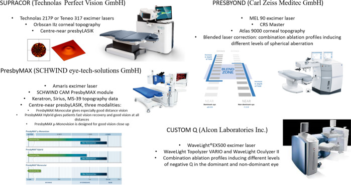
PresbyLASIK is a refractive technique in which the corneal shape is ablated with a multifocal profile, that is with a multiaspheric ablation to provide acceptable focus for distance, intermediate and near vision [2]. Different laser manufacturers have developed their own algorithms to create this multifocal profile, and accordingly different trade names have been assigned depending on the manufacturer: Supracor (Technolas Perfect Vision GmbH), Presbyond (Carl Zeiss Meditec GmbH), PresbyMax (SCHWIND eye-tech-solutions GmbH), or Custom Q (Alcon Laboratories Inc.) (Fig. 1) [3]. Some of these algorithms are programmed based on the target near addition or planned according to the subject’s age, but the effective change on refraction comes from the controlled change in the corneal asphericity from the center to the periphery, depending not only on the magnitude of near addition but also on the distance vision ablation profile (if myopic or hyperopic). Additionally, two more variants have to be considered since presbyLASIK can be central when the near vision profile is applied in the central cornea, or peripheral, when it is applied peripherally [4]. All these combinations provide a wide range of refractive options for presbyopic patients, with the additional possibility of combining a multiaspheric profile in one eye and a conventional profile in the fellow eye, generating some level of myopia and consequently some level of monovision. This wide range of options makes difficult to know the real impact of a specific multiaspheric ablation profile without the interference of factors such as the induction of micro-monovision. The aim of the current investigation was to review the scientific literature about the efficacy of presbyLASIK in myopic and hyperopic presbyopes, analysing the quality of the scientific evidence associated to this technique, the bias sources of the studies revised and to determine the requirements of future studies evaluating the clinical outcomes of this surgical option of correction of presbyopia.
Fig. 1.
Main characteristics of the four main platforms of presbyLASIK that are commercially available.
Methods
A search equation including the following terms was conducted in PubMed database: PresbyLASIK OR PresbyMax OR Presbyond OR Presbyone OR Custom Q. Additionally, the following specific selection criteria were applied as a search filter:
-
(i)
Original articles.
-
(ii)
Articles in English.
-
(iii)
Articles since 2010.
Titles and abstracts were reviewed from this first search, and only those articles whose aim was to evaluate the visual outcomes of presbyLASIK technique in presbyopic subjects were considered. Optical simulations or case reports were excluded. Duplicates were also excluded. In a second step, complete texts were reviewed to confirm the selection criteria applied. Manual search brought us back an additional article that was included afterwards.
Quality assessment of publications was performed by two methods depending on the type of study [5, 6]. The higher level of scientific evidence was represented by non-randomized clinical trials, that is prospective interventional studies, using for this type of studies the tool ROBINS-I to assess the risk of bias, as recommended [7]. At protocol stage, the review question was to evaluate presbyopic subjects (participants) operated on with presbyLASIK (experimental intervention) in comparison to monovision (comparator) for visual acuity (VA), refraction, quality of vision, spectacle independence, aberrations, or contrast sensitivity (outcomes). The aim for these studies was to assess the effect of assignment to intervention. The ROBINS-I is a tool developed to assess risk of bias in the results of non-randomized studies that compare health effects of two or more interventions. Specifically, the following bias domains are evaluated:
Pre-intervention: bias due to confounding and bias in selection of participants into the study.
At intervention: bias in classification of interventions.
Post-intervention: bias due to deviations from intended interventions, bias due to missing data, bias in measurement of outcomes, and bias in selection of the reported result.
After answering the different questions used to evaluate these domains, an overall evaluation is provided as follows: low risk of bias (the study is judged to be at low risk of bias for all domains), moderate risk of bias (he study is judged to be at low or moderate risk of bias for all domains), serious risk of bias (the study is judged to be at serious risk of bias in at least one domain, but not at critical risk of bias in any domain), and critical risk of bias (the study is judged to be at critical risk of bias in at least one domain).
The main body of the scientific evidence recruited in this search was represented by case series, that is prospective or retrospective observational studies with limited groups of participants in most of cases. For these studies, the 20-criterion quality appraisal checklist described by Moga et al. [8] was applied to evaluate this type of evidence. This tool examines different items evaluating the execution of the study (aim, recruitment, description of characteristics of subjects, inclusion criteria, definition of the intervention, blinding of assessors, follow-up, methodology, adverse events, conclusions, or competing interests) but also the quality of the reporting (clear definition of main outcomes measures and statistical tests) [9]. Depending on the number of positive responses (YES in front of PARTIAL/UNCLEAR or NO), a score is assigned. Before using the checklist, the relevant aspects should be addressed by the assessors. Summary of the applied criteria in this study is represented in Fig. 2.
Fig. 2. Criteria used for quality assessment with the 20-criterion quality appraisal checklist defined by Moga et al. [8].
Studies may provide this specific information to receive the positive rating (YES).
Results
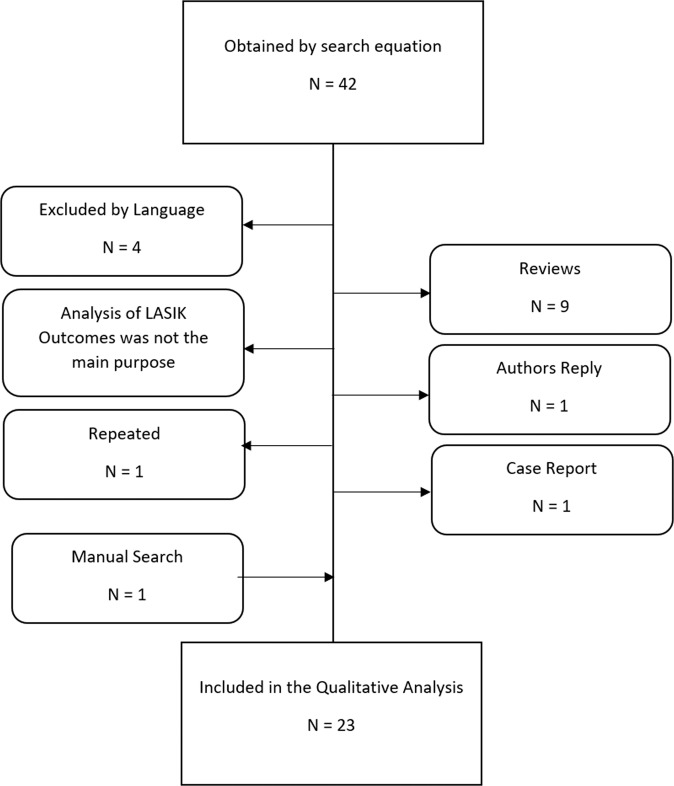
The search was conducted on 13th of May, 2021. Following the search equation described, a total of 42 articles were identified, including four articles in other languages that were excluded. After applying the selection criteria, 22 articles were selected for a more comprehensive evaluation with the selected tools. A manual search included an additional article, and therefore 23 articles were finally included: 2 non-randomized controlled clinical trials (NRCT) [10, 11] and 21 case series studies [12–32]. Figure 3 shows the flow chart followed during the search. The main causes of exclusion were nine articles that were reviews of the previous literature, and eight articles with no access to full text. Main characteristics of the articles finally included in the systematic analysis are summarized in Tables 1, 2. As shown in Table 2, different types of ablation profiles of different excimer laser platforms have been used in the articles revised.
Fig. 3.
Flow chart of the selection process of relevant articles that were included in the current systematic review.
Table 1.
Summary of main studies characteristics.
| Study | Type of Study | Prospective/retrospective | Subjects |
|---|---|---|---|
| Kohnen et al. [10] | NRCT | Prospective |
Group hybrid: 14 Group μ-monovision: 15 |
| Leray et al. [11] | NRCT | Prospective | 76 |
| Avila and Vivas [14] | Case Series | ¿? | 15 |
| Rahmania et al. [12] | Case Series | Prospective | 28 |
| Boucenna et al. [13] | Case Series | Retrospective | 23 |
| Fu et al. [17] | Case Series | Prospective | 18 |
| Fu et al. [18] | Case Series | Prospective | 22 |
| Ganesh et al. [15] | Case Series | Retrospective | 101 |
| Liu et al. [16] | Case Series | Prospective | 37 |
| Luger et al. [19] | Case Series | Retrospective | 19 |
| Villanueva et al. [20] | Case Series | Retrospective | 12 |
| Chan et al. [22] | Case Series | Retrospective | 36 |
| Pajic et al. [21] | Case Series | Prospective | 36 |
| Courtin et al. [24] | Case Series | Prospective | 65 |
| Vastardis et al. [23] | Case Series | ¿? | 19 |
| Wang et al. [25] | Case Series | Prospective | 69 |
| Luger et al. [28] | Case Series | Retrospective | 32 |
| Saib et al. [26] | Case Series | Retrospective | 37 |
| Soler et al. [27] | Case Series | Prospective |
Group symmetrical: 16 Group asymmetrical: 14 |
| Gifford et al. [29] | Case Series | Retrospective | 31 |
| Baudu et al. [31] | Case Series | Retrospective | 358 |
| Luger et al. [30] | Case Series | ¿? | 33 |
| Uthoff et al. [32] | Case Series | Prospective | 30 |
Table 2.
Characteristics of the laser ablation profile or mode used in the different studies revised in the current systematic review.
| Study | Laser | Algorithm | Uni/bilateral | Symmetry | Design |
|---|---|---|---|---|---|
| Kohnen et al. [10] | Schwind | PresbyMax Multifocal mode | Bilateral | Asymmetrical | Group Hybrid: DE Multifocal profile A; NDE Multifocal profile B/Group μ-monovision: DE Multifocal profile target Neutro; NDE Multifocal profile target μ-monovision |
| Leray et al. [11] | Allegretto | Custom Q | Unilateral | Asymmetrical | DE: Monofocal profile; NDE: Multifocal profile target μ-monovision |
| Avila and Vivas [14] | Wave Light EX 500 | Custom Q | Bilateral | Asymmetrical | DE: Multifocal profile target Neutro/NDE: Multifocal profile target μ-monovision |
| Rahmania et al. [12] | Wave Light EX 500 | Custom Q | Bilateral | Asymmetrical | DE: Multifocal profile target Neutro/NDE: Multifocal profile target Monovision |
| Boucenna et al. [13] | Technolas Teneo TM 317 | Supracor | Bilateral | Asymmetrical | DE: Multifocal profile target Neutro/NDE: Multifocal profile target μ-monovision |
| Fu et al. [17] | Amaris Schwind 1050RS | PresbyMax Schwind monocular mode | Unilateral | DE: Monofocal profile target Neutro/NDE: Multifocal profile target Neutro | |
| Fu et al. [18] | Amaris Schwind 1050RS | PresbyMax Schwind monocular mode | Unilateral | DE: Monofocal profile target Neutro/NDE: Multifocal profile target Neutro | |
| Ganesh et al. [15] | MEL 90 platform | CRS Master | Bilateral | Asymmetrical | DE: Multifocal profile target Neutro/NDE: Multifocal profile target Monovision |
| Liu et al. [16] | Amaris Schwind 750S | PresbyMax Schwind Hybrid mode | Bilateral | Asymmetrical | DE: Multifocal profile target Neutro/NDE: Multifocal profile target μ-monovision |
| Luger et al. [19] | Amaris Schwind 500 | PresbyMax Schwind Aspheric mode | Bilateral | Asymmetrical | DE: Multifocal profile target Neutro/NDE: Multifocal profile target μ-monovision |
| Villanueva et al. [20] | Amaris Schwind 500 | PresbyMax Schwind v3.01 | No information | ||
| Chan et al. [22] | Amaris Schwind 500 | PresbyMax Schwind Aspheric mode | Unilateral | DE: Monofocal profile target Neutro/NDE: Multifocal profile target Neutro | |
| Pajic et al. [21] | Technolas 217P | Supracor | Bilateral | Asymmetrical | DE: Multifocal profile target Neutro/NDE: Multifocal profile target μ-monovision |
| Courtin et al. [24] | Wave Light EX 500 | Custom Q | Unilateral | DE: Monofocal profile target Neutro/NDE: Multifocal profile target Neutro | |
| Vastardis et al. [23] | Technolas 217Z | European Conformity marked | Bilateral | Asymmetrical | DE: Multifocal profile target Neutro/NDE: Multifocal profile target μ-monovision |
| Wang et al [25] | Wave Light EX 500 | Custom Q | Bilateral | Asymmetrical | DE: Multifocal profile target Neutro/NDE: Multifocal profile target μ-monovision |
| Luger et al. [28] | Amaris Schwind 500 | PresbyMax Schwind Aspheric mode | Bilateral | Asymmetrical | DE: Multifocal profile target Neutro/NDE: Multifocal profile target μ-monovision |
| Saib et al. [26] | Technolas 217P | Supracor | Bilateral | Asymmetrical | DE: Multifocal profile target Neutro/NDE: Multifocal profile target μ-monovision |
| Soler et al. [27] | Technolas 217P | Supracor Zyoptix Tissue Saving Algorithm | Bilateral | Symmetrical Asymmetrical | Group Symmetrical: Multifocal profile target μ-monovision in both eyes; Group Asymmetrical: DE: Multifocal profile target Neutro/NDE: Multifocal profile target μ-monovision |
| Gifford et al. [29] | MEL 80 platform | CRS Master | Bilateral | Asymmetrical | DE: Multifocal profile target Neutro/NDE: Multifocal profile target Monovision |
| Baudu et al. [31] | Amaris Schwind 500 | PresbyMax Schwind Aspheric mode | Bilateral | Symmetrical | Multifocal profile target Neutro in both eyes |
| Luger et al. [30] | Amaris Schwind 500 | PresbyMax Schwind Aberration-Free mode | Bilateral | Symmetrical | Multifocal profile target Neutro in both eyes |
| Uthoff et al. [32] | Amaris Schwind 500 | PresbyMax Schwind Aberration-Free mode | Bilateral | Symmetrical | Multifocal profile target μ-monovision in both eyes |
The quality and risk of bias of the revised articles was obtained and summarized following the guidelines of each evaluation tool in Table 3 (for ROBINS-I) and Table 4 (for Moga tool). For NRCT, the risk of bias was moderate in one study [10] and serious in the other NRCT [11], being the main source of risk the domains related to confounding, selection of participants and measurement of outcomes (Table 3). For case series studies [12–32], the main sources of risk of bias was subjects not entering the study at the same point of the conditions (different levels of presbyopia) (14 “No” answers from the 21 articles evaluated) (Table 4), and the inclusion of cases collected from different centers (17 “No” answers from the 21 articles evaluated). The answer “Partial” was reported for half of the articles revised or more in the following items: establishment a priori of relevant outcome measures (10/21) and report of both competing interests and sources of support of the study (13/21). Likewise, there was a significant level of uncertainty for a large proportion of the studies revised for the following items: consecutive recruitment of patients (12 unclear answers/21), blinding of outcome assessors to the intervention that the patient received (21 unclear answers/21), and conclusions of the study not supported by the results (6 unclear answers/21).
Table 3.
Summary of the results obtained by ROBINS-I tool for risk of bias assessment for non-randomized clinical trials.
| Author, ref. | Bias due to confounding | Bias in selection of participants | Bias in classification of interventions | Bias due to deviations from intervention | Bias due to missing data | Bias in measurement of outcomes | Bias in selection of reportedresults | Overall bias |
|---|---|---|---|---|---|---|---|---|
| Kohnen et al. [10] | Low | Moderate | Low | Low | Low | Moderate | Low | Moderate |
| Leray et al. [11] | Serious | Low | Low | Low | Low | Low | Low | Serious |
Table 4.
Summary of the results of 20-criterion quality appraisal checklist tool for case series assessment developed by Mogaetal.
| Was the aim of the study clearly stated? | Was the study conducted prospectively? | Were the cases collected in more than one center? | Were the patients recruited consecutively? | Were the characteristics of the subjects included in the study described? | Were the eligibility criteria for entry into the study clearly stated? | Did patients enter the study at a similar point in the disease? | Was the intervention of interest clearly described? | Were relevant outcome measures established a priori? | Were outcome assessors blinded to the intervention that patients received? | Were the relevant outcomes measured using appropriate objective/subjective methods? | Were the relevant outcome measures made before and after the intervention? | Were the statistical tests used to assess the relevant outcomes appropriate? | Was follow-up long enough for important events and outcomes tooccur? | Were losses to follow-up reported? | Did the study provide estimates of random variability in the data analysis of relevant outcomes? | Were the adverse events reported? | Were the conclusions of the study supported by the results? | Were both competing interests and sources of support for the study reported? | |
|---|---|---|---|---|---|---|---|---|---|---|---|---|---|---|---|---|---|---|---|
| Avila and Vivas [14] | Yes | Unclear | No | Unclear | Partial | Partial | No | Yes | Yes | Unclear | Yes | Yes | Yes | Yes | Yes | Yes | Partial | Unclear | Yes |
| Baudu et al. [31] | Yes | No | No | Unclear | Yes | Yes | No | Yes | Yes | Unclear | Yes | Yes | Yes | Yes | No | Yes | Partial | Unclear | Partial |
| Boucenna et al. [13] | Yes | No | Yes | Unclear | Partial | Yes | Unclear | Yes | Yes | Unclear | Yes | Yes | Yes | Yes | No | Yes | Yes | Yes | Partial |
| Chan et al. [22] | Yes | No | No | Yes | Partial | Yes | Unclear | Yes | Yes | Unclear | Yes | Yes | Yes | Yes | Yes | Yes | Partial | Unclear | Partial |
| Courtin et al. [24] | Yes | Yes | No | Yes | Yes | Yes | No | Yes | Partial | Unclear | Yes | Yes | Yes | Yes | Yes | Yes | Yes | Yes | Partial |
| Fu et al. [17] | Partial | Yes | No | Unclear | Yes | Yes | No | Yes | Yes | Unclear | Yes | Yes | Yes | Yes | Yes | Yes | Yes | Yes | Yes |
| Fu et al. [18] | Partial | Yes | No | Yes | Yes | Yes | No | Yes | Partial | Unclear | Yes | Yes | Yes | Yes | No | Yes | Yes | Yes | Partial |
| Ganesh et al. [15] | Partial | No | No | Unclear | Partial | Partial | No | Yes | Yes | Unclear | Yes | Yes | Yes | Yes | Yes | Yes | Partial | Yes | Partial |
| Gifford et al. [29] | Partial | No | No | Unclear | Partial | Partial | No | Yes | Yes | Unclear | Yes | Yes | Yes | No | No | Yes | No | Yes | Yes |
| Liu et al. [16] | Yes | Yes | No | Yes | Yes | Yes | Yes | Yes | Partial | Unclear | Yes | Yes | Yes | Yes | Yes | Yes | Yes | Yes | Partial |
| Luger et al. [28] | Partial | No | No | Yes | Yes | Yes | Yes | Yes | Partial | Unclear | Yes | Yes | Yes | Yes | Yes | Partial | Partial | Yes | Yes |
| Luger et al. 2013 [30] | Yes | Unclear | Unclear | Unclear | Yes | Yes | No | Yes | Partial | Unclear | Yes | Yes | Yes | Yes | Yes | Yes | No | Unclear | Yes |
| Luger et al. [19] | Partial | No | No | Yes | Yes | Partial | Yes | No | Partial | Unclear | Yes | Yes | Yes | Yes | Yes | Partial | Partial | Yes | Yes |
| Pajic et al. [21] | Yes | Yes | Unclear | Unclear | No | No | Unclear | Yes | Partial | Unclear | Yes | Yes | Unclear | Yes | Yes | Yes | Partial | Yes | Yes |
| Rahmania et al. [12] | Yes | Yes | No | Yes | Partial | Yes | No | Yes | Partial | Unclear | Partial | No | Yes | Yes | Yes | Partial | Yes | Unclear | Partial |
| Saib et al. [26] | Yes | No | No | Yes | Yes | Yes | No | Yes | Partial | Unclear | Yes | Yes | Yes | Yes | Yes | Yes | Yes | Yes | Partial |
| Soler et al. [27] | Yes | Yes | No | Unclear | Yes | Partial | No | Yes | Yes | Unclear | Yes | Yes | Yes | Yes | Yes | Yes | Partial | Yes | Yes |
| Uthoff et al. [32] | Yes | Yes | No | Unclear | Yes | Yes | No | Yes | Yes | Unclear | Yes | Yes | Unclear | Yes | Yes | Yes | No | Unclear | Partial |
| Vastardis et al. [23] | Partial | Unclear | No | Unclear | Partial | Yes | No | Yes | Yes | Unclear | Yes | Yes | Yes | Yes | Yes | Partial | Partial | Yes | Partial |
| Villanueva et al. [20] | Partial | No | Unclear | Yes | Yes | Partial | Yes | Partial | Yes | Unclear | Yes | No | Yes | Yes | Yes | Yes | No | Yes | Partial |
| Wang et al. [25] | Partial | Yes | No | Unclear | Partial | Partial | No | Yes | Partial | Unclear | Yes | Yes | Yes | Yes | Yes | Yes | Yes | Yes | Partial |
Question 9 of the tool about co-interventions was not considered in the analysis.
Discussion
The induction of controlled amounts of primary spherical aberration has been shown to be an effective option to expand the depth of focus (DOF) of the eye and consequently to provide a functional correction of vision across different distances for presbyopes [33]. Likewise, the induction of some level of other types of high order aberrations (HOAs), such as secondary spherical aberration or primary coma, has also shown a positive impact on the enlargement of the DOF [34, 35]. This aberrometric induction can be generated at the corneal plane by means of an excimer laser which was the initial concept of presbyLASIK [4]. However, this technique has been modified continuously with aim of maximizing the level of DOF achieved and minimizing the level of spectacle independence [5]. This evolution has led to the combination of a multiaspheric profile inducing a controlled amount of HOAs with the concept of micro-monovision, which consists of leaving a small myopic residual refraction in the non-dominant eye. The question is if this procedure can be still considered or called presbyLASIK or if it is modified version of the monovision concept. Likewise, there are several combinations that have been proposed but their real clinical benefit is still unclear: multiaspheric profile in both eyes (classical or symmetric presbyLASIK), multiaspheric profile in both eyes with additional micro-monovision in the non-dominant eye (bilateral presbyLASIK combined with µ-monovision), different multiaspheric profiles (different addition) in both eyes (asymmetric bilateral hybrid presbyLASIK), a conventional monofocal ablation profile in the dominant eye and multiaspheric profile in the non-dominant eye (asymmetric monocular mode presbyLASIK). The aim of the current study was to revise systematically the published research on presbyLASIK to know the real evidence associated to this technique, its risk of bias, if there are sufficient evidence of the benefit of one specific combination over the other ones, and how the scientific evidence on this technique can be consistently improved.
In the bibliographic search performed, only two NRCTs were found in which central presbyLASIK was applied. Kohnen et al. [10] performed a comparison of two types of presbyLASIK techniques: 15 patients treated with bilateral presbyLASIK combined with µ-monovision and 15 patients treated with asymmetric bilateral hybrid presbyLASIK. These authors found that the hybrid and µ-monovision groups did not differ significantly except for distance-corrected near visual acuity, with a better outcome in those eyes treated with presbyLASIK combined with µ-monovision (0.21 ± 0.15 logMAR vs. 0.34 ± 0.17 logMAR). This trial showed a moderate bias due to selection of participants and measurement of outcomes, with no inclusion of a control group using a conventional ablation profile with or without monovision approach. The second NRCT included in the current systematic review was conducted by Leray et al. [11] in which the dominant eye was operated using a conventional profile and the non-dominant eye was programmed with an aspheric ablation profile and −0.75 D of residual refractive error. These authors concluded that aspheric hyperopic LASIK could increase the DOF without impairing far vision, but this conclusion should be taken with care as the increase of DOF cannot be attributed exclusively to the induction of spherical aberration because a myopic residual refractive error was present in all patients. This leads to a serious level of bias due to confounding variables.
Concerning the case series, as shown in Table 4, all studies showed one or more sources of bias, being subjects not entering the study at the same point of the conditions (different levels of presbyopia) and no inclusion of cases collected from different centers among the most detected. Likewise, some conditions to avoid bias was partially accomplished in most of the case series revised, especially in terms of establishment a priori of relevant outcome measures and report of both competing interests and sources of support of the study. Furthermore, some conditions leading to bias were not clearly identified in these case series, such as a non-consecutive recruitment of patients, not blinding of outcome assessors to the intervention that the patient received, and the description of some conclusions not fully supported by the results. It should be considered that all these series reported an improvement in intermediate and near visual performance after the laser intervention, but without a control group to compare.
Most of articles on presbyLASIK are focused on reporting the outcomes of central presbyLASIK [10–32], with very limited research on peripheral presbyLASIK [36, 37]. As most of articles on this technique were published several years ago and the current research on this issue is focused on central presbyLASIK, it can be hypothesized that some limitations could be present in peripheral presbyLASIK. However, there are no comparative studies of both techniques confirming this issue. Pinelli et al. [36] reported a mean binocular uncorrected decimal visual acuity of 1.06 ± 0.13 for distance and 0.84 ± 0.14 for near at 6 months after peripheral presbyLASIK, with a decrease of contrast sensitivity for the spatial frequencies of 3, 6, 12, and 18 cycles/degree. Epstein and Gurgos [37] investigated the outcomes of monocular peripheral presbyLASIK on the non-dominant eye with distance-directed monofocal refractive surgery on the dominant eye, reporting the achievement of complete spectacle independence in 89% of hyperopes and 92% of myopes. However, these authors did not measure the defocus curve to assess the level of visual functionality achieved and the impact on visual quality was not fully investigated.
The research on presbyLASIK shows significant weaknesses and biases that should be avoided in future trials. First, more controlled comparative studies are needed to obtain consistent conclusions about the real clinical benefit of this technique over the use of a conventional ablation profile to correct the refractive error inducing a specific level of monovision. Second, as is commonly performed in articles reporting the outcomes of presbyopia-correcting IOLs, some results and analyses should be mandatory for reporting the outcomes of any type of presbyLASIK technique, including the defocus curve, the measurement on uncorrected and distance-corrected intermediate and near visual acuity and the analysis of contrast sensitivity to confirm the real impact on visual quality. Furthermore, the evaluation of the binocular vision, with specific analysis of the impact on sensorial fusion and stereopsis, must be performed in any study evaluating the results of an asymmetric presbyLASIK approach with different types of correction in each eye. Likewise, comparative studies of the results of presbyLASIK and the outcomes with different types of presbyopia-correcting IOLs, including trifocal or extended depth of focus IOLs, should be also performed in the near future to understand better the role of this therapeutic option on presbyopia.
In conclusion, research on presbyLASIK to this date is mainly focused on case series generating a limited level of scientific evidence. The two non-randomized clinical trials identified only demonstrated the potential benefit of combining the multiaspheric profile with some level of monovision in the non-dominant eye for inducing an enhanced near visual performance. Future controlled clinical trials are needed to provide more consistent evidence of the benefit or equivalence of this therapeutic option over other options for compensation of presbyopia.
Summary
What was known before
PresbyLASIK is a refractive technique in which the corneal shape is ablated with a multifocal profile, that is with a multiaspheric ablation to provide acceptable focus for distance, intermediate and near vision in presbyopia.
Different laser manufacturers have developed their own algorithms to create their own presbyLASIK multifocal profile.
Different studies have been conducted to show the results of this option of presbyopia correction with different laser platforms.
What this study adds
Research on presbyLASIK to this date is provided a limited level of scientific evidence as it is mainly focused on case series.
Two non-randomized clinical trials have been performed that have demonstrated the combination of the multiaspheric profile with some level of monovision in the non-dominant eye generates a satisfactory outcomes.
More controlled comparative studies are needed to obtain consistent conclusions about the real clinical benefit of this technique over a monofocal ablation as well as about the real differences between the different presbyLASIK options that are currently available.
Funding
The author DPP has been supported by the Ministry of Economy, Industry and Competitiveness of Spain within the program Ramón y Cajal, RYC-2016-20471.
Competing interests
The authors declare no competing interests.
Footnotes
Publisher’s note Springer Nature remains neutral with regard to jurisdictional claims in published maps and institutional affiliations.
References
- 1.Balgos MD, Vargas V, Alió J. Correction of presbyopia: an integrated update for the practical surgeon. Taiwan J Ophthalmol. 2018;8:121–40. doi: 10.4103/tjo.tjo_53_18. [DOI] [PMC free article] [PubMed] [Google Scholar]
- 2.Vargas-Fragoso V, Alió JL. Corneal compensation of presbyopia: PresbyLASIK: an updated review. Eye Vis. 2017;4:11. doi: 10.1186/s40662-017-0075-9. [DOI] [PMC free article] [PubMed] [Google Scholar]
- 3.Shetty R, Brar S, Sharma M, Dadachanji Z, Lalgudi VG. PresbyLASIK: a review of PresbyMAX, Supracor, and laser blended vision: Principles, planning, and outcomes. Indian J Ophthalmol. 2020;68:2723–31. doi: 10.4103/ijo.IJO_32_20. [DOI] [PMC free article] [PubMed] [Google Scholar]
- 4.Alió JL, Amparo F, Ortiz D, Moreno L. Corneal multifocality with excimer laser for presbyopia correction. Curr Opin Ophthalmol. 2009;20:264–71. doi: 10.1097/ICU.0b013e32832a7ded. [DOI] [PubMed] [Google Scholar]
- 5.Zeng X, Zhang Y, Kwong JSW, Zhang C, Li S, Sun F, et al. The methodological quality assessment tools for preclinical and clinical studies, systematic review and meta-analysis, and clinical practice guideline: a systematic review. J Evid Based Med. 2015;8:2–10. doi: 10.1111/jebm.12141. [DOI] [PubMed] [Google Scholar]
- 6.Kelly RE. Biliary problems in infancy and childhood. Probl Gen Surg. 1999;16:101–7. [Google Scholar]
- 7.Sterne JA, Hernán MA, Reeves BC, Savović J, Berkman ND, Viswanathan M, et al. ROBINS-I: a tool for assessing risk of bias in non- randomised studies of interventions. BMJ. 2016;355:i4919. doi: 10.1136/bmj.i4919. [DOI] [PMC free article] [PubMed] [Google Scholar]
- 8.Moga C, Guo B, Harstall C. Development of a quality appraisal tool for case series studies using a modified delphi technique. IHE Publ. 2012:1–71. https://www.ihe.ca/advanced-search/development-of-a-quality-appraisal-tool-for-case-series-studies-using-a-modified-delphi-technique. Accessed 2 June 2021.
- 9.Guo B, Moga C, Harstall C, Schopflocher D. A principal component analysis is conducted for a case series quality appraisal checklist. J Clin Epidemiol. 2016;69:199–207.e2. doi: 10.1016/j.jclinepi.2015.07.010. [DOI] [PubMed] [Google Scholar]
- 10.Kohnen T, Böhm M, Herzog M, Hemkeppler E, Petermann K, Lwowski C. Near visual acuity and patient-reported outcomes in presbyopic patients after bilateral multifocal aspheric laser in situ keratomileusis excimer laser surgery. J Cataract Refract Surg. 2020;46:944–52. doi: 10.1097/j.jcrs.0000000000000198. [DOI] [PubMed] [Google Scholar]
- 11.Leray B, Cassagne M, Soler V, Villegas EA, Triozon C, Perez GM, et al. Relationship between induced spherical aberration and depth of focus after hyperopic LASIK in presbyopic patients. Ophthalmology. 2015;122:233–43. doi: 10.1016/j.ophtha.2014.08.021. [DOI] [PubMed] [Google Scholar]
- 12.Rahmania N, Salah I, Rampat R, Gatinel D. Clinical effectiveness of laser-induced increased depth of field for the simultaneous correction of hyperopia and presbyopia. J Refract Surg. 2021;37:16–24. doi: 10.3928/1081597X-20201013-03. [DOI] [PubMed] [Google Scholar]
- 13.Boucenna W, Hagège A, Lussato M, Morfeq H, Kochbati E, Jany B, et al. PresbyPRK vs presbyLASIK using the SUPRACOR algorithm and micromonovision in presbyopic hyperopic patients: visual and refractive results at 12 months. J Cataract Refract Surg. 2021;47:878–85. doi: 10.1097/j.jcrs.0000000000000544. [DOI] [PubMed] [Google Scholar]
- 14.Avila MY, Vivas PR. Visual outcomes in hyperopic myopic and emmetropic patients with customized aspheric ablation (Q factor) and micro-monovision. Int Ophthalmol. 2021;41:2179–85. doi: 10.1007/s10792-021-01775-4. [DOI] [PubMed] [Google Scholar]
- 15.Ganesh S, Brar S, Gautam M, Sriprakash K. Visual and refractive outcomes following laser blended vision using non-linear aspheric micro-monovision. J Refract Surg. 2020;36:300–7. doi: 10.3928/1081597X-20200407-02. [DOI] [PubMed] [Google Scholar]
- 16.Liu F, Zhang T, Liu Q. One-year results of presbyLASIK using hybrid bi-aspheric micro-monovision ablation profile in correction of presbyopia and myopic astigmatism. Int J Ophthalmol. 2020;13:271–7. doi: 10.18240/ijo.2020.02.11. [DOI] [PMC free article] [PubMed] [Google Scholar]
- 17.Fu D, Zhao J, Zhou XT. Objective optical quality and visual outcomes after the PresbyMAX monocular ablation profile. Int J Ophthalmol. 2020;13:1060–5. doi: 10.18240/ijo.2020.07.07. [DOI] [PMC free article] [PubMed] [Google Scholar]
- 18.Fu D, Zhao J, Zeng L, Zhou X. One year outcome and satisfaction of presbyopia correction using the PresbyMAX® monocular ablation profile. Front Med. 2020;7:589275. doi: 10.3389/fmed.2020.589275. [DOI] [PMC free article] [PubMed] [Google Scholar]
- 19.Luger MHA, McAlinden C, Buckhurst PJ, Wolffsohn JS, Verma S, Arba-Mosquera S. Long-term outcomes after LASIK using a hybrid bi-aspheric micro-monovision ablation profile for presbyopia correction. J Refract Surg. 2020;36:89–96. doi: 10.3928/1081597X-20200102-01. [DOI] [PubMed] [Google Scholar]
- 20.Villanueva A, Vargas V, Mas D, Torky M, Alió JL. Long-term corneal multifocal stability following a presbyLASIK technique analysed by a light propagation algorithm. Clin Exp Optom. 2019;102:496–500. doi: 10.1111/cxo.12883. [DOI] [PubMed] [Google Scholar]
- 21.Pajic B, Pajic-Eggspuehler B, Mueller J, Cvejic Z, Studer H. A Novel laser refractive surgical treatment for presbyopia: optics-based customization for improved clinical outcome. Sensors. 2017;17:1367. doi: 10.3390/s17061367. [DOI] [PMC free article] [PubMed] [Google Scholar]
- 22.Chan TCY, Kwok PSK, Jhanji V, Woo VCP, Ng ALK. Presbyopic correction using monocular bi-aspheric ablation profile (PresbyMAX) in hyperopic eyes: 1-year outcomes. J Refract Surg. 2017;33:37–43. doi: 10.3928/1081597X-20161006-03. [DOI] [PubMed] [Google Scholar]
- 23.Vastardis I, Pajic-Eggspühler B, Müller J, Cvejic Z, Pajic B. Femtosecond laser-assisted in situ keratomileusis multifocal ablation profile using a mini-monovision approach for presbyopic patients with hyperopia. Clin Ophthalmol. 2016;10:1245–56.. doi: 10.2147/OPTH.S102008. [DOI] [PMC free article] [PubMed] [Google Scholar]
- 24.Courtin R, Saad A, Grise-Dulac A, Guilbert E, Gatinel D. Changes to corneal aberrations and vision after monovision in patients with hyperopia after using a customized aspheric ablation profile to increase corneal asphericity (Q-factor) J Refract Surg. 2016;32:734–41. doi: 10.3928/1081597X-20160810-01. [DOI] [PubMed] [Google Scholar]
- 25.Wang Yin GH, McAlinden C, Pieri E, Giulardi C, Holweck G, Hoffart L. Surgical treatment of presbyopia with central presbyopic keratomileusis: one-year results. J Cataract Refract Surg. 2016;42:1415–23. doi: 10.1016/j.jcrs.2016.07.031. [DOI] [PubMed] [Google Scholar]
- 26.Saib N, Abrieu-Lacaille M, Berguiga M, Rambaud C, Froussart-Maille F, Rigal-Sastourne JC. Central presbyLASIK for hyperopia and presbyopia using micro-monovision with the Technolas 217P platform and SUPRACOR algorithm. J Refract Surg. 2015;31:540–6. doi: 10.3928/1081597X-20150727-04. [DOI] [PubMed] [Google Scholar]
- 27.Soler Tomás JR, Fuentes-Páez G, Burillo S. Symmetrical versus asymmetrical presbyLASIK: results after 18 months and patient satisfaction. Cornea. 2015;34:651–7. doi: 10.1097/ICO.0000000000000339. [DOI] [PubMed] [Google Scholar]
- 28.Luger MHA, McAlinden C, Buckhurst PJ, Wolffsohn JS, Verma S, Arba Mosquera S. Presbyopic LASIK using hybrid bi-aspheric micro-monovision ablation profile for presbyopic corneal treatments. Am J Ophthalmol. 2015;160:493–505. doi: 10.1016/j.ajo.2015.05.021. [DOI] [PubMed] [Google Scholar]
- 29.Gifford P, Kang P, Swarbrick H, Versace P. Changes to corneal aberrations and vision after Presbylasik refractive surgery using the MEL 80 platform. J Refract Surg. 2014;30:598–603. doi: 10.3928/1081597X-20140709-01. [DOI] [PubMed] [Google Scholar]
- 30.Luger MH, Ewering T, Arba-Mosquera S. One-year experience in presbyopia correction with biaspheric multifocal central presbyopia laser in situ keratomileusis. Cornea. 2013;32:644–52.. doi: 10.1097/ICO.0b013e31825f02f5. [DOI] [PubMed] [Google Scholar]
- 31.Baudu P, Penin F, Arba, Mosquera S. Uncorrected binocular performance after biaspheric ablation profile for presbyopic corneal treatment using AMARIS with the PresbyMAX module. Am J Ophthalmol. 2013;155:636–47. doi: 10.1016/j.ajo.2012.10.023. [DOI] [PubMed] [Google Scholar]
- 32.Uthoff D, Pölzl M, Hepper D, Holland D. A new method of cornea modulation with excimer laser for simultaneous correction of presbyopia and ametropia. Graefes Arch Clin Exp Ophthalmol. 2012;250:1649–61. doi: 10.1007/s00417-012-1948-1. [DOI] [PubMed] [Google Scholar]
- 33.Fernández J, Rodríguez-Vallejo M, Burguera N, Rocha-de-Lossada C, Piñero DP. Spherical aberration for expanding the depth of focus: a review for the anterior segment surgeon. J Cataract Refract Surg. 2021;47:1587–95. doi: 10.1097/j.jcrs.0000000000000713. [DOI] [PubMed] [Google Scholar]
- 34.Legras R, Benard Y, Lopez-Gil N. Effect of coma and spherical aberration on depth-of-focus measured using adaptive optics and computationally blurred images. J Cataract Refract Surg. 2012;38:458–69. doi: 10.1016/j.jcrs.2011.10.032. [DOI] [PubMed] [Google Scholar]
- 35.Benard Y, Lopez-Gil N, Legras R. Optimizing the subjective depth-of-focus with combinations of fourth- and sixth-order spherical aberration. Vis Res. 2011;51:2471–7. doi: 10.1016/j.visres.2011.10.003. [DOI] [PubMed] [Google Scholar]
- 36.Pinelli R, Ortiz D, Simonetto A, Bacchi C, Sala E, Alió JL. Correction of presbyopia in hyperopia with a center-distance, paracentral-near technique using the Technolas 217z platform. J Refract Surg. 2008;24:494–500. doi: 10.3928/1081597X-20080501-07. [DOI] [PubMed] [Google Scholar]
- 37.Epstein RL, Gurgos MA. Presbyopia treatment by monocular peripheral presbyLASIK. J Refract Surg. 2009;25:516–23. doi: 10.3928/1081597X-20090512-05. [DOI] [PubMed] [Google Scholar]


