Abstract
The Dictyostelid social amoebas represent one of nature’s several inventions of multicellularity. Though normally feeding as single cells, nutrient stress triggers collection of amoebas into colonies that form delicately shaped fruiting structures in which the cells differentiate into spores and up to three cell types to support the spore mass. Cyclic AMP (cAMP) plays a very dominant role in controlling morphogenesis and cell differentiation in the model species D. discoideum. As a secreted chemoattractant cAMP coordinates cell movement during aggregation and fruiting body morphogenesis. Secreted cAMP also controls gene expression at different developmental stages, while intracellular cAMP is extensively used to transduce the effect of other stimuli that control the developmental programme. In this review, I present an overview of the different roles of cAMP in the model D. discoideum and I summarize studies aimed to resolve how these roles emerged during Dictyostelid evolution.
Keywords: Amoebozoa, Cyclic AMP, Dictyostelium, Evolution of multicellularity, Encystation
INTRODUCTION
Dictyostelia are soil-dwelling protists that exist both as unicellular predators and as gregarious community members. They start life as amoebas that feed on bacteria in decaying vegetation. Lack of food triggers social behaviour and the amoebas move together to form a colony that proceeds to build a fruiting structure. Here the best fed amoebas enter a dormant spore stage and the rest construct a pedestal to bear the spore mass aloft. Over a hundred species of Dictyostelia have currently been identified, but research has mostly been focussed on the model species Dictyostelium discoideum. This is largely a consequence of the fact that this species uses the well-known intracellular messenger cyclic AMP (cAMP) as a secreted signal to coordinate the aggregation process (Konijn et al., 1967).
In addition to forming fruiting bodies, many species can encyst individually when starved and form dormant microcysts (Raper, 1984; Kessin, 2001). Microcysts are less dehydrated than spores and have a thinner two-layered cell wall instead of the thick three-layered spore coat (Hohl et al., 1970). This process of encystation in response to stress is used by most if not all amoebozoan ancestors of the Dictyostelia and is also common among other unicellular protists (Eichinger, 2001). Alternatively, Dictyostelia can form sexual macrocysts (Urushihara & Muramoto, 2006). This often requires the presence of cells with opposite mating types, although homothallic mating also occurs. Macrocyst formation begins when two cells fuse to form a zygote. The zygote then chemotactically attracts other starving cells and cannibalizes them in order to synthesize a highly resistant thick cell wall, whereupon the cyst enters a long period of dormancy. More information on the sexual cycle and the recent identification of the Dictyostelium mating type loci (Bloomfield et al., 2010) can be found in Chapter 15 of this issue. The formation of zygotic cysts or zygospores is also fairly common, occurring e.g. in some fungi (Valle & Santamaria, 2005) and in the Volvocales (Gilbert, 2006). However, the cannabalistic embellishment appears thus far to be unique for the Dictyostelia. In Dictyostelia, macrocysts and microcysts are more usually formed under dark and wet conditions that are not favorable for fruiting body formation, which requires an air-water interface (Raper, 1984; Kessin, 2001).
In this review, I will firstly discuss the taxonomic position of the Dictyostelia and the phenotypic distinctions that characterize the major groups. I will then present an overview of the roles of cAMP in controlling morphogenesis and cell differentiation in the model organism D. discoideum and finally discuss recent studies aimed to reconstruct the evolutionary history of cAMP signalling in the Dictyostelia and its contribution to the evolution of multicellular complexity in the group.
TAXONOMY
Dictyostelia are also known as ‘cellular slime molds’, due to the fact that their fruiting structures resemble those of small species of fungi. However, it is now clear that this morphology-based classification does not reflect the genetic relationships between different groups. Until about 15 years ago, all amoeba-like protists that formed a spore-bearing structure were classified as Myxomycota, placed in the kingdom Fungi. The Myxomycota were subdivided into the Protostelids with fruiting structures of 1–4 cells, the true slime molds or Myxogastrids that form fruiting bodies from a multinucleate syncytium, and the Acrasiomycetes that form fruiting bodies from cell aggregates. The Dictyostelia were placed as a subclass of the Acrasiomycetes, and subdivided into three genera: Dictyostelium with simple or branched cellular stalks, Acytostelium with acellular stalks, and Polysphondylium with regular whorls of side branches. The other subclass of Acrasiomycetes – the Acrasids – differ from the Dictyostelids in the morphology of their amoebas and aggregates, and the lack of cellulose in spore-bearing structures [Raper, 1984 #4574].
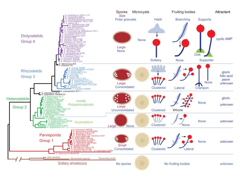
Modern taxonomy based on gene or protein sequence comparisons shows an entirely different view. The Protostelia, Myxogastrids, and Dictyostelia are members of the supergroup Amoebozoa, and this group is separate from but closely related to the Opisthokonts, the group containing the animals and fungi (Baldauf & Doolittle, 1997, Baldauf et al., 2000). Most importantly, most Acrasiomycetes are members of the unrelated supergroup Discicristates, but one species, Fonticula alba is a member of the Opisthokonts (Brown et al., 2009). The Protostelia are polyphyletic and emerged several times independently across the Amoebozoa (Shadwick et al., 2009). The Dictyostelia are monophyletic and are subdivided into four major groups (Figure 1), which, in order of divergence from their last common ancestor, are called the Parvisporids (group 1), Heterostelids (group 2), Rhizostelids (group 3) and Dictyostelids (group 4). None of these groups correspond to the three traditional genera. In fact, group 2, the Heterostelids, includes members of all three (Dictyostelium, Polysphondylium, and Acytostelium). Evidently, similarity of fruiting body morphology is not a good marker for genetic similarity.
Figure 1. Phenotypic evolution in the Dictyostelia.
A molecular phylogeny based on small subunit ribosomal RNA sequences subdivides most known species of Dictyostelia into four major groups (Schaap et al., 2006). A mapping of species characters onto the phylogeny revealed a number of trends in the evolution of phenotype. The Parvisporids take their name from the fact that their spores are smaller than those in the other groups. These spores carry clusters of consolidated granules at their poles, a feature that is also common to the larger spores of the group 3 Rhizostelids. The genum Acytostelium forms a clade within the Heterostelids. It contains the only species with an acellular stalk and except for one species, A. ellipticum, their spores are globose. The other Heterostelids have mainly elliptical spores with unconsolidated granules. The group 4 Dictyostelids have no polar granules. Species in groups 1 to 3 usually form multiple organizing tips on their aggregates, which give rise to multiple clustered fruiting bodies. Additional tips can also be formed later, initiating formation of side branches. Group 4 aggregates tend to form single tips giving rise to large solitary unbranched fruiting bodies, which also have a third cellular structure, the basal disk or supporter, to buttress the stalk. Group 4 species furthermore stand out by having lost the ability to encyst individually and by using cAMP as chemoattractant (Schaap et al., 2006).
There are however some morphological characters that are group-specific (Figure 1). Species in groups 1–3 generally form small clustered and branched fruiting structures from a single aggregate, while group 4 species tend to form a single robust unbranched structure. Many species in groups 1–3 can still form microcysts, but group 4 species have lost this survival strategy. On the other hand, sexual macrocyst formation occurs in all taxon groups. Group 4 species also stand out by using cAMP as chemoattractant, with a variety of other compounds being used by the other groups (Schaap et al., 2006).
ROLES OF cAMP IN DICTYOSTELIUM DISCOIDEUM
From growth to aggregation
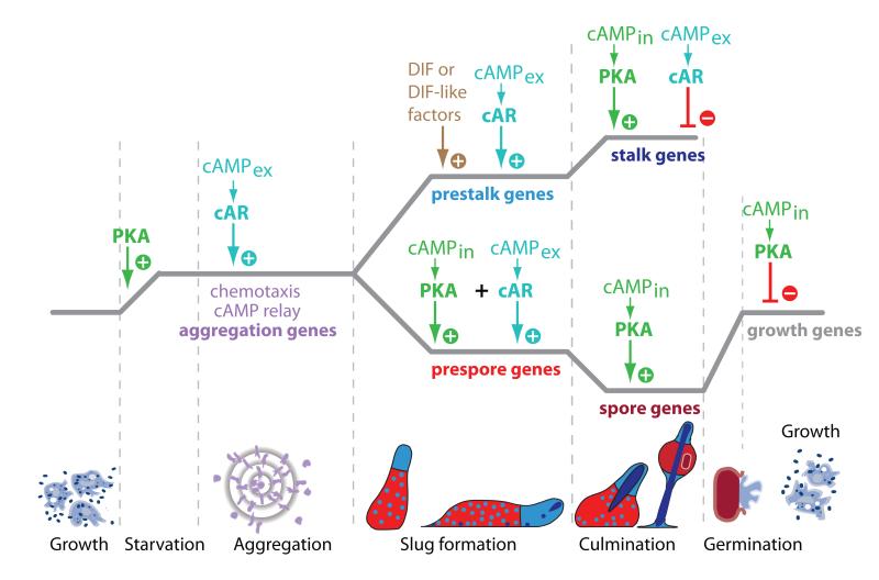
One of the most remarkable aspects of D. discoideum development is that so much of it is regulated by cAMP (Figure 2). As a secreted signal, cAMP controls cell movement and differentiation throughout the developmental programme, but in its more common role as intracellular messenger, it mediates the effect of many other developmental signals. The main intracellular target for cAMP is cAMP-dependent protein kinase or PKA, which similar to fungal PKAs, consist of a single regulatory (PKA-R) and a single catalytic subunit (PKA-C) (Mutzel et al., 1987). During canonical PKA activation, cAMP binds to PKA-R, which causes PKA-R to dissociate from PKA-C, leaving PKA-C in its active form. However, because PKA-C is active on its own, the ratio of inhibitory PKA-R to active PKA-C molecules is also an important determinant for PKA-C activity.
Figure 2. Roles of intracellular and secreted cAMP during D. discoideum development.
The transition from growth to aggregation requires release of PKA from translational repression. At this stage, PKA triggers a basal level of expression of genes required for aggregation, such as cAR1, PdsA and ACA, which enable the cells to synthesize and secrete cAMP pulses. In addition to inducing chemotaxis and cell aggregation, the cAMP pulses further upregulate the expression of aggregation genes. After aggregation, ACG is translationally upregulated in the posterior region of the emerging slug. ACG synthesizes cAMP that acts both on cARs and PKA to induce prespore gene expression. The prespore cells produce DIF and DIF-like factors that cause prestalk cell differentiation in combination with cAMP acting on cARs. Later, extracellular cAMP (cAMPex) becomes an inhibitor of stalk cell differentiation. During fruiting body formation intracellular cAMP (cAMPin) acting on PKA is essential for terminal maturation of spores and stalk cells. In the spores, active PKA blocks the transition from dormancy to germination.
This is evident in early development, where PKA-C is regulated at the translational level. PKA activity is not required for growth, but it is essential for the transition from growth to aggregation (Simon et al., 1989). In feeding cells, PKA-C translation is inhibited by binding of the translational repressor PufA to the 3′ untranslated region (UTR) of PKA-C mRNA (Souza et al., 1999). Upon starvation, this repression is relieved by YakA, a member of a deeply conserved protein kinase family that also regulates the decision between growth and differentiation in animals and fungi (Souza et al., 1998, Hartley et al., 1994, Mercer & Friedman, 2006). Once synthesized, PKA-C activates full expression of early genes such as discoidin I, and a basal level of expression of genes that are required for aggregation, such as the cAMP receptor cAR1, the extracellular cAMP phosphodiesterase PdsA, and the adenylate cyclase ACA (Schulkes & Schaap, 1995).
cAR1, PdsA, ACA and several other proteins, amongst which PKA and the intracellular cAMP phosphodiesterase RegA, form a biochemical network that can generate cAMP in an oscillatory manner (Laub & Loomis, 1998). The cAMP pulses are initially secreted by a few starving cells and elicit three responses: 1. cAMP-induced cAMP secretion, also called cAMP relay, which results in propagation of the cAMP pulse throughout the cell population (Roos et al., 1975) 2. Chemotactic movement of cells towards the cAMP source, resulting in cell aggregation (Konijn et al., 1967). 3. Upregulation of aggregation genes, causing all cells to become rapidly competent for aggregation (Gerisch et al., 1975).
Once aggregation is completed, cAMP waves continue to be emitted from the top of cell mounds (Siegert & Weijer, 1995), causing continued cell movement towards the top and emergence of a slug-shaped structure. The slug next falls over and starts to migrate over the substratum guided by light and warmth, which in nature bring it to the top level of the soil, where in response to incident light, it will initiate fruiting body formation. cAMP waves originating from slug tip continue to guide and shape the organism during migration and fruiting body formation by coordinating the movement of its component cells (Dormann & Weijer, 2001). After aggregation, secreted cAMP gains other roles in cell type-specification, and new layers of complexity in cAMP signalling become apparent.
From aggregate to fruiting body
Once aggregated, overt phenotypic differences between the cells become evident, which mark their entry into either the spore or stalk cell differentiation pathway. At first these prestalk and prespore cells are interspersed with each other (Figure 2). However, the prespore cells become less sensitive to cAMP pulses, which causes the prestalk cells to move selectively to the oscillating tip and take up an anterior position in the emerging slug (Matsukuma & Durston, 1979, Traynor et al., 1992). During fruiting body formation, the prestalk cells first lay down a central cellulose tube. They then crawl into the tube and differentiate into stalk cells. This differentiation process involves massive vacuolization of the cells and construction of a cellulose cell wall (Raper & Fennell, 1952). The prespore cells have meanwhile synthesized the first layer of the spore wall and further spore wall precursors in Golgi-derived prespore vesicles. They climb up the newly formed stalk and initiate spore formation by rapid fusion of the prespore vesicles with the plasma membrane (West, 2003).
The differentiation of prespore cells requires both extracellular cAMP acting on cARs (Schaap & Van Driel, 1985, Wang et al., 1988), and intracellular cAMP acting on PKA (Hopper et al., 1993b). In slugs, ACA is mainly expressed in the tip region (Verkerke-van Wijk et al., 2001, Galardi-Castilla et al., 2010), but a second adenylate cyclase, ACG, becomes translationally upregulated in the posterior prespore region. ACG is localized in the membrane of prespore vesicles and synthesizes cAMP that remains both intracellular to activate PKA and is secreted to activate cARs (Alvarez-Curto et al., 2007).
The prestalk cells consist of several subpopulations that are destined to either form the stalk, the basal disk and lower cup that support the stalk and spore head, respectively, and the upper cup that caps the spore head (Williams, 2006) (Yamada et al., 2010). A chlorinated polyketide, called DIF-1 (Differentiation Inducing Factor 1) is responsible for inducing differentiation of basal disk cells (Saito et al., 2008), while related molecules most likely induce the other prestalk cell types (Saito et al., 2006, Serafimidis & Kay, 2005). Secreted cAMP promotes prestalk differentiation at its earlier stage (Verkerke-Van Wijk et al., 1998).
The transition of prestalk into stalk cells requires intracellular cAMP acting on PKA, but is inhibited by extracellular cAMP acting on cARs (Hopper et al., 1993a, Harwood et al., 1992). Prestalk cells express a third adenylate cyclase, ACR, which is localized on the nuclear envelope and endoplasmic reticulum (Alvarez-Curto et al., 2007, Chen et al., 2010).
Spore maturation and germination
Fruiting body formation or culmination requires coordinated movement of cells up to the very end, when spores mature after reaching the top of the stalk. Because both stalk and spore cells become immobilized by formation of a rigid cell wall, their final differentiation stages require very accurate regulation. Largely through the work of Anjard and coworkers, a range of signals were identified, which are exchanged between the maturing prestalk and prespore cells, and control their terminal differentiation.
Culmination initiates when the migrating slug projects its tip upward. This normally occurs in response to incident light and allows loss of ammonia from the tip by gaseous diffusion. Ammonia is the end-product of protein degradation in the starving cells and acts as a signal to block both initiation of fruiting body formation and the maturation of stalk cells (Schindler & Sussman, 1977, Wang & Schaap, 1989). Both processes are dependent on PKA activity (Harwood et al., 1992, Hopper et al., 1993b), and ammonia indirectly inhibits PKA by promoting cAMP hydrolysis. Ammonia is one of several signals that regulate the activity of sensor histidine kinases/phosphatases, which, upon ligand binding initiate either forward or reverse phosphoryltransfer that ultimately leads to phosphorylation or dephosphorylation of a conserved aspartate in the response regulator of the intracellular cAMP phosphodiesterase RegA (Thomason et al., 1998, Shaulsky et al., 1998). Ammonia activates forward phosphoryl transfer by acting on the histidine kinase DhkC, resulting in RegA activation, hydrolysis of cAMP and inhibition of PKA (Singleton et al., 1998). Loss of ammonia from the tip therefore allows PKA activation and stalk maturation at this position.
At the onset of culmination, the steroid SDF-3 (spore differentiation factor 3) is released, which triggers the production of γ-amino butyric acid (GABA) by prespore cells (Anjard et al., 2009). GABA has two effects: it triggers the secretion of Acyl-CoA binding protein (AcbA) from prespore cells, and it causes exposure of the TagC serine protease at surface of prestalk cells. TagC cleaves secreted AcbA to form SDF-2 (Anjard & Loomis, 2005, Anjard & Loomis, 2006, Cabral et al., 2006), which in turn activates the histidine phosphatase DhkA of prespore cells, leading to dephosphorylation, and thereby inactivation of RegA. The resulting increase in intracellular cAMP then causes PKA activation and spore maturation (Wang et al., 1999). In addition to this cascade, two other signals are required for spores to mature: i, the secreted peptide SDF-1, which promotes cAMP production by ACG leading to PKA activation and ii, the cytokinin discadenine, which acts on the histidine kinase DhkB and ACR to upregulate PKA (Anjard & Loomis, 2008).
The germination of spores is also under very tight regulation and here cAMP acting on PKA is the predominant control mechanism, acting in this case to maintain dormancy. Large amounts of ammonium phosphate are present in the spore head generating an ambient osmolarity of about 0.2 osmolar (Cotter et al., 1999). During spore maturation, prespore vesicles fuse with the plasma membrane, and ACG, which harbours an intramolecular osmosensor domain, now becomes localized on the plasmamembrane (Saran & Schaap, 2004). Here it is activated by the high osmolarity, resulting in PKA activation and maintenance of dormancy (Van Es et al., 1996). Discadenine, which is also present in the spore head (Abe et al., 1981), continues to act on DhkB to activate PKA and maintain dormancy. Spore can only germinate after being dispersed and freed from these inhibitory agents and even then only germinate under conditions that favor growth of the emerging amoeba (Dahlberg & Cotter, 1978).
When taking a broad overview of Dictyostelium development, it appears that the roles of both intracellular and secreted cAMP are to bring starving amoebas in an encapsulated dormant state and to keep them there until conditions improve.
THE EVOLUTION OF cAMP SIGNALING IN THE DICTYOSTELIA
A study of developmental signalling in D. discoideum development naturally leads to the question: why does cAMP play such a dominant role? The rationale for any multilayered biological process can only be derived from the order in which its component parts evolved. Studies were therefore initiated to reconstruct the evolutionary history of cAMP signalling in the Dictyostelia.
The deep origins of ACG and PKA
The purpose of Dictyostelium development is to produce resilient dispersable spores in response to stress. While all Dictyostelia form spores, not all of them form stalk cells. The spore differentiation pathway is therefore the most likely ancestral differentiation pathway. In this pathway, ACG plays a central role, firstly by inducing the differentiation of prespore cells and secondly by regulating the process of spore germination (Van Es et al., 1996, Alvarez-Curto et al., 2007). ACG has a highly conserved catalytic domain, which makes it a good candidate for phylogeny-wide identification of orthologs by screening of DNA libraries and/or amplification by PCR. A full-length ACG cDNA clone was retrieved from the group 3 species D. minutum, while PCR fragments of the catalytic domain were amplified from the group 1 species D. fasciculatum and the group 2 species P. pallidum. D. minutum ACG was also activated by high osmolarity and this condition was found to universally inhibit spore germination in species from all four taxon groups (Ritchie et al., 2008).
Many species in groups 1-3, have retained the ancestral stress survival strategy of encystation (Figure 1). Cyst germination is also inhibited by high osmolarity, but remarkably, high osmolarity actively triggers encystation, while the cells are still actively feeding. Osmolarity-induced encystation is accompanied by an increase in cAMP levels, indicating that ACG mediates this process. Membrane-permeant PKA analogs also induce encystation, while inhibition of PKA prevents osmolarity-induced encystation, suggesting that similar to prespore induction, ACG acts on PKA (Ritchie et al., 2008). However, in constrast to prespore differentiation cAMP acting on cARs is not required.
These studies and earlier work (Toama & Raper, 1967) established high osmolarity as an independent trigger for encystation. Free-living soil amoebas are not only exposed to starvation, but also to drought. Increased osmolarity due to increasing mineral concentrations in drying soil is most likely a natural environmental trigger for encystation. The roles of ACG and PKA in prespore differentiation and spore germination are homologous to those in the more ancestral process of cyst formation and germination, which strongly suggests that ACG and PKA regulation of sporulation is evolutionary derived from ACG and PKA regulation of encystation.
The emerging roles of secreted cAMP
Another important function of cAMP is its role as chemoattractant, coordinating both aggregation and fruiting body formation in D. discoideum, while secreted cAMP has additional roles in the induction of aggregation genes and prespore genes and the repression of stalk genes (Figure 2). In D. discoideum, all roles of secreted cAMP are mediated by G-protein coupled cAMP receptors and the presence of such receptors across the Dictyostelid phylogeny is therefore most diagnostic for identification of conserved roles of secreted cAMP.
Orthologs of the D. discoideum cAMP receptor cAR1 could by identified by PCR or screening of genomic libraries in representative species of all four taxon groups. A number of species have only a single cAR, but independent gene duplications occurred in the different groups yielding up to 4 cARs in group 4, and 2 or 3 cARs in group 2 (Alvarez-Curto et al., 2005, Kawabe et al., 2009). In D. discoideum, cAR1 is expressed from separate early and late promoters during aggregation and post-aggregative development respectively (Louis et al., 1993) and car1 null mutants loose the ability to produce cAMP pulses and to aggregate and form fruiting bodies (Sun & Devreotes, 1991). D. minutum uses folic acid instead of cAMP for aggregation (De Wit & Konijn, 1983). Nevertheless, its cAR gene fully restores cAMP binding, oscillatory cAMP signalling, aggregation and fruiting body formation of a D. discoideum car1 null mutant, indicating that the D. minutum cAR is functionally identical to D. discoideum cAR1 (Alvarez-Curto et al., 2005).
The non-hydrolysable cAMP analog Sp-cAMPS inhibits oscillatory cAMP signalling by binding to cAR1 and causing permanent cAR desensitization (Van Haastert & Van der Heijden, 1983). Similar to a cAR1 gene deletion, it prevents cells from forming aggregates and developing into fruiting bodies (Rossier et al., 1978). Species in groups 1-3 do not use cAMP for aggregation and mainly express cAR1 after aggregation. In these species, SpcAMPS has no effect on aggregation, but it completely disrupts the process of fruiting body formation (Alvarez-Curto et al., 2005). This suggests that in groups 1-3 oscillatory cAMP signalling is universally required to coordinate cell movement during fruiting body morphogenesis.
The group 2 species Polysphondylium pallidum has two cAR1 orthologs. Disruption of the first gene, called TasA, causes loss of the whorls of side branches from the fruiting body that are typical for this species (Kawabe et al., 2002). Additional loss of the second gene, TasB, causes severe disruption of fruiting body morphogenesis (Kawabe et al., 2009). However, the double cAR null mutant still aggregates normally, confirming that in basal species cARs are required for fruiting body morphogenesis, but not for aggregation.
The P. pallidum cAR double null mutant displays another remarkable phenotype. Its stunted fruiting bodies consist of a disorganized mass of vacuolated stalk cells, indicating that secreted cAMP is not required for stalk cell differentiation per se. However, instead of elliptical spores, the top of the structure contains round encapsulated cells with the same ultrastructure and physiology as microcysts, which are normally only formed from unaggregated cells. The cAR null mutant no longer expresses prespore genes in response to cAMP stimulation (Kawabe et al., 2009).
When including the data discussed in the previous section, it becomes clear why microcyst formation occurs. Both spore formation and encystation require intracellular cAMP acting on PKA, but spore formation also requires extracellular cAMP acting on cARs. Because this pathway is no longer present in the P. pallidum cAR null mutant, the cells revert to encystation. This confirms that spore formation is evolutionary derived from encystation and points to what might be the most ancestral role for secreted cAMP. The Dictyostelid ancestor already used intracellular cAMP to mediate stress-induced encystation. Dictyostelids secrete most of the cAMP that they produce and accumulation of cAMP in aggregates may have acted to inform cells of their aggregated state and cause them to form spores and not cysts. Such a mechanism provides a rationale for the observation that prespore induction requires much higher (micromolar) cAMP concentrations (Schaap & Van Driel, 1985, Oyama & Blumberg, 1986) than chemotaxis and cAMP relay (0.1–30 nM) that take place before cells have collected in aggregates (Van Haastert & Konijn, 1982). cAMP can only accumulate to micromolar concentrations between the closely packed aggregated cells.
A MODEL FOR THE EVOLUTION OF cAMP SIGNALLING
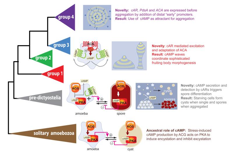
We can now tentatively reconstruct how cAMP signalling evolved in the Dictyostelia and understand why cAMP has so many different roles in this organism (Figure 3). It appears that cAMP acting on PKA triggers encystation not only in the Dictyostelia but also in the distantly related solitary amoebozoa Hartmannella culbertsoni and Entamoeba invadens (Raizada & Murti, 1972, Coppi et al., 2002), while osmolyte-induced encystation was observed in Acanthamoeba castellani and Hartmannella rhysodes (Band, 1963, Cordingley et al., 1996). Although these phenomena have not been causally linked before, they very likely indicate that cAMP and PKA are universal intermediate for osmolyte-induced encystation in amoebozoa. Basal dictyostelids do not use cAMP to aggregate, and the first colonial amoebas may have adapted their food-seeking strategy for aggregation, while still using cAMP intracellularly to trigger encystation. Our data suggest that accumulation of passively secreted cAMP in aggregates was probably used next as a signal to prompt the starving cells to form spores and not cysts. Oscillatory cAMP signaling would likely have evolved later, firstly to coordinate cell movement in the aggregated cell masses, thus allowing the amoebas to form well-structured fruiting bodies, and finally to coordinate the aggregation process in the most recently diverged group 4 (Kawabe et al., 2009). The three genes, ACA, cAR1 and PdsA that are absolutely essential for oscillatory cAMP production (Kriebel & Parent, 2004) all have multiple promoters. In all three, the promoter that directs expression after aggregation is closest to the coding sequence, whereas the promoter that directs expression before and during aggregation is at a more distal location (Louis et al., 1993, Faure et al., 1990, Alvarez-Curto et al., 2005, Galardi-Castilla et al., 2010). This suggests that the novel role for cAMP as the chemoattractant that mediates aggregation in group 4 was achieved by adding distal promoters to the existing cAMP signalling genes.
Figure 3. A model for the evolution of cAMP signaling in the Dictyostelia.
Starting from the solitary ancestors of the Dictyostelia, which used intracellular cAMP acting on PKA to mediate stress-induced encystation, the first colonial amoebas may have used accumulated levels of passively secreted cAMP as a signal for the aggregated state, which informed them to proceed with spore rather than cyst formation. Pulsatile cAMP secretion probably evolved next when ACA activity came under cAR mediated positive and negative feedback. This allowed the sophisticated coordination of fruiting body morphogenesis that marks all modern Dictyostelia. Expression of cAMP signalling genes at an earlier developmental stage finally led to the use of cAMP as chemoattractant for aggregation in the group 4 species.
CONCLUSIONS
The complexity and apparent redundancy of the signalling networks that control the development and other functions of multicellular organisms can often appear entirely baffling. In D. discoideum, one set of the pathways that is well characterized is that controlling the chemotactic response. At the latest count, there were four different, but interlinked pathways, that translate an extracellular cAMP gradient into directional cell polarization (Veltman et al., 2008). Another set, outlined in this review, are the pathways that control spore maturation and germination. These processes involve a broad spectrum of signals, ranging from solute stress, a catabolite, steroid, and cytokinin to a neurotransmitter and a neuropeptide, detected by a variety of receptors ranging from a sensor-linked adenylate cyclase to G-protein coupled receptors and sensor-linked histidine kinases. Quite remarkably, all these signals ultimately act to regulate one event: the activation of PKA by cAMP. As I hope to have demonstrated in this review, the evolutionary reconstruction of signalling pathways reveals why this is the case; cAMP activation of PKA originally mediated stress-induced encapsulation of the unicellular ancestor. Interestingly, PKA activation not only triggers encapsulation of the viable spores, but also of the stalk cells, suggesting that the stalk pathway is also derived from the encystation pathway. This is somewhat evident from the fact that the two-layered microcyst cell wall resembles the two-layered stalk cell wall more closely than the three-layered spore wall (Kawabe et al., 2009), but requires information about shared gene products for further confirmation. The origin of the third encysted cell type, the macrocyst, is still unclear. Kessin argues that the relatively simple developmental programme of the macrocysts indicates that they evolved before fruiting bodies (Kessin, 2001). However, analysis of character evolution in the Dictyostelia showed that the apparent complexity of morphological characters can be a poor indicator of the order and timing of their emergence (Schaap et al., 2006). In the absence of ancestral taxa that form macrocysts, but not fruiting bodies, any statement about their evolutionary origin remains speculative.
Another striking outcome of the evolutionary studies is the apparent correlation between the use of cAMP as chemoattractant for aggregation in group 4 and the increase in fruiting body size and cell-type diversification in this group, combined with the loss of microcysts and the loss of granules from the spores (figure 1). Because the increased fruiting body size in group 4 taxa is a consequence of the fact that they form only a single dominant oscillating tip on their aggregate, causality between earlier use of oscillatory cAMP signalling and size is not impossible to envisage. However, how and why this should be linked to loss of microcysts and spore granules is as yet obscure.
Through the combined efforts of Japanese (Urushihara), US (Kuspa, Queller, Strassman) and EU (Glöckner, Schaap) teams, the genome sequences of one or two species in each taxon group are now available. The species in question are D. fasciculatum in group 1, A. subglobosum and P. pallidum in group 2, D. lacteum in group 3 and D. purpureum in group 4. Combined with the D. discoideum genome, which was sequenced five years ago (Eichinger et al., 2005), the newly sequenced genomes will enable us to investigate these and many other intriguing questions about the evolution of developmental signalling at the molecular genetic level.
ACKNOWLEDGMENT
Pauline Schaap is funded by Wellcome Trust grant WT090276MA and BBSRC grant BB/G020426/1.
REFERENCES
- Abe H, Hashimoto K, Uchiyama M. Discadenine distribution in cellular slime molds and its inhibitory activity on spore germination. Agric.Biol.Chem. 1981;45:1295–1296. [Google Scholar]
- Alvarez-Curto E, Rozen DE, Ritchie AV, Fouquet C, Baldauf SL, Schaap P. Evolutionary origin of cAMP-based chemoattraction in the social amoebae. Proc. Natl. Acad. Sci. USA. 2005;102:6385–6390. doi: 10.1073/pnas.0502238102. [DOI] [PMC free article] [PubMed] [Google Scholar]
- Alvarez-Curto E, Saran S, Meima M, Zobel J, Scott C, Schaap P. cAMP production by adenylyl cyclase G induces prespore differentiation in Dictyostelium slugs. Development. 2007;134:959–966. doi: 10.1242/dev.02775. [DOI] [PMC free article] [PubMed] [Google Scholar]
- Anjard C, Loomis WF. Peptide signaling during terminal differentiation of Dictyostelium. Proc. Natl. Acad. Sci. USA. 2005;102:7607–7611. doi: 10.1073/pnas.0501820102. [DOI] [PMC free article] [PubMed] [Google Scholar]
- Anjard C, Loomis WF. GABA induces terminal differentiation of Dictyostelium through a GABA(B) receptor. Development. 2006;133:2253–2261. doi: 10.1242/dev.02399. [DOI] [PubMed] [Google Scholar]
- Anjard C, Loomis WF. Cytokinins induce sporulation in Dictyostelium. Development. 2008;135:819–827. doi: 10.1242/dev.018051. [DOI] [PubMed] [Google Scholar]
- Anjard C, Su Y, Loomis WF. Steroids initiate a signaling cascade that triggers rapid sporulation in Dictyostelium. Development. 2009;136:803–812. doi: 10.1242/dev.032607. [DOI] [PMC free article] [PubMed] [Google Scholar]
- Baldauf SL, Doolittle WF. Origin and evolution of the slime molds (mycetozoa) Proc. Natl. Acad. Sci. USA. 1997;94:12007–12012. doi: 10.1073/pnas.94.22.12007. [DOI] [PMC free article] [PubMed] [Google Scholar]
- Baldauf SL, Roger AJ, Wenk-Siefert I, Doolittle WF. A kingdom-level phylogeny of eukaryotes based on combined protein data. Science. 2000;290:972–977. doi: 10.1126/science.290.5493.972. [DOI] [PubMed] [Google Scholar]
- Band RN. Extrinsic requirements for encystation by soil amoeba, Hartmannella rhysodes. J Protozool. 1963;10:101–106. doi: 10.1111/j.1550-7408.1963.tb01642.x. [DOI] [PubMed] [Google Scholar]
- Bloomfield G, Skelton J, Ivens A, Tanaka Y, Kay R. Sex determination in the social amoeba Dictyostelium discoideum. Science. 2010;330:1533–1536. doi: 10.1126/science.1197423. [DOI] [PMC free article] [PubMed] [Google Scholar]
- Brown MW, Spiegel FW, Silberman JD. Phylogeny of the “forgotten” cellular slime mold, Fonticula alba, reveals a key evolutionary branch within Opisthokonta. Mol Biol Evol. 2009;26:2699–2709. doi: 10.1093/molbev/msp185. [DOI] [PubMed] [Google Scholar]
- Cabral M, Anjard C, Loomis WF, Kuspa A. Genetic evidence that the acyl coenzyme A binding protein AcbA and the serine protease/ABC transporter TagA function together in Dictyostelium discoideum cell differentiation. Eukaryot. Cell. 2006;5:2024–2032. doi: 10.1128/EC.00287-05. [DOI] [PMC free article] [PubMed] [Google Scholar]
- Chen ZH, Schilde C, Schaap P. Functional dissection of adenylate cyclase R, an inducer of spore encapsulation. J Biol Chem. 2010;285:41724–41731. doi: 10.1074/jbc.M110.156380. [DOI] [PMC free article] [PubMed] [Google Scholar]
- Coppi A, Merali S, Eichinger D. The enteric parasite Entamoeba uses an autocrine catecholamine system during differentiation into the infectious cyst stage. J Biol Chem. 2002;277:8083–8090. doi: 10.1074/jbc.M111895200. [DOI] [PubMed] [Google Scholar]
- Cordingley JS, Wills RA, Villemez CL. Osmolarity is an independent trigger of Acanthamoeba castellanii differentiation. J. Cell. Biochem. 1996;61:167–171. doi: 10.1002/(SICI)1097-4644(19960501)61:2%3C167::AID-JCB1%3E3.0.CO;2-S. [DOI] [PubMed] [Google Scholar]
- Cotter DA, Dunbar AJ, Buconjic SD, Wheldrake JF. Ammonium phosphate in sori of Dictyostelium discoideum promotes spore dormancy through stimulation of the osmosensor ACG. Microbiology. 1999;145:1891–1901. doi: 10.1099/13500872-145-8-1891. [DOI] [PubMed] [Google Scholar]
- Dahlberg KR, Cotter DA. Activators of Dictyostelium discoideum spore germination released by bacteria. Microbios Lett. 1978;9:139–146. [PubMed] [Google Scholar]
- De Wit RJW, Konijn TM. Identification of the acrasin of Dictyostelium minutum as a derivative of folic acid. Cell Differ. 1983;12:205–210. doi: 10.1016/0045-6039(83)90029-5. [DOI] [PubMed] [Google Scholar]
- Dormann D, Weijer CJ. Propagating chemoattractant waves coordinate periodic cell movement in Dictyostelium slugs. Development. 2001;128:4535–4543. doi: 10.1242/dev.128.22.4535. [DOI] [PubMed] [Google Scholar]
- Eichinger D. Encystation in parasitic protozoa. Curr Opin Microbiol. 2001;4:421–426. doi: 10.1016/s1369-5274(00)00229-0. [DOI] [PubMed] [Google Scholar]
- Eichinger L, Pachebat JA, Glockner G, et al. The genome of the social amoeba Dictyostelium discoideum. Nature. 2005;435:43–57. doi: 10.1038/nature03481. [DOI] [PMC free article] [PubMed] [Google Scholar]
- Faure M, Franke J, Hall AL, Podgorski GJ, Kessin RH. The cyclic nucleotide phosphodiesterase gene of Dictyostelium discoideum contains three promoters specific for growth, aggregation, and late development. Mol. Cell. Biol. 1990;10:1921–1930. doi: 10.1128/mcb.10.5.1921. [DOI] [PMC free article] [PubMed] [Google Scholar]
- Galardi-Castilla M, Garciandia A, Suarez T, Sastre L. The Dictyostelium discoideum acaA gene is transcribed from alternative promoters during aggregation and multicellular development. PLoS One. 2010;5:e13286. doi: 10.1371/journal.pone.0013286. [DOI] [PMC free article] [PubMed] [Google Scholar]
- Gerisch G, Fromm H, Huesgen A, Wick U. Control of cell-contact sites by cyclic AMP pulses in differentiating Dictyostelium cells. Nature. 1975;255:547–549. doi: 10.1038/255547a0. [DOI] [PubMed] [Google Scholar]
- Gilbert SF. Developmental Biology. Sinauer Associates; Sunderland, MA: 2006. [Google Scholar]
- Hartley AD, Ward MP, Garrett S. The Yak1 protein kinase of Saccharomyces cerevisiae moderates thermotolerance and inhibits growth by an Sch9 protein kinase-independent mechanism. Genetics. 1994;136:465–474. doi: 10.1093/genetics/136.2.465. [DOI] [PMC free article] [PubMed] [Google Scholar]
- Harwood AJ, Hopper NA, Simon M-N, Driscoll DM, Veron M, Williams JG. Culmination in Dictyostelium is regulated by the cAMP-dependent protein kinase. Cell. 1992;69:615–624. doi: 10.1016/0092-8674(92)90225-2. [DOI] [PubMed] [Google Scholar]
- Hohl HR, Miura-Santo LY, Cotter DA. Ultrastuctural changes during formation and germination of microcysts in Polysphondylium pallidum, a cellular slime mould. J.Cell Sci. 1970;7:285–306. doi: 10.1242/jcs.7.1.285. [DOI] [PubMed] [Google Scholar]
- Hopper NA, Anjard C, Reymond CD, Williams JG. Induction of terminal differentiation of Dictyostelium by cAMP- dependent protein kinase and opposing effects of intracellular and extracellular cAMP on stalk cell differentiation. Development. 1993a;119:147–154. doi: 10.1242/dev.119.1.147. [DOI] [PubMed] [Google Scholar]
- Hopper NA, Harwood AJ, Bouzid S, Véron M, Williams JG. Activation of the prespore and spore cell pathway of Dictyostelium differentiation by cAMP-dependent protein kinase and evidence for its upstream regulation by ammonia. EMBO J. 1993b;12:2459–2466. doi: 10.1002/j.1460-2075.1993.tb05900.x. [DOI] [PMC free article] [PubMed] [Google Scholar]
- Kawabe Y, Kuwayama H, Morio T, Urushihara H, Tanaka Y. A putative serpentine receptor gene tasA required for normal morphogenesis of primary stalk and branch structure in Polysphondylium pallidum. Gene. 2002;285:291–299. doi: 10.1016/s0378-1119(01)00887-3. [DOI] [PubMed] [Google Scholar]
- Kawabe Y, Morio T, James JL, Prescott AR, Tanaka Y, Schaap P. Activated cAMP receptors switch encystation into sporulation. Proc Natl Acad Sci U S A. 2009;106:7089–7094. doi: 10.1073/pnas.0901617106. [DOI] [PMC free article] [PubMed] [Google Scholar]
- Kessin RH. Dictyostelium: Evolution, cell biology and the development of multicellularity. Cambridge University Press; Cambridge, UK: 2001. [Google Scholar]
- Konijn TM, Van De Meene JG, Bonner JT, Barkley DS. The acrasin activity of adenosine-3′,5′-cyclic phosphate. Proc. Natl. Acad. Sci. USA. 1967;58:1152–1154. doi: 10.1073/pnas.58.3.1152. [DOI] [PMC free article] [PubMed] [Google Scholar]
- Kriebel PW, Parent CA. Adenylyl cyclase expression and regulation during the differentiation of Dictyostelium discoideum. IUBMB Life. 2004;56:541–546. doi: 10.1080/15216540400013887. [DOI] [PubMed] [Google Scholar]
- Laub MT, Loomis WF. A molecular network that produces spontaneous oscillations in excitable cells of Dictyostelium. Mol. Biol. Cell. 1998;9:3521–3532. doi: 10.1091/mbc.9.12.3521. [DOI] [PMC free article] [PubMed] [Google Scholar]
- Louis JM, Saxe CL, Iii, Kimmel AR. Two transmembrane signaling mechanisms control expression of the cAMP receptor gene cAR1 during Dictyostelium development. Proc. Natl. Acad. Sci. USA. 1993;90:5969–5973. doi: 10.1073/pnas.90.13.5969. [DOI] [PMC free article] [PubMed] [Google Scholar]
- Matsukuma S, Durston AJ. Chemotactic cell sorting in Dictyostelium discoideum. J Embryol Exp Morphol. 1979;50:243–251. [PubMed] [Google Scholar]
- Mercer SE, Friedman E. Mirk/Dyrk1B: a multifunctional dual-specificity kinase involved in growth arrest, differentiation, and cell survival. Cell Biochem Biophys. 2006;45:303–315. doi: 10.1385/CBB:45:3:303. [DOI] [PubMed] [Google Scholar]
- Mutzel R, Lacombe M-L, Simon M-N, De Gunzburg J, Veron M. Cloning and cDNA sequence of the regulatory subunit of cAMP- dependent protein kinase from Dictyostelium discoideum. Proc. Natl. Acad. Sci. USA. 1987;84:6–10. doi: 10.1073/pnas.84.1.6. [DOI] [PMC free article] [PubMed] [Google Scholar]
- Oyama M, Blumberg DD. Interaction of cAMP with the cell-surface receptor induces cell-type specific mRNA accumulation in Dictyostelium discoideum. Proc. Natl. Acad. Sci. USA. 1986;83:4819–4823. doi: 10.1073/pnas.83.13.4819. [DOI] [PMC free article] [PubMed] [Google Scholar]
- Raizada MK, Krishna Murti CR. Transformation of trophic Hartmannella culbertsoni into viable cysts by cyclic 3′5′-adenosine monophosphate. J. Cell Biol. 1972;52:743–748. doi: 10.1083/jcb.52.3.743. [DOI] [PMC free article] [PubMed] [Google Scholar]
- Raper KB. The Dictyostelids. Princeton University Press; Princeton, New Jersey: 1984. [Google Scholar]
- Raper KB, Fennell DI. Stalk formation in Dictyostelium. Bull. Torrey Canyon Botanical Club. 1952;79:25–51. [Google Scholar]
- Ritchie AV, Van Es S, Fouquet C, Schaap P. From drought sensing to developmental control: evolution of cyclic AMP signaling in social amoebas. Mol Biol Evol. 2008;25:2109–2118. doi: 10.1093/molbev/msn156. [DOI] [PMC free article] [PubMed] [Google Scholar]
- Roos W, Nanjundiah V, Malchow D, Gerisch G. Amplification of cyclic-AMP signals in aggregating cells of Dictyostelium discoideum. FEBS Lett. 1975;53:139–142. doi: 10.1016/0014-5793(75)80005-6. [DOI] [PubMed] [Google Scholar]
- Rossier C, Gerisch G, Malchow D, Eckstein F. Action of a slowly hydrolysable cyclic AMP analogue on developing cells of Dictyostelium discoideum. J. Cell Sci. 1979;35:321–338. doi: 10.1242/jcs.35.1.321. [DOI] [PubMed] [Google Scholar]
- Saito T, Kato A, Kay RR. DIF-1 induces the basal disc of the Dictyostelium fruiting body. Dev Biol. 2008;317:444–453. doi: 10.1016/j.ydbio.2008.02.036. [DOI] [PMC free article] [PubMed] [Google Scholar]
- Saito T, Taylor GW, Yang JC, et al. Identification of new differentiation inducing factors from Dictyostelium discoideum. Biochim Biophys Acta. 2006;1760:754–761. doi: 10.1016/j.bbagen.2005.12.006. [DOI] [PubMed] [Google Scholar]
- Saran S, Schaap P. Adenylyl cyclase G is activated by an intramolecular osmosensor. Mol. Biol. Cell. 2004;15:1479–1486. doi: 10.1091/mbc.E03-08-0622. [DOI] [PMC free article] [PubMed] [Google Scholar]
- Schaap P, Van Driel R. Induction of post-aggregative differentiation in Dictyostelium discoideum by cAMP. Evidence of involvement of the cell surface cAMP receptor. Exp. Cell Res. 1985;159:388–398. doi: 10.1016/s0014-4827(85)80012-4. [DOI] [PubMed] [Google Scholar]
- Schaap P, Winckler T, Nelson M, et al. Molecular phylogeny and evolution of morphology in the social amoebas. Science. 2006;314:661–663. doi: 10.1126/science.1130670. [DOI] [PMC free article] [PubMed] [Google Scholar]
- Schindler J, Sussman M. Ammonia determines the choice of morphogenetic pathways in Dictyostelium discoideum. J.Mol.Biol. 1977;116:161–169. doi: 10.1016/0022-2836(77)90124-3. [DOI] [PubMed] [Google Scholar]
- Schulkes C, Schaap P. cAMP-dependent protein kinase activity is essential for preaggegative gene expression in Dictyostelium. FEBS Lett. 1995;368:381–384. doi: 10.1016/0014-5793(95)00676-z. [DOI] [PubMed] [Google Scholar]
- Serafimidis I, Kay RR. New prestalk and prespore inducing signals in Dictyostelium. Dev Biol. 2005;282:432–441. doi: 10.1016/j.ydbio.2005.03.023. [DOI] [PubMed] [Google Scholar]
- Shadwick LL, Spiegel FW, Shadwick JD, Brown MW, Silberman JD. Eumycetozoa = Amoebozoa?: SSUrDNA phylogeny of protosteloid slime molds and its significance for the amoebozoan supergroup. PLoS One. 2009;4:e6754. doi: 10.1371/journal.pone.0006754. [DOI] [PMC free article] [PubMed] [Google Scholar]
- Shaulsky G, Fuller D, Loomis WF. A cAMP-phosphodiesterase controls PKA-dependent differentiation. Development. 1998;125:691–699. doi: 10.1242/dev.125.4.691. [DOI] [PubMed] [Google Scholar]
- Siegert F, Weijer CJ. Spiral and concentric waves organize multicellular Dictyostelium mounds. Curr. Biol. 1995;5:937–943. doi: 10.1016/s0960-9822(95)00184-9. [DOI] [PubMed] [Google Scholar]
- Simon M-N, Driscoll D, Mutzel R, Part D, Williams J, Véron M. Overproduction of the regulatory subunit of the cAMP-dependent protein kinase blocks the differentiation of Dictyostelium discoideum. EMBO J. 1989;8:2039–2043. doi: 10.1002/j.1460-2075.1989.tb03612.x. [DOI] [PMC free article] [PubMed] [Google Scholar]
- Singleton CK, Zinda MJ, Mykytka B, Yang P. The histidine kinase dhkC regulates the choice between migrating slugs and terminal differentiation in Dictyostelium discoideum. Dev Biol. 1998;203:345–357. doi: 10.1006/dbio.1998.9049. [DOI] [PubMed] [Google Scholar]
- Souza GM, Da Silva AM, Kuspa A. Starvation promotes Dictyostelium development by relieving PufA inhibition of PKA translation through the YakA kinase pathway. Development. 1999;126:3263–3274. doi: 10.1242/dev.126.14.3263. [DOI] [PubMed] [Google Scholar]
- Souza GM, Lu SJ, Kuspa A. YakA, a protein kinase required for the transition from growth to development in Dictyostelium. Development. 1998;125:2291–2302. doi: 10.1242/dev.125.12.2291. [DOI] [PubMed] [Google Scholar]
- Sun TJ, Devreotes PN. Gene targeting of the aggregation stage cAMP receptor cAR1 in Dictyostelium. Genes Dev. 1991;5:572–582. doi: 10.1101/gad.5.4.572. [DOI] [PubMed] [Google Scholar]
- Thomason PA, Traynor D, Cavet G, Chang W-T, Harwood AJ, Kay RR. An intersection of the cAMP/PKA and two-component signal transduction systems in Dictyostelium. EMBO J. 1998;17:2838–2845. doi: 10.1093/emboj/17.10.2838. [DOI] [PMC free article] [PubMed] [Google Scholar]
- Toama MA, Raper KB. Microcysts of the cellular slime mold Polysphondylium pallidum. I. Factors influencing microcyst formation. J Bacteriol. 1967;94:1143–1149. doi: 10.1128/jb.94.4.1143-1149.1967. [DOI] [PMC free article] [PubMed] [Google Scholar]
- Traynor D, Kessin RH, Williams JG. Chemotactic sorting to cAMP in the multicellular stage of Dictyostelium development. Proc.Natl.Acad.Sci.USA. 1992;89:8303–8307. doi: 10.1073/pnas.89.17.8303. [DOI] [PMC free article] [PubMed] [Google Scholar]
- Urushihara H, Muramoto T. Genes involved in Dictyostelium discoideum sexual reproduction. Eur. J. Cell Biol. 2006;85:961–968. doi: 10.1016/j.ejcb.2006.05.012. [DOI] [PubMed] [Google Scholar]
- Valle LG, Santamaria S. Zygospores as evidence of sexual reproduction in the genus Orphella. Mycologia. 2005;97:1335–1347. doi: 10.3852/mycologia.97.6.1335. [DOI] [PubMed] [Google Scholar]
- Van Es S, Virdy KJ, Pitt GS, et al. Adenylyl cyclase G, an osmosensor controlling germination of Dictyostelium spores. J. Biol. Chem. 1996;271:23623–23625. doi: 10.1074/jbc.271.39.23623. [DOI] [PubMed] [Google Scholar]
- Van Haastert PJM, Konijn TM. Signal transduction in the cellular slime molds. Mol. Cell. Endocrinol. 1982;26:1–17. doi: 10.1016/0303-7207(82)90002-8. [DOI] [PubMed] [Google Scholar]
- Van Haastert PJM, Van Der Heijden PR. Excitation, adaptation, and deadaptation of the cAMP-mediated cGMP response in Dictyostelium discoideum. J.Cell Biol. 1983;96:347–353. doi: 10.1083/jcb.96.2.347. [DOI] [PMC free article] [PubMed] [Google Scholar]
- Veltman DM, Keizer-Gunnik I, Van Haastert PJ. Four key signaling pathways mediating chemotaxis in Dictyostelium discoideum. J Cell Biol. 2008;180:747–753. doi: 10.1083/jcb.200709180. [DOI] [PMC free article] [PubMed] [Google Scholar]
- Verkerke-Van Wijk I, Brandt R, Bosman L, Schaap P. Two distinct signaling pathways mediate DIF induction of prestalk gene expression in Dictyostelium. Exp. Cell Res. 1998;245:179–185. doi: 10.1006/excr.1998.4248. [DOI] [PubMed] [Google Scholar]
- Verkerke-Van Wijk I, Fukuzawa M, Devreotes PN, Schaap P. Adenylyl cyclase A expression is tip-specific in Dictyostelium slugs and directs StatA nuclear translocation and CudA gene expression. Dev Biol. 2001;234:151–160. doi: 10.1006/dbio.2001.0232. [DOI] [PubMed] [Google Scholar]
- Wang M, Schaap P. Ammonia depletion and DIF trigger stalk cell differentiation in intact Dictyostelium discoideum slugs. Development. 1989;105:569–574. [Google Scholar]
- Wang M, Van Driel R, Schaap P. Cyclic AMP-phosphodiesterase induces dedifferentiation of prespore cells in Dictyostelium discoideum slugs: evidence that cyclic AMP is the morphogenetic signal for prespore differentiation. Development. 1988;103:611–618. [Google Scholar]
- Wang N, Soderbom F, Anjard C, Shaulsky G, Loomis WF. SDF-2 induction of terminal differentiation in Dictyostelium discoideum is mediated by the membrane-spanning sensor kinase DhkA. Mol. Cell. Biol. 1999;19:4750–4756. doi: 10.1128/mcb.19.7.4750. [DOI] [PMC free article] [PubMed] [Google Scholar]
- West CM. Comparative analysis of spore coat formation, structure, and function in Dictyostelium. Int. Rev. Cytol. 2003;222:237–293. doi: 10.1016/s0074-7696(02)22016-1. [DOI] [PubMed] [Google Scholar]
- Williams JG. Transcriptional regulation of Dictyostelium pattern formation. EMBO Rep. 2006;7:694–698. doi: 10.1038/sj.embor.7400714. [DOI] [PMC free article] [PubMed] [Google Scholar]
- Yamada Y, Kay RR, Bloomfield G, Ross S, Ivens A, Williams JG. A new Dictyostelium prestalk cell sub-type. Dev Biol. 2010;339:390–397. doi: 10.1016/j.ydbio.2009.12.045. [DOI] [PMC free article] [PubMed] [Google Scholar]


