Abstract
Aims
Implantable cardiac vagal nerve stimulators are a promising treatment for ventricular arrhythmia in patients with heart failure. Animal studies suggest the antifibrillatory effect may be nitric oxide (NO) dependent, although the exact site of action is controversial. We investigated whether a stable analogue of acetylcholine could raise ventricular fibrillation threshold (VFT), and whether this was dependent on NO generation and/or muscarinic/nicotinic receptor stimulation.
Methods and Results
VFT was determined in Langendorff perfused rat hearts by burst pacing until sustained VF was induced. Carbamylcholine (CCh, 200nmol/L, n=9) significantly (p<0.05) reduced heart rate from 292±8 to 224±6bpm. Independent of this heart rate change, CCh caused a significant increase in VFT (control 1.5±0.3, CCh 2.4±0.4, wash 1.1±0.2 mA) and flattened the restitution curve (n=6,) derived from optically mapped action potentials. The effect of CCh on VFT was abolished by a muscarinic (atropine, 0.1μmol/L, n=6) or a nicotinic receptor antagonist (mecamylamine, 10μmol/L, n=6). CCh significantly increased NOx content in coronary effluent (n=8), but not in the presence of mecamylamine (n=8). The nNOS inhibitor AAAN (10μmol/L, n=6) or soluble guanylate cyclase (sGC) inhibitor ODQ (10μmol/L, n=6) prevented the rise in VFT with CCh. The NO donor sodium nitrprusside (SNP 10μmol/L, n=8) mimicked the action of CCh on VFT, an effect that was also blocked by atropine (n=10).
Conclusions
These data demonstrate a protective effect of CCh on VFT that depends upon both muscarinic and nicotinic receptor stimulation, where the generation of NO is likely to be via a neuronal nNOS/sGC dependent pathway.
Keywords: ventricular fibrillation, cholinergic, muscarinic, nicotinic, neuronal nitric oxide synthase
Introduction
The anti-arrhythmic action of the vagus on the ventricle is well established (Einbrodt, 1859; Kent et al., 1973; Kolman et al., 1975; De Ferrari et al., 1991; Nash et al., 2001). Early studies demonstrated that vagal nerve stimulation reduces the occurrence of ventricular arrhythmia post coronary artery occlusion (CAO) in canine models, and raised ventricular fibrillation (VF) threshold (VFT) independent of heart rate (HR) changes (Kent et al., 1973). Conversely, bilateral vagotomy in the setting of CAO increased mortality (Corr & Gillis, 1974). There are a range of mechanisms by which stimulation of the vagus may prevent the initiation of ventricular ectopics, re-entry and subsequent wavebreak, that leads to VF. These include reducing myocyte calcium load (Levy & Zieske, 1969; Levy & Blattberg, 1976), prolonging action potential duration (APD) and reducing dynamic APD shortening (electrical restitution) (Ng et al., 2001), prolonging refractory period (Martins & Zipes, 1980b, a; Ito & Zipes, 1994),increasing conduction velocity (Ando et al., 2005; Sabbah, 2011), and reducing the spatial dispersion of these variables (Levy & Zieske, 1969; Martin et al., 1969). In the long term, chronic vagal stimulation also improves conduction velocity by modifying gap junction expression (Ando et al., 2005).
Despite these observations, the pathways that link cardiac vagal nerve stimulation to these mechanisms remain controversial. Early studies demonstrated that the anti-fibrillatory effect of the vagus on the ventricle was abolished by the muscarinic receptor antagonist atropine (Corr & Gillis, 1974; Yoon et al., 1977; Vanoli et al., 1991). Muscarinic agonists such as oxotremorine and methacholine can also reduce ventricular arrhythmias following myocardial infarction (De Ferrari et al., 1992; De Ferrari et al., 1993). More recently, in an isolated Langendorff perfused rabbit heart, the anti-fibrillatory action of vagal stimulation appears to be preserved in the presence of atropine (Brack et al., 2011), but rather dependent on the generation of nitric oxide by neuronal nitric oxide synthase (nNOS) acting in a paracrine fashion (Brack et al., 2007; Brack et al., 2009). This seemed surprising given previous observations with atropine and muscarinic agonists in-vivo, and the fact that NO generated by nNOS can facilitate acetylcholine release (Herring & Paterson, 2001). We therefore sought to determine whether a stable acetylcholine analogue, carbamylcholine (CCh) directly raises VFT in an isolated Langendorff perfused heart. Moreover we investigated whether the VFT was dependent on muscarinic and/or nicotinic receptor stimulation and the generation of NO.
Methods
Animals
Male Sprague-Dawley rats (300-350g, n=92) were sourced from Harlan (Bicester, UK) and kept under standard laboratory conditions in accordance with the Animals (Scientific Procedures) Act 1986 (UK) and the Guide for the Care and Use of Laboratory Animals published by the US National Institutes of Health (NIH Publication No. 85-23, revised 1996). Experiments were performed under British Home Office Project License PPL 30/2630. Animals were killed in accordance with the Animals (Scientific Procedures) Act (1986) using a schedule one method. Surgical depth anaesthesia was induced with inhaled isoflurane (3%) followed by cervical dislocation.
Langendorff perfused rat heart
The heart was removed following thoractomy and placed in ice-cold heparinised Tyrode’s solution (50 units/mL) before mounting on to a cannula in order to establish retrograde perfusion via the ascending aorta with constant flow (10 ml/minute). An in line pressure transducer was used to record perfusion pressure (PP). The heart was left to stabilize for 15 minutes before the left atrial appendage was removed and a custom-made fluid-filled, balloon introduced into the left ventricle to measure left ventricular developed pressure (LVDP). The balloon was inflated to obtain an end diastolic pressure of 5-10mmHg. An electrocardiogram (ECG) was recorded via custom-made silver chloride contact electrodes. HR was calculated from ECG and the signals displayed in real time. All signals were amplified by a data acquisition system (Biopac System MP150) and were recorded using Acknowledge 4.0 software (Biopac systems, USA). Experimental protocols commenced after a 30-minute equilibration period by which time measurements had stabilized (±5 bpm/mmHg respectively). Only one experimental protocol was run on an individual heart and these were a maximum duration of 120 minutes.
Ventricular fibrillation threshold testing
Electrical pacing protocols were designed on Acknowledge 4.0. A custom-made bipolar platinum electrode, approximately 2mm apart, was inserted into the apex of the right ventricle. This was connected to a constant current stimulator (Digitimer DS7A). VFT was obtained with pacing using a fixed 20 beat drive train at a cycle length of 150msec followed by a 5 sec 50Hz burst scanning the refractory period. The current delivered was increased by 0.5mA, from 0.5mA, until VF was induced. This is a well- established protocol for the induction of VF and testing arrhythmogenecity (Brack et al., 2011). Ventricular fibrillation was defined as chaotic, fractionated electrical activity persisting for >5 seconds, and was cardioverted to sinus rhythm with a bolus injection of 1ml potassium chloride (50mmol/L) within 10 seconds (Choi & Salama, 2000). Hearts were given 15 minutes to re-stabilise following VF induction, until HR and LVDP were back to pre-stimulation levels (±5bpm/mmHg respectively).
Optical mapping of voltage
Following a stabilization period of 20 minutes, hearts were perfused with the excitation-contraction uncoupler blebbistatin (10μmol/L, Invitrogen) and loaded with voltage-sensitive dye RH237 (15μL of 5mg/mL over two minutes) (Choi & Salama, 2000; Efimov et al., 2004; Herron et al., 2012). The anterior surface of the heart was illuminated by two 530nm green lights using the left anterior descending artery as a landmark. A Photometrics Evolve optical imaging camera was used to acquire 32×32 pixel frames for voltage mapping (Bishop et al., 2014). Acquisition and data analysis was performed using custom-written software (courtesy of Dr Gil Bub and Jakub Tomek). Action potential duration restitution curves were generated by applying an exponential function to the data (OriginPro 9, Northampton, MA, USA) and their maximum gradients determined using a tangent function.
NOx chemiluminescence assay
Perfusate samples were collected from the stabilized preparation before and after administration of CCh (200nmol/L), or CCh (200nmol/L) and mecamylamine (10μmol/L) every 2 minutes and immediately snap frozen in liquid nitrogen. After thawing, samples were injected into a vanadium chloride solution heated to 95°C to ensure complete conversion of NO metabolites to NO. Values were derived from a gas-phase chemiluminescence reaction between NO and ozone in which an intermediary radical dissociated to yield a single photon, and compared across the two groups (Pinder et al., 2008). Blank Tyrode solution samples and a nitrite standard curve provided negative, and positive controls, respectively.
Solutions and drugs
The Tyrode’s solution contained (mmol/L) NaCl 120, KCl 4, MgSO4*7H2O 1.3, NaH2PO4*2H2O 1.2, CaCl2 1.2, NaHCO3 25.2, Glucose 11, and was constantly aerated with carbogen (95% O2, 5% CO2), to maintain pH 7.35-7.45. All solutions were filtered before passing through two oxygenators and a bubble trap as part of the Langendorff apparatus. All glassware was water-jacketed to maintain a coronary perfusate temperature of 37±0.5°C. Drugs were prepared in Tyrode’s solution at the desired concentration. The concentration of CCh (200nmol/L, Sigma) was chosen to produce a stable physiological change in HR (~20%), whilst the muscarinic receptor antagonist atropine (0.1μmol/L, Sigma) was used at a concentration that completely blocked the CCh mediated bradycardia. Given the poor solubility of atropine, solutions were made immediately prior to each experiment and checked to make sure it was fully dissolved. Mecamylamine (10μmol/L, Sigma) was used at a concentration that had no effect on the CCh mediated bradycardia but that has previously been shown to block the nicotinic receptor (Beker et al., 2003). The sodium nitroprusside (SNP) concentration used (10μmol/L, Sigma) had previously been shown to produce a maximal tachycardia in cardiac tissue (Musialek et al., 1997). Concentrations of the non-specific NOS inhibitor L-Nitro Arginine (L-NA, 100μmol/L, Sigma) (Herring et al., 2000), the nNOS inhibitor N-(4S)-4-amino-5-[aminoethyl]aminopentyl-N’-nitroguanidine (AAAN; 10μmol/L, Tocris) (Wang et al., 2009), the soluble guanylyl cyclase (sGC) inhibitor 1H-[1,2,4]oxadiazolo[4,3-a]quinoxalin-1-one (ODQ; 10μmol/L, Tocris) (Herring & Paterson, 2001) and the NOS substrate L-Arginine (L-Arg, 5mmol/L, Sigma) (Herring et al., 2000) are above the reported K50 for the isolated enzymes and similar to concentrations used in previous studies of cardiac tissue.
Statistical analysis
Data are presented as mean ± standard error of the mean. All significance tests are two-tailed and all data were assessed using a normality test (Shapiro-Wilk). A paired t-test was used to compare grouped data with 2 measures whilst a one-way ANOVA was applied to grouped data with more than 2 measures, with post-hoc analysis to determine significance (Neuman-Keuls, p<0.05). An unpaired t-test assuming unequal variance was used to determine significance between independent groups. Independent non-parametric groups were compared using a Mann Whitney U test.
Results
After the equilibration period the physiological parameters of HR (>240bpm), LVDP (>45mmHg) and PP (>50mmHg) had stabilized and remained constant throughout each protocol unless otherwise stated (n=92). A series of time control experiments demonstrated that VFT also remained constant over successive inductions (VFT1 2.21±0.39 vs. VFT2 2.21±0.40 vs. VFT3 2.35±0.45mA, n=7) with no deterioration in Langendorff physiology or epicardial scarring (n=7).
The effect of carbamylcholine on ventricular electrophysiology and VFT
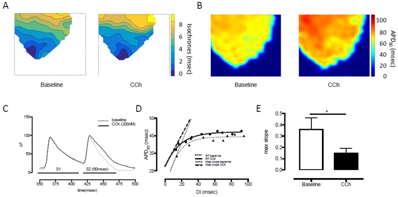
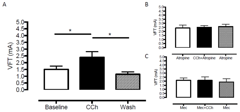
CCh (200nmol/L) significantly reduced HR (baseline 292±8 vs. CCh 224±6 vs. wash out 285±9bpm, n=9) without altering LVDP or perfusion pressure (Figure 1A-C). There was no significant change in conduction velocity with fixed rate pacing at a range of frequencies (5-9Hz, n=6) as can be seen by the isochronal map for pacing at 5Hz in Figure 2A (conduction velocity: baseline 4.2±0.4 v’s CCh 4.9±0.4 frames per second). Constant pacing for 20 beats at 150msec (400bpm) followed by an extra-stimulus (S2, reducing from 140-40msec in 10msec increments) during CCh perfusion significantly (p<0.05) flattened the maximal gradient of the action potential restitution curve driven by global action potential prolongation in the presence of CCh (Figure 2B-E). In keeping with these observations, VFT was significantly increased with application of CCh, and this effect was reversed with wash out of the drug (baseline 1.5±0.25 vs. CCh 2.4±0.4, vs. wash out 1.14±0.18mA, n=9) as shown in Figure 3A.
Figure 1. Carbamylcholine perfusion and haemodynamic responses.
A. Carbamylcholine (CCh, 200nmol/L) perfusion results in a significant bradycardia (****p<0.0001, n=9) in the absence of changes in B. left ventricular developed pressure (LVDP) or C. perfusion pressure.
Figure 2. Carbamylcholine perfusion and effects on ventricular electrophysiology.
A. Conduction velocity during CCh perfusion and apical pacing remains unchanged while there is B. a significant increase in median APD80 as determined by optical mapping of the anterior wall of the left ventricle. C. CCh perfusion results in prolongation of the APD in response to a closely coupled extra-stimulus following a drive train compared to baseline conditions (ΔF: fractional change in RH237 fluorescence). D/E. This results in a significant (*p<0.05) flattening of the electrical restitution slope (RT: restitution, max slope: steepest part of the restitution curve).
Figure 3. The anti-fibrillatory effect of carbamylcholine is dependent upon stimulation of the nicotinic and muscarinic receptors.
A. Carbamylcholine (CCh, 200nmol/L) results in a significant increase in ventricular fibrillation threshold (VFT), which is reversed upon washout of the drug (*p<0.05, n=9) B. This effect is abolished by blockade of the muscarinic receptor with atropine (0.1μmol/L, n=6) and C. nicotinic receptor with mecamylamine (Mec, 10μmol/L, n=6).
Role of muscarinic and nicotinic receptors
The bradycardia in response to CCh was completely abolished by the muscarinic antagonist atropine (0.1 μmol/L atropine 276±7 vs. atropine+CCh 286±8bpm, n=6) but remained intact in the presence of the nicotinic antagonist mecamylamine (10μmol/L mecamylamine 258±87 vs. mecamylamine+CCh 222±11bpm, n=6, p<0.05) confirming the specificity of the respective drugs. However, both atropine and mecamylamine were able to abolished the effect of CCh on VFT as can be seen in Figure 3B and C. A higher dose of atropine (10μmol/L) was also effective at preventing the rise in VFT to CCh (atropine 1.9±.0.19 vs. atropine+CCh 1.4±0.29 vs. atropine 1.38±0.55, n=5).
Role of nitric oxide
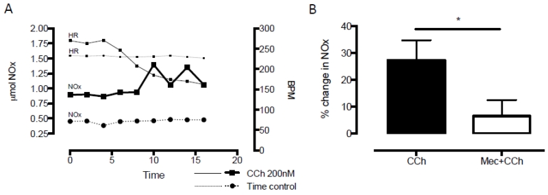
To directly measure NO production we collected coronary perfusate samples before and after the application of CCh, and measured the concentration of NO metabolites (NOx) using an ozone chemiluminescence assay. Samples collected from the stabilized preparation displayed no significant temporal variation in their NOx content, whereas CCh perfusion evoked a significant increase (n=8) in perfusate NOx content which became manifest at peak bradycardia (after 10 minutes perfusion; see Figures 4A and B). There was no significant difference in baseline NOx in the time control or CCh groups (0.72±0.26 vs. 1.05±0.18μmol, p=0.89, Mann Whitney U test). The increase in NOx production was abolished by the nicotinic receptor antagonist mecamylamine (10μmol/L, n=8) despite a similar bradycardia from muscarinic receptor stimulation.
Figure 4. Carbamylcholine perfusion increase the nNOS derived NO metabolite content in coronary perfusate.
A. Experimental data comparing NO metabolite (NOx) levels in coronary perfusate sampled every 2 minutes in time control and during carbamylcholine (CCh, 200nmol/L) perfusion. There is no change in heart rate (HR) or NOx during the time control while CCh results in bradycardia with corresponding increase in NOx levels compared to baseline. B. Summary data demonstrating the increase in NOx levels during CCh perfusion (n=8) and significant reduction if co-perfused with the nicotinic receptor antagonist mecamylamine (Mec, 10μmol/L, *p<0.05, n=8).
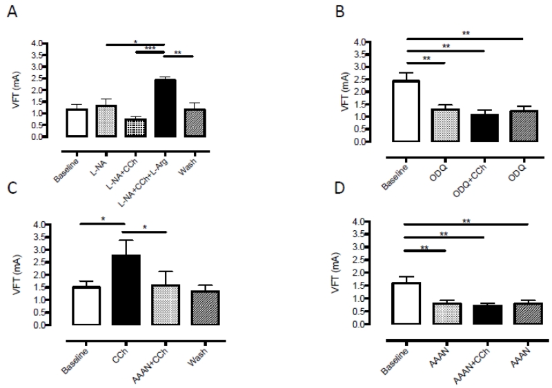
To investigate if the increase in VFT with CCh was also dependent on NO generation, CCh was perfused during NOS inhibition with the competitive non-specific NOS inhibitor L-NA (100μmol/L, n=6). L-NA prevented an increase in VFT with perfusion of CCh and this was significantly reversed upon addition of NOS substrate L-arginine as shown in Figure 5A. A similar result was observed with the nNOS inhibitor AAAN (10μmol/L) that prevented an increase in VFT when administered before CCh (n=6), and abolished the increase in VFT when added after CCh (n=6) as shown in Figure 5C and D. The sGC inhibitor ODQ (10μmol/L, n=6) also prevented the rise in VFT with CCh (Figure 5B). Neither L-NA, AAAN nor ODQ altered baseline HR or LVDP. CCh produced a similar bradycardia in the presence of all 3 compounds. Perfusion pressure was significantly increased by both L-NA (baseline 60±7 vs. L-NA 107±9mmHg) and ODQ (baseline 102 vs. ODQ 136±6 mmHg) but not by AAAN (baseline 73±12 vs AAAN 74±13 mmHg). AAAN and ODQ (but not L-NA) also significantly reduced baseline VFT prior to the addition of CCh, suggesting that baseline endogenous nNOS-NO-sGC signalling may also be anti-fibrillatory.
Figure 5. Generation of nitric oxide is required for the anti-fibrillatory effect of carbamylcholine.
A. The anti-fibrillatory action of carbamylcholine (CCh, 200nmol/L) is abolished by the non-specific nitric oxide synthase (NOS) inhibitor L-NA (10μmol/L) and reversed by addition of L-arginine (L-Arg 5mmol/L, ***p<0.001). This effect is lost upon wash out of all drugs. B. Soluble guanylate cyclase (sGC) inhibition by ODQ (10μmol/L, n=6) results in a significant reduction in ventricular fibrillation threshold (VFT) which can not be increased by the addition of CCh. C/D. Neuronal NOS inhibition by AAAN (10μmol/L) is able to abolish and prevent the increase in VFT in response to CCh perfusion (n=6/each series, *p<0.05, **p<0.01)
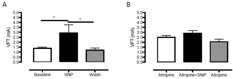
The NO donor SNP (10μmol/L, n=8) mimicked the action of CCh on VFT despite causing a small increase in HR (+19.3±3.1 bpm, p<0.0001) and reduction in PP (−13.1±5.7mmHg, p=0.013) as can be seen in Figure 6A. This action on HR is caused by stimulation of If (Musialek et al., 1997) and the reduction in perfusion pressure is caused by the vasodilatory effect of NO (Palmer et al., 1987; Furchgott, 1996). The effect of SNP could be blocked in the presence of atropine (n=10, Figure 6B).
Figure 6. Sodium nitroprusside exerts a muscarinic receptor dependent anti-fibrillatory effect on the ventricle.
A. Perfusion of sodium nitroprusside (SNP, 10μmol/L, n=8) results in haemodynamic changes consistent with an increase in NO (see text) and increases ventricular fibrillation threshold (VFT). B. This effect is abolished by co-perfusion of atropine (10μmol/L, n=10)
Discussion
The main findings of our study are
In the isolated rat heart the acetylcholine analogue, carbamylcholine raises ventricular fibrillation threshold and flattens the electrical restitution curve.
The anti-fibrillatory action of carbamylcholine was dependent upon stimulation of nicotinic receptors, and the generation of NO from neuronal NOS. This was mimicked by the NO donor sodium nitroprusside.
The anti-fibrillatory action of both carbamylcholine and sodium nitroprusside was ultimately dependent on muscarinic receptor stimulation since all effects were blocked by atropine.
These data add to the growing evidence base supporting the role of cholinergic signalling in exerting an anti-fibrillatory effect on the ventricle. Whilst this observation was described over 100 years ago by Einbrodt (1859), and later supported through the seminal experiments of Kolman (1975), Kent (1973) and De Ferrari (1991a), the mechanistic basis of this effect has recently been questioned by Brack et al (2011). In contrast to the previous evidence that described an obligatory role for muscarinic receptor activation in the mechanism of vagal protection, their innervated rabbit heart model demonstrated preservation of the anti-fibrillatory effect of vagus stimulation in the presence of atropine. The mechanism proposed was a paracrine effect by nNOS derived NO from parasympathetic ganglia acting directly on the ventricular myocardium. This observation was supported by evidence of NO release by DAF2 flourescence and blocking the VNS associated rise in VFT with hexamethonium, a nicotinic AChR antagonist (Brack et al., 2007). Although, stimulation of the cervical vagus activates efferent preganglionic parasympathetic neurons to lower HR and reduce inotropy via stimulation of muscarinic receptors on myocytes, up to 70% of the fibres are sensory afferents (Berthoud & Neuhuber, 2000) that can be retrogradely stimulated. The NO generated during these conditions may therefore originate from nNOS in sensory fibres, postganglionic parasympathetic neurons or indeed myocytes (Xu et al 1999). By using an acetylcholine analogue that is resistant to cholinesterase and produces reliable and persistent responses at both muscarinic receptors on ventricular myocytes and nicotinic receptors on postganglionic parasympathetic ganglia, we therefore tried to localize the source of NO in terms of its anti-fibrillatory action.
Role and source of NO as a cholinergic antiarrhythmic modulator
Using an ozone chemilumiscence method to quantify the absolute concentration of NO metabolites, perfusion of CCh in our model resulted in release of NO as evidenced by a rise in NOx in the coronary perfusate. The source of NO as a result of CCh perfusion could have been via endothelial NOS (eNOS) through its coupling to the M2 receptor (Balligand et al., 1995) or via nNOS activation from nicotinic receptors on cholinergic ganglia. Therefore we used mecamylamine, a nictonic receptor antagonist without the cross reactivity for cardiac muscarinic receptors that has been reported for hexamethonium (Eglen et al., 1989), to dissect this pathway. Our results support Brack et al’s (Brack et al., 2011) findings that the source of NO during vagus nerve stimulation signalling is neuronal since mecamylamine prevented the increase in NOx seen during CCh perfusion, whilst the muscarinic receptor activated bradycardia remained intact.
Our data support a direct neuronal NO mechanism rather than an indirect paracrine effect of NO as a co-transmitter on the ventricular myocardium (Brack et al., 2011). If NO was acting in a paracrine manner, the addition of atropine would have had no influence on this effect. However, we clearly show that atropine was able to block the anti-fibrillatory action of CCh. Moreover, the anti-fibrillatory effect of SNP was also not seen in the presence of atropine indicating that NO is acting upstream of the muscarinic receptor. We propose that the most likely unifying mechanistic explanation of these observations is that neuronal NO generated by nNOS facilitates acetylcholine release from release sites of the ganglionic projections via a cyclic guanosine monophosphate (cGMP) – phosphodiesterase-3 dependent pathway increasing protein kinase A dependent phosphorylation of N-type calcium channels as we have demonstrated previously (Herring & Paterson, 2001).
Recent evidence from anatomical and immunohistochemical studies have moved us away from the dogma that there is an absence of parasympathetic innervation of the ventricles. A variety of techniques have demonstrated significant epicardial and endocardial innervation of atria and ventricles in multiple mammalian species, albeit with some variation in numbers of parasympathetic ganglia (Coote, 2013) and stimulation of the vagus nerve can reduce ventricular contractility in humans (Lewis et al., 2001). These studies have supported the anti-fibrillatory role of the vagus on the ventricle.
The antiarrhythmic action of muscarinic receptor stimulation
Our data establishes the critical pathway in the anti-fibrillatory action of cholinergic signaling played by the muscarinic receptor, an effect that can be enhanced by nNOS driven parasympathetic transmission (Mohan et al., 2002; Dawson et al., 2008). De Ferrari et al (1991b) demonstrated the mechanistic importance of muscarinic stimulation in their elegant canine model of coronary artery ligation and assessment of vagal tone. They found that muscarinic blockade with atropine was associated with an increase in ventricular arrhythmia compared with control. Ando et al (2005) reported a similar observation in a rat model of vagus nerve stimulation during coronary artery occlusion where the pre-conditioning protective effect of vagus stimulation was lost in the presence of atropine. More recently, vagus nerve stimulation has been shown to be anti-arrhythmic in the clinically relevant setting of ischaemia-perfusion via effects on mitochondrial function with atropine abolishing this effect (Shinlapawittayatorn et al., 2014).
Ventricular arrhythmias are a major source of morbidity and mortality in patients with myocardial infarction and heart failure. The mechanisms underlying this increased propensity include changes in action potential duration due to alterations in ion currents including ICaL (Shorofsky & January, 1992; Zeng & Rudy, 1995) and IK (Maltsev et al., 1998; Wickenden et al., 1998), shortening of refractory period, steepening of the electrical restitution curve, changes in SR Ca2+ release (Pogwizd et al., 2001) and slowing of conduction velocity. These changes lead to early and delayed after depolarisations (EADs/DADs) which act as major initiators of ventricular tacharrythmias (Pogwizd et al., 1998). In addition, structural changes post myocardial infarction and in heart failure leads to fibrosis, reductions in connnexin 43 (Akar et al., 2004; Kostin et al., 2004) and INa availability resulting in areas of slower conduction. This contributes to regional heterogeneity in conduction velocity, which promotes local conduction block and re-entry (Yan et al., 2001; Akar & Rosenbaum, 2003).
Cholinergic stimulation via the muscarinic receptor counters pathophysiological changes by inhibiting adrenergic and cyclic adenosine monophosphate (cAMP)-protein kinase A dependent increases in ICaL activity, initially termed ‘accenuated antagonism’ by Levy in 1971 (Levy, 1971). Mechanistically, direct and indirect pathways have been proposed for this effect. Direct inhibition of β adrenergic stimulation occurs via direct interaction of adenylate cyclase with the α subunit of PTX-sensitive Gi/Go proteins of the M2 receptor (Sunahara et al., 1996; Smit & Iyengar, 1998). Indirect effects may also be dependent upon the generation of NO by eNOS, leading to cGMP production and increased phosphodiesterase-2 activity leading to breakdown of cAMP and subsequent reduction in ICaL (Balligand et al., 1995; Han et al., 1996; Han et al., 1998), although this is controversial (Vandecasteele et al., 1998; Vandecasteele et al., 1999). nNOS is also present in ventricular myocytes associated with the sarcoplasmic reticulum (Casadei, 2006), although there is no direct link between muscarinic receptor stimulation and generation of NO by nNOS (Martin et al., 2006). These cellular mechanisms contribute to global changes in ventricular physiology as evidenced by APD prolongation and flattening of the electrical restitution curve by our data and direct vagus stimulation in the isolated rabbit heart (Ng et al., 2001). Furthermore, recent evidence has also shown an increase in connexin-43 expression in response to chronic vagus nerve stimulation promoting homogeneity in conduction velocity (Ando et al., 2005; Sabbah, 2011).
Limitations
There is evidence that parasympathetic innervation of the ventricle varies regionally and therefore perfusion of CCh is unlikely to mimic the localised release of acetylcholine from efferent post-ganglionic fibres. In addition, vagus nerve stimulation has the potential to elicit a complex profile of mediators and modulators from efferent and afferent fibers. It also engages multiple levels of the cardiac neuraxis to alter peripheral neural processing and central-peripheral neural interactions, including central drive (Ardell et al., 2015). However, the direct physiological effects of CCh perfusion in terms of heart rate and developed pressure closely matched the physiological parameters seen in studies of direct vagus nerve stimulation across species. In-vitro preparations are also removed from the haemodynamic and neuro-humoral perturbations that contribute to arrhythmogenesis, and therefore the in-vitro preparation may not give a full quantitative mechanistic account of the pathways involved.
Whilst several studies highlight prolongation of refectory period as being an additional anti-arrhythmic mechanism of vagus stimulation that results from action potential prolongation, we were unable to effectively measure this in the rat heart as our S1-S2 coupling interval only went down to 40msec where we still had ventricular capture. Nevertheless the antiarrhythmic properties of cholinergic stimulation appear consistent across a range of species with differing ion channel expression and electrophysiology. Optical mapping is a powerful tool in imaging global electrophysiology although there is conflicting evidence regarding the influence of electro-mechanical uncoupling with blebbistatin on electrophysiology (Fedorov et al., 2007; Brack et al., 2013). Conversely, the use of electrode mapping gives poorer spatial resolution and variation in signal magnitude over time. In our data, blebbistatin was applied at a fixed concentration during control and CCh perfusion, which should therefore obviate any confounding effect.
Testing VFT may not reflect the mechanism of VT and VF initiation in-vivo but it does allow for reproducible testing of arrhythmogenecity removed from the stochastic nature of arrhythmia induction seen with ischaemia-reperfusion or infarct models. Baseline VFT will also vary depending on the size of the heart and position and contact of the pacing electrode, as we observe between our different protocols. However, all comparisons in the experimental protocols are within experiments, paired analysis (not unpaired comparisons). Many similar papers report VFT in terms of percentage change but we have chosen to present the raw data for clarity. The ability of CCh to raise VFT does not appear to be dependent on the baseline VFT however. For example, CCh was unable to raise VFT in the presence of 0.1μmol/L atropine when baseline VFT was 2.4±0.35mA or in the presence of 10μmol/L atropine when baseline VFT was. 1.9±0.19mA.
The real time measurement of NO and its metabolites is challenging and we employed ozone chemiluminscence on the collected perfusate. There is temporal variation in the onset of bradycardia and peak of NOx seen and this observation is likely to be related to both the cellular mechanisms by which CCh brings about both effects and the method of perfusate collection. The action of CCh on the sinoatrial node will occur shortly after application of the drug with rapid changes in ion channel activity on stimulation of muscarinic receptors. Stimulation of nNOS activity and diffusion of NO from dispersed groups of cholinergic ganglia throughout the heart is likely to lead to a slow NOx accumulation in the perfusate, especially when this is only being sampled every 2 minutes. A more acute temporal relationship may have been achieved by utilizing a localized microdialysis approach.
Clinical significance
Cardiac sympathetic over activity increases the risk of lethal ventricular arrhythmias (Shen & Zipes, 2014). Conversely large randomized human studies have established markers of parasympathetic activity, including increased baroreflex sensitivity and HR variability as positive prognostic indicators in patients post-myocardial infarction (ATRAMI, (La Rovere et al., 1998)), and with chronic heart failure (UK HEART, (Nolan et al., 1998)).
Whilst β adrenergic receptor blockade is the mainstay of therapy in these conditions, surgically implanted vagal nerve stimulators have emerged as a promising adjunctive treatment strategy (Schwartz et al., 2008). Whilst these devices may improve ejection fraction and quality of life in chronic heart failure (De Ferrari et al., 2011), a long-term reduction in ventricular arrhythmias or sudden cardiac death in patients has yet to be demonstrated. Moreover, this approach stimulates both cholinergic efferent and afferent fibers and surgical implantation of such devices is not without risk.
Our study demonstrates that muscarinic receptor activation and neuronal generation of NO are critical steps for the anti-fibrillatory effect of cholinergic signalling. A nicotinic receptor based NO mediated mechanism that converges on the muscarinic receptor is conceivably amenable to a more targeted gene therapy or pharmacologic treatment strategy. For example, percutaneous cardiac gene transfer of nNOS increases vagal neurotransmission and bradycardia (Mohan et al., 2002) and reduces mortality 3 days post myocardial infarction in the guinea pig (Dawson et al., 2008). Whether gene transfer of nNOS into cardiac cholinergic ganglia provides a direct anti-fibrillatory action on the ventricle that could be exploited therapeutically remains to be established.
Key points summary.
Animal studies suggest an anti-fibrillatory action of the vagus nerve on the ventricle, although the exact mechanism is controversial.
Using a Langendorff perfused rat heart, we show that the acetylcholine analogue, carbamylcholine raises ventricular fibrillation threshold (VFT) and flattens the electrical restitution curve.
The anti-fibrillatory action of carbamylcholine was prevented by the nicotinic receptor antagonist mecamylamine, inhibitors of neuronal nitric oxide synthase (nNOS) and soluble guanylyl cyclase (sGC), and can be mimicked by the NO donor sodium nitroprusside.
Carbamylcholine increased NO metabolite content in the coronary effluent and this was prevented by mecamylamine.
The anti-fibrillatory action of both carbamylcholine and sodium nitroprusside was ultimately dependent on muscarinic receptor stimulation since all effects were blocked by atropine.
These data demonstrate a protective effect of carbamylcholine on VFT that depends upon both muscarinic and nicotinic receptor stimulation, where the generation of NO is likely to be via a neuronal nNOS-sGC dependent pathway.
Acknowledgements
This work is supported by a British Heart Foundation project grant (PG/11/6/28660) and British Heart Foundation Centre for Research Excellence Award (RE/08/004)
Abbreviations
- AAAN
N-(4S)-4-amino-5-[aminoethyl]aminopentyl-N’-nitroguanidine
- APD
action potential duration
- cAMP
cyclic adenosine monophosphate
- CAO
coronary artery occlusion
- cGMP
cyclic guanosine monophosphate
- ECG
electrocardiogram
- eNOS
endothelial nitric oxide synthase
- HR
heart rate
- L-Arg
L-Arginine
- L-NA
L-Nitro Arginine
- LVDP
left ventricular developed pressure
- nNOS
neuronal nitric oxide synthase
- NO
nitric oxide
- ODQ
1H-[1,2,4]oxadiazolo[4,3-a]quinoxalin-1-one
- PP
perfusion pressure
- sGC
soluble guanylate cyclase
- SNP
sodium nitroprusside
- VFT
ventricular fibrillation threshold
Footnotes
Publisher's Disclaimer: This is an Accepted Article that has been peer-reviewed and approved for publication in the The Journal of Physiology, but has yet to undergo copy-editing and proof correction. Please cite this article as an ‘Accepted Article’; doi: 10.1113/JP271588.
Competing interests
All authors have no disclosures or conflicts of interest with respect to the manuscript submitted.
References
- Akar FG, Rosenbaum DS. Transmural electrophysiological heterogeneities underlying arrhythmogenesis in heart failure. Circulation research. 2003;93:638–645. doi: 10.1161/01.RES.0000092248.59479.AE. [DOI] [PubMed] [Google Scholar]
- Akar FG, Spragg DD, Tunin RS, Kass DA, Tomaselli GF. Mechanisms underlying conduction slowing and arrhythmogenesis in nonischemic dilated cardiomyopathy. Circulation research. 2004;95:717–725. doi: 10.1161/01.RES.0000144125.61927.1c. [DOI] [PubMed] [Google Scholar]
- Ando M, Katare RG, Kakinuma Y, Zhang D, Yamasaki F, Muramoto K, Sato T. Efferent vagal nerve stimulation protects heart against ischemia-induced arrhythmias by preserving connexin43 protein. Circulation. 2005;112:164–170. doi: 10.1161/CIRCULATIONAHA.104.525493. [DOI] [PubMed] [Google Scholar]
- Ardell JL, Rajendran PS, Nier HA, KenKnight BH, Armour JA. Central-peripheral neural network interactions evoked by vagus nerve stimulation: functional consequences on control of cardiac function. American journal of physiology Heart and circulatory physiology. 2015;309:H1740–1752. doi: 10.1152/ajpheart.00557.2015. [DOI] [PMC free article] [PubMed] [Google Scholar]
- Balligand JL, Kobzik L, Han X, Kaye DM, Belhassen L, O’Hara DS, Kelly RA, Smith TW, Michel T. Nitric oxide-dependent parasympathetic signaling is due to activation of constitutive endothelial (type III) nitric oxide synthase in cardiac myocytes. The Journal of biological chemistry. 1995;270:14582–14586. doi: 10.1074/jbc.270.24.14582. [DOI] [PubMed] [Google Scholar]
- Beker F, Weber M, Fink RH, Adams DJ. Muscarinic and nicotinic ACh receptor activation differentially mobilize Ca2+ in rat intracardiac ganglion neurons. J Neurophysiol. 2003;90:1956–1964. doi: 10.1152/jn.01079.2002. [DOI] [PubMed] [Google Scholar]
- Berthoud HR, Neuhuber WL. Functional and chemical anatomy of the afferent vagal system. Auton Neurosci. 2000;85:1–17. doi: 10.1016/S1566-0702(00)00215-0. [DOI] [PubMed] [Google Scholar]
- Bishop MJ, Burton RA, Kalla M, Nanthakumar K, Plank G, Bub G, Vigmond EJ. Mechanism of reentry induction by a 9-V battery in rabbit ventricles. American journal of physiology Heart and circulatory physiology. 2014;306:H1041–1053. doi: 10.1152/ajpheart.00591.2013. [DOI] [PMC free article] [PubMed] [Google Scholar]
- Brack KE, Coote JH, Ng GA. Vagus nerve stimulation protects against ventricular fibrillation independent of muscarinic receptor activation. Cardiovasc Res. 2011;91:437–446. doi: 10.1093/cvr/cvr105. [DOI] [PubMed] [Google Scholar]
- Brack KE, Narang R, Winter J, Ng GA. The mechanical uncoupler blebbistatin is associated with significant electrophysiological effects in the isolated rabbit heart. Experimental physiology. 2013;98:1009–1027. doi: 10.1113/expphysiol.2012.069369. [DOI] [PMC free article] [PubMed] [Google Scholar]
- Brack KE, Patel VH, Coote JH, Ng GA. Nitric oxide mediates the vagal protective effect on ventricular fibrillation via effects on action potential duration restitution in the rabbit heart. J Physiol. 2007;583:695–704. doi: 10.1113/jphysiol.2007.138461. [DOI] [PMC free article] [PubMed] [Google Scholar]
- Brack KE, Patel VH, Mantravardi R, Coote JH, Ng GA. Direct evidence of nitric oxide release from neuronal nitric oxide synthase activation in the left ventricle as a result of cervical vagus nerve stimulation. J Physiol. 2009;587:3045–3054. doi: 10.1113/jphysiol.2009.169417. [DOI] [PMC free article] [PubMed] [Google Scholar]
- Casadei B. The emerging role of neuronal nitric oxide synthase in the regulation of myocardial function. Experimental physiology. 2006;91:943–955. doi: 10.1113/expphysiol.2006.035493. [DOI] [PubMed] [Google Scholar]
- Choi BR, Salama G. Simultaneous maps of optical action potentials and calcium transients in guinea-pig hearts: mechanisms underlying concordant alternans. The Journal of physiology. 2000;529(Pt 1):171–188. doi: 10.1111/j.1469-7793.2000.00171.x. [DOI] [PMC free article] [PubMed] [Google Scholar]
- Coote JH. Myths and realities of the cardiac vagus. The Journal of physiology. 2013;591:4073–4085. doi: 10.1113/jphysiol.2013.257758. [DOI] [PMC free article] [PubMed] [Google Scholar]
- Corr PB, Gillis RA. Role of the vagus nerves in the cardiovascular changes induced by coronary occlusion. Circulation. 1974;49:86–97. doi: 10.1161/01.cir.49.1.86. [DOI] [PubMed] [Google Scholar]
- Dawson TA, Li D, Woodward T, Barber Z, Wang L, Paterson DJ. Cardiac cholinergic NO-cGMP signaling following acute myocardial infarction and nNOS gene transfer. Am J Physiol Heart Circ Physiol. 2008;295:H990–H998. doi: 10.1152/ajpheart.00492.2008. [DOI] [PMC free article] [PubMed] [Google Scholar]
- De Ferrari GM, Crijns HJ, Borggrefe M, Milasinovic G, Smid J, Zabel M, Gavazzi A, Sanzo A, Dennert R, Kuschyk J, Raspopovic S, Klein H, Swedberg K, Schwartz PJ, CardioFit Multicenter Trial I Chronic vagus nerve stimulation: a new and promising therapeutic approach for chronic heart failure. Eur Heart J. 2011;32:847–855. doi: 10.1093/eurheartj/ehq391. [DOI] [PubMed] [Google Scholar]
- De Ferrari GM, Salvati P, Grossoni M, Ukmar G, Vaga L, Patrono C, Schwartz PJ. Pharmacologic modulation of the autonomic nervous system in the prevention of sudden cardiac death. A study with propranolol, methacholine and oxotremorine in conscious dogs with a healed myocardial infarction. Journal of the American College of Cardiology. 1993;22:283–290. doi: 10.1016/0735-1097(93)90845-r. [DOI] [PubMed] [Google Scholar]
- De Ferrari GM, Vanoli E, Curcuruto P, Tommasini G, Schwartz PJ. Prevention of life-threatening arrhythmias by pharmacologic stimulation of the muscarinic receptors with oxotremorine. Am Heart J. 1992;124:883–890. doi: 10.1016/0002-8703(92)90968-2. [DOI] [PubMed] [Google Scholar]
- De Ferrari GM, Vanoli E, Stramba-Badiale M, Hull SS, Jr., Foreman RD, Schwartz PJ. Vagal reflexes and survival during acute myocardial ischemia in conscious dogs with healed myocardial infarction. The American journal of physiology. 1991;261:H63–69. doi: 10.1152/ajpheart.1991.261.1.H63. [DOI] [PubMed] [Google Scholar]
- Efimov IR, Nikolski VP, Salama G. Optical imaging of the heart. Circulation research. 2004;95:21–33. doi: 10.1161/01.RES.0000130529.18016.35. [DOI] [PubMed] [Google Scholar]
- Eglen RM, Michel AD, Cornett CM, Kunysz EA, Whiting RL. The interaction of hexamethonium with muscarinic receptor subtypes in vitro. Br J Pharmacol. 1989;98:499–506. doi: 10.1111/j.1476-5381.1989.tb12623.x. [DOI] [PMC free article] [PubMed] [Google Scholar]
- Einbrodt UeberHerzreizungundihrVerhaeltniszum Blutdruck. Akademie der Wissenschaften (Vienna) Sitzungsberichte. 1859;38:345. [Google Scholar]
- Fedorov VV, Lozinsky IT, Sosunov EA, Anyukhovsky EP, Rosen MR, Balke CW, Efimov IR. Application of blebbistatin as an excitation-contraction uncoupler for electrophysiologic study of rat and rabbit hearts. Heart Rhythm. 2007;4:619–626. doi: 10.1016/j.hrthm.2006.12.047. [DOI] [PubMed] [Google Scholar]
- Furchgott RF. The 1996 Albert Lasker Medical Research Awards. The discovery of endothelium-derived relaxing factor and its importance in the identification of nitric oxide. Jama. 1996;276:1186–1188. [PubMed] [Google Scholar]
- Han X, Kobzik L, Balligand JL, Kelly RA, Smith TW. Nitric oxide synthase (NOS3)-mediated cholinergic modulation of Ca2+ current in adult rabbit atrioventricular nodal cells. Circulation research. 1996;78:998–1008. doi: 10.1161/01.res.78.6.998. [DOI] [PubMed] [Google Scholar]
- Han X, Kubota I, Feron O, Opel DJ, Arstall MA, Zhao YY, Huang P, Fishman MC, Michel T, Kelly RA. Muscarinic cholinergic regulation of cardiac myocyte ICa-L is absent in mice with targeted disruption of endothelial nitric oxide synthase. Proceedings of the National Academy of Sciences of the United States of America. 1998;95:6510–6515. doi: 10.1073/pnas.95.11.6510. [DOI] [PMC free article] [PubMed] [Google Scholar]
- Herring N, Golding S, Paterson DJ. Pre-synaptic NO-cGMP pathway modulates vagal control of heart rate in isolated adult guinea pig atria. J Mol Cell Cardiol. 2000;32:1795–1804. doi: 10.1006/jmcc.2000.1214. [DOI] [PubMed] [Google Scholar]
- Herring N, Paterson DJ. Nitric oxide-cGMP pathway facilitates acetylcholine release and bradycardia during vagal nerve stimulation in the guinea-pig in vitro. J Physiol. 2001;535:507–518. doi: 10.1111/j.1469-7793.2001.00507.x. [DOI] [PMC free article] [PubMed] [Google Scholar]
- Herron TJ, Lee P, Jalife J. Optical imaging of voltage and calcium in cardiac cells & tissues. Circulation research. 2012;110:609–623. doi: 10.1161/CIRCRESAHA.111.247494. [DOI] [PMC free article] [PubMed] [Google Scholar]
- Ito M, Zipes DP. Efferent sympathetic and vagal innervation of the canine right ventricle. Circulation. 1994;90:1459–1468. doi: 10.1161/01.cir.90.3.1459. [DOI] [PubMed] [Google Scholar]
- Kent KM, Smith ER, Redwood DR, Epstein SE. Electrical stability of acutely ischemic myocardium. Influences of heart rate and vagal stimulation. Circulation. 1973;47:291–298. doi: 10.1161/01.cir.47.2.291. [DOI] [PubMed] [Google Scholar]
- Kolman BS, Verrier RL, Lown B. The effect of vagus nerve stimulation upon vulnerability of the canine ventricle: role of sympathetic-parasympathetic interactions. Circulation. 1975;52:578–585. doi: 10.1161/01.cir.52.4.578. [DOI] [PubMed] [Google Scholar]
- Kostin S, Dammer S, Hein S, Klovekorn WP, Bauer EP, Schaper J. Connexin 43 expression and distribution in compensated and decompensated cardiac hypertrophy in patients with aortic stenosis. Cardiovascular research. 2004;62:426–436. doi: 10.1016/j.cardiores.2003.12.010. [DOI] [PubMed] [Google Scholar]
- La Rovere MT, Bigger JT, Jr., Marcus FI, Mortara A, Schwartz PJ, ATRAMI (Autonomic Tone and Reflexes After Myocardial Infarction) Investigators Baroreflex sensitivity and heart-rate variability in prediction of total cardiac mortality after myocardial infarction. Lancet. 1998;351:478–484. doi: 10.1016/s0140-6736(97)11144-8. [DOI] [PubMed] [Google Scholar]
- Levy MN. Sympathetic-parasympathetic interactions in the heart. Circulation research. 1971;29:437–445. doi: 10.1161/01.res.29.5.437. [DOI] [PubMed] [Google Scholar]
- Levy MN, Blattberg B. Effect of vagal stimulation on the overflow of norepinephrine into the coronary sinus during cardiac sympathetic nerve stimulation in the dog. Circulation research. 1976;38:81–84. doi: 10.1161/01.res.38.2.81. [DOI] [PubMed] [Google Scholar]
- Levy MN, Zieske H. Effect of enhanced contractility on the left ventricular response to vagus nerve stimulation in dogs. Circulation research. 1969;24:303–311. doi: 10.1161/01.res.24.3.303. [DOI] [PubMed] [Google Scholar]
- Lewis ME, Al-Khalidi AH, Bonser RS, Clutton-Brock T, Morton D, Paterson D, Townend JN, Coote JH. Vagus nerve stimulation decreases left ventricular contractility in vivo in the human and pig heart. The Journal of physiology. 2001;534:547–552. doi: 10.1111/j.1469-7793.2001.00547.x. [DOI] [PMC free article] [PubMed] [Google Scholar]
- Maltsev VA, Sabbah HN, Higgins RS, Silverman N, Lesch M, Undrovinas AI. Novel, ultraslow inactivating sodium current in human ventricular cardiomyocytes. Circulation. 1998;98:2545–2552. doi: 10.1161/01.cir.98.23.2545. [DOI] [PubMed] [Google Scholar]
- Martin PJ, Levy MN, Zieske H. Analysis and simulation of the left ventricular response to autonomic nervous activity. Cardiovascular research. 1969;3:396–410. doi: 10.1093/cvr/3.4.396. [DOI] [PubMed] [Google Scholar]
- Martin SR, Emanuel K, Sears CE, Zhang YH, Casadei B. Are myocardial eNOS and nNOS involved in the beta-adrenergic and muscarinic regulation of inotropy? A systematic investigation. Cardiovascular research. 2006;70:97–106. doi: 10.1016/j.cardiores.2006.02.002. [DOI] [PubMed] [Google Scholar]
- Martins JB, Zipes DP. Effects of sympathetic and vagal nerves on recovery properties of the endocardium and epicardium of the canine left ventricle. Circulation research. 1980a;46:100–110. doi: 10.1161/01.res.46.1.100. [DOI] [PubMed] [Google Scholar]
- Martins JB, Zipes DP. Epicardial phenol interrupts refractory period responses to sympathetic but not vagal stimulation in canine left ventricular epicardium and endocardium. Circulation research. 1980b;47:33–40. doi: 10.1161/01.res.47.1.33. [DOI] [PubMed] [Google Scholar]
- Mohan RM, Heaton DA, Danson EJ, Krishnan SP, Cai S, Channon KM, Paterson DJ. Neuronal nitric oxide synthase gene transfer promotes cardiac vagal gain of function. Circulation research. 2002;91:1089–1091. doi: 10.1161/01.res.0000047531.75030.b5. [DOI] [PubMed] [Google Scholar]
- Musialek P, Lei M, Brown HF, Paterson DJ, Casadei B. Nitric oxide can increase heart rate by stimulating the hyperpolarization-activated inward current, I(f) Circulation research. 1997;81:60–68. doi: 10.1161/01.res.81.1.60. [DOI] [PubMed] [Google Scholar]
- Nash MP, Thornton JM, Sears CE, Varghese A, O’Neill M, Paterson DJ. Ventricular activation during sympathetic imbalance and its computational reconstruction. J Appl Physiol (1985) 2001;90:287–298. doi: 10.1152/jappl.2001.90.1.287. [DOI] [PubMed] [Google Scholar]
- Ng GA, Brack KE, Coote JH. Effects of direct sympathetic and vagus nerve stimulation on the physiology of the whole heart--a novel model of isolated Langendorff perfused rabbit heart with intact dual autonomic innervation. Exp Physiol. 2001;86:319–329. doi: 10.1113/eph8602146. [DOI] [PubMed] [Google Scholar]
- Nolan J, Batin PD, Andrews R, Lindsay SJ, Brooksby P, Mullen M, Baig W, Flapan AD, Cowley A, Prescott RJ, Neilson JM, Fox KA. Prospective study of heart rate variability and mortality in chronic heart failure: results of the United Kingdom heart failure evaluation and assessment of risk trial (UK-heart) Circulation. 1998;98:1510–1516. doi: 10.1161/01.cir.98.15.1510. [DOI] [PubMed] [Google Scholar]
- Palmer RM, Ferrige AG, Moncada S. Nitric oxide release accounts for the biological activity of endothelium-derived relaxing factor. Nature. 1987;327:524–526. doi: 10.1038/327524a0. [DOI] [PubMed] [Google Scholar]
- Pinder AG, Rogers SC, Khalatbari A, Ingram TE, James PE. The measurement of nitric oxide and its metabolites in biological samples by ozone-based chemiluminescence. Methods Mol Biol. 2008;476:11–28. doi: 10.1007/978-1-59745-129-1_2. [DOI] [PubMed] [Google Scholar]
- Pogwizd SM, McKenzie JP, Cain ME. Mechanisms underlying spontaneous and induced ventricular arrhythmias in patients with idiopathic dilated cardiomyopathy. Circulation. 1998;98:2404–2414. doi: 10.1161/01.cir.98.22.2404. [DOI] [PubMed] [Google Scholar]
- Pogwizd SM, Schlotthauer K, Li L, Yuan W, Bers DM. Arrhythmogenesis and contractile dysfunction in heart failure: Roles of sodium-calcium exchange, inward rectifier potassium current, and residual beta-adrenergic responsiveness. Circulation research. 2001;88:1159–1167. doi: 10.1161/hh1101.091193. [DOI] [PubMed] [Google Scholar]
- Sabbah HN. Electrical vagus nerve stimulation for the treatment of chronic heart failure. Cleve Clin J Med. 2011;78(Suppl 1):S24–29. doi: 10.3949/ccjm.78.s1.04. [DOI] [PMC free article] [PubMed] [Google Scholar]
- Schwartz PJ, De Ferrari GM, Sanzo A, Landolina M, Rordorf R, Raineri C, Campana C, Revera M, Ajmone-Marsan N, Tavazzi L, Odero A. Long term vagal stimulation in patients with advanced heart failure: first experience in man. Eur J Heart Fail. 2008;10:884–891. doi: 10.1016/j.ejheart.2008.07.016. [DOI] [PubMed] [Google Scholar]
- Shen MJ, Zipes DP. Role of the autonomic nervous system in modulating cardiac arrhythmias. Circulation research. 2014;114:1004–1021. doi: 10.1161/CIRCRESAHA.113.302549. [DOI] [PubMed] [Google Scholar]
- Shinlapawittayatorn K, Chinda K, Palee S, Surinkaew S, Kumfu S, Kumphune S, Chattipakorn S, KenKnight BH, Chattipakorn N. Vagus nerve stimulation initiated late during ischemia, but not reperfusion, exerts cardioprotection via amelioration of cardiac mitochondrial dysfunction. Heart Rhythm. 2014;11:2278–2287. doi: 10.1016/j.hrthm.2014.08.001. [DOI] [PubMed] [Google Scholar]
- Shorofsky SR, January CT. L- and T-type Ca2+ channels in canine cardiac Purkinje cells. Single-channel demonstration of L-type Ca2+ window current. Circulation research. 1992;70:456–464. doi: 10.1161/01.res.70.3.456. [DOI] [PubMed] [Google Scholar]
- Smit MJ, Iyengar R. Mammalian adenylyl cyclases. Adv Second Messenger Phosphoprotein Res. 1998;32:1–21. doi: 10.1016/s1040-7952(98)80003-7. [DOI] [PubMed] [Google Scholar]
- Sunahara RK, Dessauer CW, Gilman AG. Complexity and diversity of mammalian adenylyl cyclases. Annu Rev Pharmacol Toxicol. 1996;36:461–480. doi: 10.1146/annurev.pa.36.040196.002333. [DOI] [PubMed] [Google Scholar]
- Vandecasteele G, Eschenhagen T, Fischmeister R. Role of the NO-cGMP pathway in the muscarinic regulation of the L-type Ca2+ current in human atrial myocytes. The Journal of physiology. 1998;506(Pt 3):653–663. doi: 10.1111/j.1469-7793.1998.653bv.x. [DOI] [PMC free article] [PubMed] [Google Scholar]
- Vandecasteele G, Eschenhagen T, Scholz H, Stein B, Verde I, Fischmeister R. Muscarinic and beta-adrenergic regulation of heart rate, force of contraction and calcium current is preserved in mice lacking endothelial nitric oxide synthase. Nat Med. 1999;5:331–334. doi: 10.1038/6553. [DOI] [PubMed] [Google Scholar]
- Vanoli E, De Ferrari GM, Stramba-Badiale M, Hull SS, Jr., Foreman RD, Schwartz PJ. Vagal stimulation and prevention of sudden death in conscious dogs with a healed myocardial infarction. Circulation research. 1991;68:1471–1481. doi: 10.1161/01.res.68.5.1471. [DOI] [PubMed] [Google Scholar]
- Wang L, Li D, Dawson TA, Paterson DJ. Long-term effect of neuronal nitric oxide synthase over-expression on cardiac neurotransmission mediated by a lentiviral vector. The Journal of physiology. 2009;587:3629–3637. doi: 10.1113/jphysiol.2009.172866. [DOI] [PMC free article] [PubMed] [Google Scholar]
- Wickenden AD, Kaprielian R, Kassiri Z, Tsoporis JN, Tsushima R, Fishman GI, Backx PH. The role of action potential prolongation and altered intracellular calcium handling in the pathogenesis of heart failure. Cardiovasc Res. 1998;37:312–323. doi: 10.1016/s0008-6363(97)00256-3. [DOI] [PubMed] [Google Scholar]
- Yan GX, Rials SJ, Wu Y, Liu T, Xu X, Marinchak RA, Kowey PR. Ventricular hypertrophy amplifies transmural repolarization dispersion and induces early afterdepolarization. American journal of physiology Heart and circulatory physiology. 2001;281:H1968–1975. doi: 10.1152/ajpheart.2001.281.5.H1968. [DOI] [PubMed] [Google Scholar]
- Yoon MS, Han J, Tse WW, Rogers R. Effects of vagal stimulation, atropine, and propranolol on fibrillation threshold of normal and ischemic ventricles. Am Heart J. 1977;93:60–65. doi: 10.1016/s0002-8703(77)80172-5. [DOI] [PubMed] [Google Scholar]
- Zeng J, Rudy Y. Early afterdepolarizations in cardiac myocytes: mechanism and rate dependence. Biophys J. 1995;68:949–964. doi: 10.1016/S0006-3495(95)80271-7. [DOI] [PMC free article] [PubMed] [Google Scholar]


