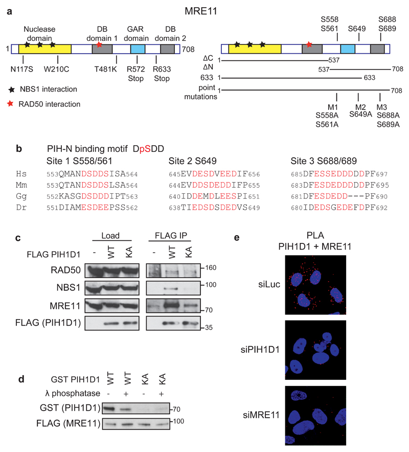
Figure 1. PIH1D1 interacts with MRN complex.
(a) Schematic representation of human MRE11 comprising of a nuclease domain, two DNA binding domains (DB1 and DB2) and Glycine-Arginine Rich domain (GAR). Positions of ATLD mutations (left panel) and putative PIH-N phospho-binding sites (right panel) are indicated. Bars below the right panel represent the MRE11 constructs used in this study. (b) Conservation of the PIH-N consensus motif in the C-terminus of MRE11. Acidic amino acids and serines that form potential PIH-N binding sites are displayed in red. (c) FLAG PIH1D1 WT interacts with MRN complex components. HEK293T cells transfected with FLAG PIH1D1 WT or phospho-binding mutant PIH1D1 KA were lysed in IP buffer (50 mM Tris-HCL pH 7.5, 150 mM NaCl, 1 % Triton X-100, 1 mM EDTA, 2.5 mM EGTA, 10 % v/v glycerol supplemented with cOmplete EDTA free protease inhibitor, PhosSTOP phosphatase inhibitor (Roche) and EtBr (50 μg/ml)) and sonicated 3x 10s. Cleared cell extracts were incubated with anti-FLAG M2 Affinity gel (Sigma-Aldrich) for 2 hours. Beads were washed 4x with IP buffer, boiled in 2x LSB buffer (100 mM Tris pH 6,8, 200 mM DTT, 4% SDS, 0,2% bromophenol blue, 20% v/v glycerol) and bound proteins were detected by immunoblotting using antibodies against RAD50 (Abcam, ab119708), MRE11 (Cell Signaling, #4895), NBS1 (Cell Signaling, #3002). (d) MRE11-PIH1D1 interaction depends on MRE11 phosphorylation and on the PIH-N domain of PIH1D1. FLAG MRE11 was purified from HEK293T using anti-FLAG M2 Affinity gel. Beads were washed with IP buffer supplemented with 1 M NaCl, treated or not with λ phosphatase for 30 min at 30°C and MRE11 was eluted with 3X FLAG peptide (30 µg, Sigma-Aldrich). GST PIH1D1 WT and phospho-binding mutant GST PIH1D1 KA were purified from BL21 E. coli and pull down with purified MRE11 was performed as described previously (20). (e) MRE11 and PIH1D1 interact in vivo. U2OS cells grown on coverslips were transfected with 40nM siRNA targeting luciferase (CGUACGCGGAAUACUUCGA, Sigma), PIH1D1 (1:1 mixture of GAAUGGAAAUGUAGUCUUA and GAGAAGAGGCUGCUGGCUU in the untranslated region of PIH1D1 as described (20)) or MRE11 (GAGCAUAACUCCAUAAGUA in the untranslated region of MRE11 mRNA) with Lipofectamine RNAiMAX reagent according to the manufacturers. Cells were permeabilized with 0.2 % Triton X-100 for 5 min at room temperature and Proximity ligation assay (PLA) was performed using MRE11 (Cell Signaling, #4895), PIH1D1 (Abcam, ab57512) antibodies and Duolink reagent according to the manufacturers protocol (Sigma-Aldrich). Red signal is PLA staining, DAPI is in blue. Representative image is shown.

