Abstract
X-linked Intellectual Disabilities (XLID) are common developmental disorders. The enzyme O-GlcNAc transferase encoded by OGT, a recently discovered XLID gene, attaches O-GlcNAc to nuclear and cytoplasmic proteins. As few missense mutations have been described, it is unclear what the aetiology of the patient phenotypes is. Here, we report the discovery of a missense mutation in the catalytic domain of OGT in an XLID patient. X-ray crystallography reveals that this variant leads to structural rearrangements in the catalytic domain. The mutation reduces in vitro OGT activity on substrate peptides/protein. Mouse embryonic stem cells carrying the mutation reveal reduced O-GlcNAcase (OGA) and global O-GlcNAc levels. These data suggest a direct link between changes in the O-GlcNAcome and intellectual disability observed in patients carrying OGT mutations.
Introduction
Intellectual disability (ID) is a neurodevelopmental disorder, with a world-wide prevalence of 1% [1], characterised by significant limitations in both intellectual functioning and adaptive behaviour [2]. ID is often syndromic, involving not only a limited IQ but also developmental delay, congenital malformations or neurological abnormalities such as autism spectrum disorder (ASD), epilepsy, and sensory impairment [3–8]. Although ID can be caused by environmental factors such as maternal alcohol abuse and birth complications [6,8,9], genetics plays a crucial role in its aetiology. Mutations, deletions or rearrangements affecting genes involved in development and neuronal function can have severe consequences for proper brain development or cognitive function [10].
To date, approximately 1000 genes have been shown to be involved in ID [11]. A disproportionate number (141) of these are located on the X-chromosome, leading to the coining of the term X-linked intellectual disability (XLID) [5]. In the last few years, next-generation sequencing (NGS) has led to enormous progress in deciphering monogenic forms of XLID [3–5,10]. This unbiased NGS approach has accelerated identification of de novo mutations in newly described XLID genes, including OGT, which encodes O- β-N-acetylglucosamine (GlcNAc) Transferase (OGT).
OGT catalyses an essential post-transitional modification, the addition of a single GlcNAc onto serine or threonine of nucleocytoplasmic proteins, using UDP-GlcNAc as donor substrate [12]. OGT is also known to be involved in the processing of the Host Cell Factor C1 (HCF1), the proteolytic products of which are involved in the activation of many genes involved in cell cycle progression [13–15]. The enzyme is divided into a glycosyltransferase catalytic domain and an N-terminal domain consisting of 13.5 tetratricopeptide repeats (TPRs) that have been shown to contribute to protein substrate binding [16–18]. Together with the hydrolase O-GlcNAcase (OGA), the homeostasis of dynamic protein O-GlcNAcylation is maintained for optimal cellular function [19]. Effects of protein O-GlcNAcylation have been linked to protein synthesis [20], stability [21] and turnover [22,23] and complex processes such as cell cycle progression [24], stress response [25,26], and transcription [27]. O-GlcNAcylation has also been implicated in pathology, including cancer [28], type II diabetes [29], cardiovascular disease [30] and neurodegeneration [31], and it is essential for proper vertebrate development [32–35]. In a range of model systems, O-GlcNAcylation has also been found to be critical for processes in early development. In mice, Ogt knockout causes lethality, with mouse embryos dying at the blastocyst stage [36].
O-GlcNAcylation also plays a key role in stem cell biology. Numerous pluripotency factors such as Oct4 and Sox2 have been shown to be O-GlcNAcylated [33,37]. Furthermore, mutations in OGT trigger alterations in stem cell differentiation and development, affecting neuronal lineages [37,38]. To date, five OGT mutations, all located to the N-terminal TPR domain, have been reported in patients with XLID [39–42]. These patients display developmental delay, facial dysmorphia, clinodactyly and microcephaly. To date, it is not clear whether the underpinning mechanisms leading to these phenotypes involve changes in the O-GlcNAc proteome.
Here, we report a patient with moderate ID, coarse facial features and a de novo mutation located in the catalytic domain of OGT. This mutation induces structural rearrangements in the catalytic domain, leading to reduced levels of protein O-GlcNAcylation in vitro and in mESCs, suggesting a direct link between perturbations in protein O-GlcNAcylation and XLID.
Results and Discussion
A patient with XLID possesses a missense mutation in the OGT catalytic domain
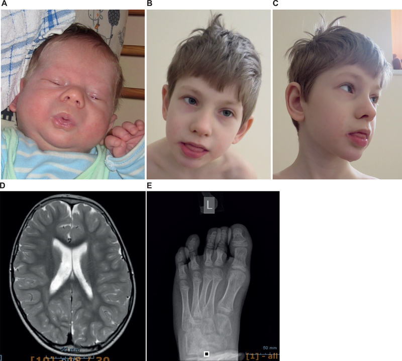
The patient, E.T., was born at term with normal birth weight; 3354 g, length; 50 cm, head circumference; 34.5 cm, and an Apgar score of 9/9. He is the second child of an Estonian non-consanguineous family. His elder brother is healthy. Facial asymmetry was noticed soon after birth (Fig. 1A). His toenails were also very small and with soft structure at birth. His development was evaluated as normal during the first months of life; he started to hold his head at 3 m and to turn at 6 m of age. Initially, he also had good weight gain, but after some months gastric reflux and gasses became very problematic.
Figure 1: Clinical pictures from patient carrying the N648Y mutation in the catalytic domain of OGT.
(a) Facial asymmetry at birth
(b) Facial view at 7 y.
(c) Profile view at 7 y. Note coarse facial features, drooling.
(d) MRI image showing brain atrophy and mega cisterna magna.
(e) X-ray image of the left foot – cone shape epiphyses.
Recurrent otitis started at 4 m and after narcosis (the shunting of middle ears) his development arrested. His muscular tone became very flaccid. He started to turn at 12 m, to crawl at 14 m, to sit at 22 m, and to walk at 3 y of age. First teeth came very late and slowly at 12 m. At the age of 16m alimentary vitamin B12 deficiency was diagnosed and treated with intramuscular injections. After that his development improved significantly. However, at the age of 19 m persistent otitis recurred and his development arrested.
He was subjected to extensive clinical investigation at 19 m due to his developmental delay. He had coarse facial features with open mouth and drooling. He suffered from hypotonia with brisk tendon reflexes and truncal ataxia. There was a suspicion of epilepsy, but EEG was normal. Brain MRI was normal for that age. On cardiac evaluation only very mild pericardial effusion was noticed, which did not affect cardiac function. Griffith scale evaluation suggested that his development corresponded to 8 m of age. He had serious problems with obstipation, but no obvious aetiology was detected. Extensive metabolic investigations were performed, which showed normal results (urinary organic acids, glycosaminoglycans, oligosaccharides, sialic acid, creatine/guanidinoacetate, and purine/pyrimidines; transferrin isoelectric focusing (TIEF) and acylcarnitine in serum; neurotransmitters in cerebrospinal fluid). Chromosomal microarray analysis showed no abnormal copy number variations.
At the age of 7 y 8 m his height was 127 cm (+1 SD), weight 23.4 kg (0 SD), head circumference 52 cm (−0.5 SD). He is moderately intellectually disabled. He had coarse facial features, convergent strabismus, large ears and intense and constant drooling (Fig. 1B–1C). He had no speech and moderately hyperactive behaviour, frequent body jerks, increased sensibility to light and sounds, and fears. In addition, he had hyper-elastic connective tissue, which manifested as frequent joint dislocations and scoliosis. He presented mild T2–3 syndactyly, inverted nipples, abnormally pale skin and body temperature fluctuations in addition to his frequent infections. X-ray investigation showed cone shape epiphyses of T2-T5 (Fig. 1D), fragmentation of the 1st rib and synostosis of 1–2 ribs. Brain MRI investigation at 5 y showed brain atrophy and mega cisterna magna (Fig. 1C). Ophthalmological investigation revealed astigmatism and myopia.
Biochemical analyses showed repeatedly increased thyroglobulin (45–48 μg/L; normal 5–43) and follicle stimulating hormone (3.5–3.9 U/L; normal < 3). All other hormonal analysis and coagulation factors were in normal range. Immunological analysis showed repeatedly low percentage of activated T lymphocytes (1.4–2.1; normal 2.3–7.0 %) and mildly low CD8 T cells % (18 %; normal 19–34). TIEF analysis of the serum has been repeatedly performed with normal profile result.
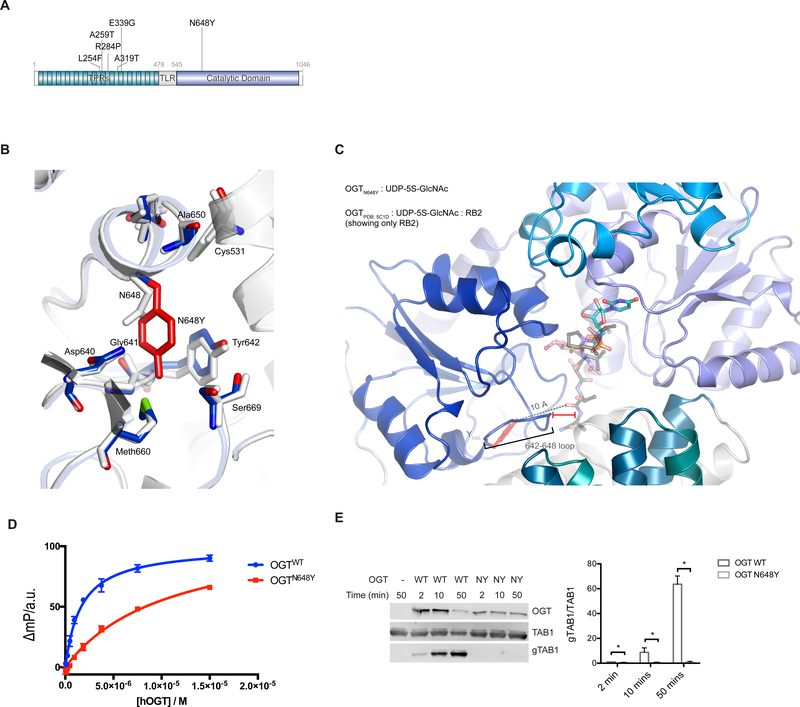
Trio whole genome sequencing was performed. After variant filtration for de novo, recessive or X-linked variants with allele frequencies below 1%, we identified a missense variant in the OGT gene ChrX (GRCh38): g.71561865A>T; NM_181672.2: c.1942A>T p.(Asn648Tyr). Both parents and sibling were healthy and did not carry this mutation. Unlike the other OGT XLID mutations identified to date, this mutation maps to the catalytic domain of OGT (Fig. 2A)
Figure 2: Effects of the N648Y mutation on OGT structure and activity.
(a) Schematic diagram of OGT highlighting the TPRs, TPR-like repeat, the Catalytic domain, the N648Y mutation (bold) and all the previous identified mutations in OGT. TPR: tetratricopeptide repeat domain; TLR; tetratricopeptide repeat-like domain.
(b) Immunoblots showing OGT glycosyltransferase activity against TAB1 and gTAB1. Quantification of gTAB1 normalised to TAB1 signal. N = 3, mean ± SD. Multiple t-test using the Holm-Sidak method. * corresponds to p = 0.021 (2 mins), p = 0.017 (5 mins) and p = 0.008 (50 mins) TAB1:TAK1-binding protein antibody; gTAB1: glycosylated TAB1 antibody.
(c) OGT superimposed complexes of OGTWT (In light gray; PDB: 5C1D; [43]) and OGTN648Y (In blue; PDB: 6Q4M) showing the mutated site and the proximal residues. N648Y mutated residue is shown in red.
(d) OGTN648Y in complex with superimposed RB2 peptide from OGTWT (PDB: 5C1D) showing the putative location of the peptide. The loop 642–648 of OGTN648Y is indicated including the distance from the superimposed peptide.
(e) FP assay showing the binding of the UDP-Peptide bisubstrate conjugate to OGTWT and OGTN648Y
The N648Y mutation leads to structural changes in the catalytic domain
Asn648 is highly conserved in OGT from C. elegans to H. sapiens (Supplementary Fig. 1). Inspection of the human OGT crystal structure reveals that Asn648 maps to the interface of the OGT TPRs with the catalytic domain. The Asn648 side chain forms van der Waals interactions with that of Tyr642, while the loop between these two interacting residues (hereafter 642–648 loop) forms part of the composite OGT acceptor substrate binding cleft. It is thus possible that this mutation could affect the TPR-catalytic domain interface and lead to changes in the stability of the protein. We first analysed the effect of the mutation on the folding stability using differential scanning fluorimetry. Using an E. coli expression system, we purified a recombinant truncated form of OGT version containing the catalytic domain and 3.5 TPR repeats, for both wild type (OGTWT) and mutant (OGTN648Y) OGT. Sigmoidal temperature-induced unfolding curves were obtained for both OGTWT and OGTN648Y with inflection points (Tm) of 44 °C, indicative of identical unfolding temperatures (Supplementary Fig S2). Given that the mutation has the potential to alter the 642–648 loop, which forms an important part of the catalytic pocket, we next investigated possible structural changes by X-ray crystallography. Recombinant OGTN648Y was crystallised in presence of the donor substrate analogue UDP-5S-GlcNAc and an acceptor peptide derived from the well-characterised OGT substrate TGF-beta-activated kinase 1 binding protein 1 (TAB1). Diffraction data were collected to 2.2 Å and initial refinement started from the published OGTWT ternary structure [43]. Mutation of Asn648 to the bulkier tyrosine appears to be accommodated without disruption of the domain organisation observed in the wild type structure (RMSD of 0.4 Å on all backbone Cα atoms) (Fig. 2B). However, the aromatic Tyr648 side chain forms a π-π stacking interaction with Tyr642 causing positional shifts of up to 0.7 Å in the 642–648 loop. Strikingly, despite the presence of an acceptor peptide in the crystallisation condition, no corresponding electron density was observed in the OGT active site. This could be because the changes in 642–648 loop caused by the mutation affects the ability of OGT to bind to acceptor substrates (Fig. 2C). To explore this possibility, we used a recently established fluorescence polarimetry assay (FP) where increasing concentrations of OGT are titrated against a fixed concentration of a fluorescent probe that incorporated elements of both peptide and nucleotide substrates [44]. The direct binding affinity of the probe to OGTWT yielded a Kd value of 1.6 μM as previously reported, whereas the Kd value for the OGTN648Y was 7 times higher (11 μM), suggesting reduced substrate binding in agreement with the structural observations (Fig. 2D). In summary, the N648Y mutation may lead to changes in the OGT acceptor substrate binding cleft and affect substrate binding.
OGTN648Y does not glycosylate the model acceptor substrate TAB1
Given the absence of effects on stability and the apparent effect of the N648Y mutation in OGT substrate-binding, it is possible that catalytic activity is affected. To investigate this, we used an enzyme assay where we incubated OGTWT or OGTN648Y with the well characterised substrate TAB1. This substrate is efficiently mono-glycosylated on Ser395 by wild type OGT [45]. Western blotting analysis using a specific antibody which binds the O-GlcNAcylated form of TAB1 [45] showed that the OGTN648Y variant was unable to glycosylate the substrate (Fig. 2E), in contrast to the efficient glycosylation observed by OGTWT. Thus, OGTN648Y does not glycosylate the model acceptor substrate TAB1.
The N648Y mutation leads to hypo-glycosylation in mES cells
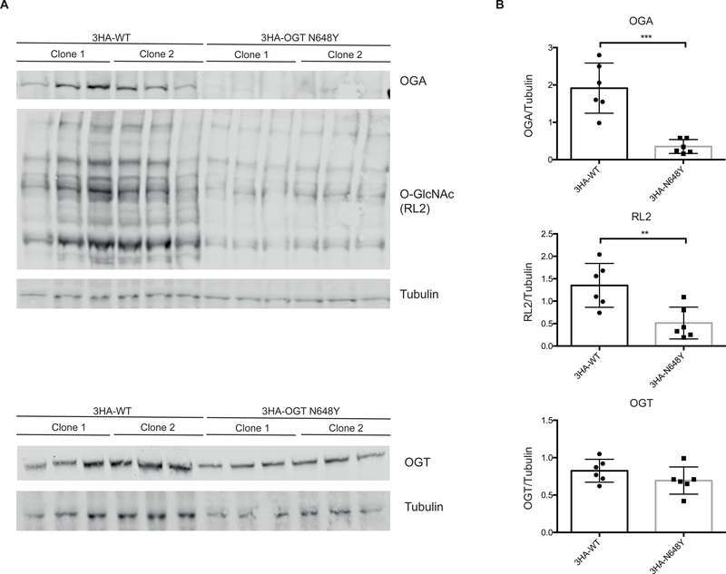
Maintenance of O-GlcNAc homeostasis is essential for optimal cellular function, and perturbations in protein O-GlcNAcylation have been implicated in pathogenesis. We next investigated the effects of reduced OGTN648Y activity on O-GlcNAc homeostasis and levels of OGA/OGT. We used CRISPR/Cas9 genome editing to introduce the N648Y mutation in the endogenous ogt locus carrying an additional N-terminal triple HA (3HA) tag (as described in detail in Materials and Methods). Two independent clones each were obtained for mESCs carrying 3HA-tagged wild type and 3HA-tagged mutant OGT, as verified by diagnostic restriction digest and sequencing. Western blotting analyses of 3HA-N648Y mESC cells was carried out to analyse OGT, OGA and protein O-GlcNAcylation levels (Fig. 3A). In agreement with loss of in vitro activity towards the TAB1 substrate (Fig. 2E) and the potential effect of the loop 642–648 on OGT substrate binding (Fig. 2D), this experiment revealed significantly reduced O-GlcNAc levels in 3HA-N648Y mESCs compared to the controls. This is evidence for a possible link between perturbation in protein O-GlcNAcylation and XLID. No reduction in OGT protein levels were detected. However, OGA levels appeared reduced in 3HA-N648Y mESCs (Fig. 3B). This hints at the existence of a compensatory mechanism that allows cells to partially maintain O-GlcNAc homeostasis by reducing OGA levels – as also observed in some of the other OGT XLID mutations in the TPR domain [39,46]. Thus, the N648Y mutation leads to hypo-glycosylation in mES cells.
Figure 3: The N648Y mutation leads to reduced protein O-GlcNAcylation in 3HA-N648Y mES cells.
(a) Immunoblots showing OGA, protein O-GlcNAcylation (RL2) and OGT levels in 3HA-WT and 3HA-N648Y undifferentiated mES cells.
(b) Quantification of Western blotting of OGA, protein O-GlcNAcylation (RL2) and OGT levels normalised to tubulin signal. n = 3, mean ± SD. Unpaired t-test. ** corresponds to p = 0.0068 (RL2); *** corresponds to p = 0.0003 (OGA)
Concluding remarks
Protein O-GlcNAcylation has emerged as an essential PTM involved in disease, and more recently in neuronal function and development [19,32,33,41]. Recent NGS studies have led to the identification of mutations in OGT that give rise to XLID, suggesting a direct link between O-GlcNAc and regulation of brain development. However, experimental evidence for a link between significant loss of OGT activity and changes in the O-GlcNAc proteome has remained elusive, presumably because of the location of these mutations in the N-terminal TPR substrate binding domain of OGT. Here, we report a patient with XLID attributable to an OGT missense mutation (Asp648Tyr), which maps to the OGT catalytic domain (Fig. 2A). In agreement with the five previously reported patients, the patient shows reduced IQ, limited speech, developmental delay, facial dysmorphia and hypotonia.
We initially delineated the effects of the mutation on the stability, structure and activity of the enzyme using in vitro approaches. Unlike the previously reported OGT XLID mutations we were not able to detect changes in folding stability induced by the mutation (Supplementary Fig. 2). Using X-ray crystallography, we revealed that the mutation did not induce large global conformational changes. However, the observed local conformational changes in the OGT acceptor substrate binding cleft could affect activity. Indeed, using in vitro analyses, the mutant led to significant reduction in the binding affinity of a bisubstrate fluorescence polarimetry probe and defects in the glycosylation of TAB1, suggesting a direct effect of the mutation on enzymatic activity (Fig. 2D and E)
To extend our investigations, we introduced the patient mutation into a male mouse embryonic stem cell line. In previous studies, examination of the steady-state global OGT, OGA and O-GlcNAc levels in different cell lines showed no significant changes in protein O-GlcNAc and OGT, while OGA levels appeared reduced in most of the analysed cell lines, suggesting a compensatory mechanism between OGT and OGA to maintain O-GlcNAc homeostasis (Fig. 3). To our surprise, the 3HA-N648Y mESC lines showed not only reduced levels of OGA, but also reduced protein O-GlcNAcylation levels. This suggests that perturbations in O-GlcNAc cycling may contribute to the patient ID phenotype. This now enables the search for specific proteins and O-GlcNAc sites, whose altered O-GlcNAcylation levels can be linked to cellular processes that could be causative for the observed phenotypes.
There is currently only a very limited number (six) of XLID patients identified that carry different mutations in ogt. It is thus not yet feasible to consider possible treatments. An interesting clue to a possible treatment is the observed changes in OGA levels. This is a mechanism that may compensate for loss in OGT activity or protein levels. This would lead to the hypothesis that (further) inhibition of OGA is a possible route towards re-establishing full O-GlcNAc homeostasis and possible alleviation of the patient phenotypes. Another route would be to increase cellular production of UDP-GlcNAc, which has been shown to directly lead to increased levels of protein O-GlcNAcylation. It has been demonstrated that this is possible to achieve by feeding cells with the UDP-GlcNAc precursor glucosamine, which enters the hexosamine biosynthetic pathway as glucosamine-6-phosphate and leads to increased UDP-GlcNAc levels [47–50]. Glucosamine, in the form of glucosamine sulphate, is a food supplement frequently taken by patients suffering from osteoarthritis, albeit with limited clinical evidence for significant benefits [51,52]. In an initial experiment, at the age of 8 y of our patient we started the treatment with glucosamine sulphate (400 mg three times a day). This treatment has been performed over a period of 13 m without any adverse effects (unpublished data). Parents have noticed that the patient is more active; he has more energy and has increased plasticity in movements. He started to use syllables and teeth development progressed. However, there are no changes in clinical biochemical analyses, and in the absence of a proper randomised trial it is not possible to attribute these improvements to the treatments. Nevertheless, this is an approach that could be evaluated once sufficient patient numbers are available.
Materials & Methods
Whole genome sequencing
Trio whole genome sequencing and data processing were performed by Genomics Platform at the Broad Institute of Harvard and MIT (Broad Institute, Cambridge, MA, USA). The variant filtration for de novo, recessive or X-linked variants with allele frequencies below 1% in ExAC and gnomAD databases revealed three high quality variants, all appearing hemizygous on X-chromosome in genes DLG3, OGT, KIAA1210. Out of them the OGT variant ChrX(GRCh38): g.71561865A>T; NM_181672.2: c.1942A>T p.(Asn648Tyr) was the only variant with no hemizygous carriers in ExAC, gnomAD and in-house database, and thus considered as the most probable candidate after variant filtration. The variant appeared as de novo in protein domain: O-GlcNAc transferase, C-terminal. The variant was validated by Sanger sequencing. The mutated p.Asn648 amino acid is highly conserved from man to C. elegans, and multiple in-silico pathogenicity predicting algorithms indicated a damaging effect. Pathogenicity classified by the American College of Medical Genetics variant interpretation guidelines is likely pathogenic (absent from population databases, multiple computational evidence, de novo).
Protein expression and purification
Truncated (323–1044) OGT constructs were expressed in E. coli BL21(DE3) RIPL cells as N-terminal GST fusion protein, as described previously [43]. Briefly, transformed E. coli cells were grown in LB broth at 37 °C with agitation until OD600 reached 0.8, at which point the temperature was lowered to 18 °C and expression was induced by addition of 100 μM IPTG. Cells were lysed using French Press in base buffer (0.1 M Tris-HCl, pH 7.5, 0.15 M NaCl, 0.5 mM TCEP) supplemented with 25 mM imidazole 0.1 mg/ml DNase I and protease inhibitor cocktail (1 mM benzamidine, 0.2 mM PMSF, 5 mM leupeptin). Then, recombinant proteins were affinity purified following manufacturers’ guidelines. After cleavage of affinity tags, dialyzed protein was loaded onto 5 ml HiTrap Q Sepharose FF anion exchange resin (GE Healthcare) and eluted with a linear gradient up to 60% of buffer B (0.1 M Tris-HCl, pH 8.5, 500 mM NaCl). Peak fractions were pooled, concentrated and further purified via size exclusion chromatography using a 300 ml Superdex™ 200 column (GE Healthcare) equilibrated with base buffer. The peak fractions were concentrated to 10 mg/ml. For crystallisation, truncated mutant OGT was used fresh at 7 mg/ml concentration. For all other purposes, proteins were concentrated to 10 mg/ml, mixed 1:1 with 50% glycerol, snap-frozen and stored at −80 ˚C until use.
Structure solution
Crystallisation of truncated OGTN648Y (residues 323–1044) was performed as described previously [43]. Briefly, experiments were performed at 22 °C using 24-well hanging drop crystallization plates, by combining 1 μl drops containing 7 mg/ml OGTN648Y (in base buffer), 3 mM UDP-5S-GlcNAc and 3 mM acceptor peptide derived from TAB1 sequence (PVSVPYSSAQSTS) with 2 μl of reservoir solution (1.45 M K2HPO4, 8 mM EDTA and 1% xylitol. Large, clear-faced crystals appeared overnight. Prior to diffraction experiments, individual crystals were cryo-protected in reservoir solution supplemented with 3.5 M maleic acid, 3 mM UDP-5S-GlcNAc and 3 mM acceptor peptide and flash-frozen in liquid nitrogen. Diffraction data were collected at the European Synchrotron Radiation Facility beamline ID23. Data were processed with XDS [53] and scaled to 2.2 Å using SCALA [54]. The structure was solved by molecular replacement using the structure for OGTWT (PDB: 5C1D;[43]) as the search model. The resulting model was manually refined using Coot [54] and REFMAC [55], respectively. Although the mutant protein was crystallised in the presence of donor and acceptor, there was no evidence for the latter in the electron density maps and was therefore excluded from the model. Coordinates and structure factors were deposited in the PDB (PDB:6Q4M). Scaling and model building statistics can be seen in Table 1.
Table 1: X-ray diffraction data processing and refinement statistics of the OGTN648Y ternary complex.
Numbers in brackets show represent the highest resolution bin.
| OGTN648Y | |
|---|---|
| Space group | F222 |
| Cell Dimensions | |
| α, β, γ (°) | 137.3, 150.7, 199.5 |
| Resolution (Å) | 45.92 – 2.27 (2.20 – 2.27) |
| Rsym or Rmerge | 0.08 (0.87) |
| I/σI | 12.0 (2.0) |
| Completeness (%) | 100 (99) |
| Redundancy | 6.7 (6.7) |
| No. reflections | 52373 (819) |
| Rwork / Rfree | 0.17 / 0.20 |
| R. M. S. DEVIATIONS | |
| Bond lengths (Å) | 0.011 |
| Bond angles (°) | 1.8 |
Fluorescence polarimetry assay.
To evaluate the expected binding differences between the OGTWT and the OGTN648Y we titrated a previously published fluorescently labelled UDP-peptide bisubstrate conjugate [56] with increasing concentrations of either OGTWT or the OGTN648Y in 0.1 M Tris-HCl pH 7.5, 0.15 M NaCl, 0.5 mM TCEP and 5% DMSO. Each sample, containing 25 μl was incubated in the dark for 30 min before the read-out with a PHERAStar plate reader (BMG LABTECH). Subsequent data analysis was performed using GraphPad Prism 6 as outlined previously [56].
Activity assays
O-GlcNAcylation assays were performed on TAB1 protein (residues 7–420). TAB1 (1 μM) was combined with full length OGTWT or OGTN648Y (0.1 μM) in the presence of 100 μM UDP-GlcNAc. The reaction mixtures were incubated at 37 °C for 2 to 50 minutes and subsequently stopped by addition of LDS loading buffer (4x, Thermo Fisher Scientific). Proteins were resolved by SDS-PAGE (4–12% acrylamide [Life Technologies]) and transferred onto nitrocellulose membranes (GE Healthcare). After antibody treatment, progress of the reaction was visualised using LI-COR Odyssey scanner and associated quantification software.
Mouse ES Cell culture
mESC E14-TG2a.IV AW2 line was acquired from the MRC Centre for Regenerative Medicine, Institute for Stem Cell Research, University of Edinburgh [57]. mESCs were cultured in an undifferentiated state in 0.1% gelatin [w/v] coated plates in GMEM BHK-21 (Gibco) supplemented with 10% FBS [v/v] (Gibco), 0.1 mM MEM non-essential amino acids (Gibco), 1 mM sodium pyruvate (Gibco), 0.1 mM 2-mercaptoethanol (Gibco) and 100 units/mL LIF (produced in house) at 37 °C in 5% CO2.
Generation of 3HA-tag N567K mES cell line
For the generation of the 3HA mES cell line, we transfected wild type male E14-TG2a.IV AW2 mES cells with pBABED puro U6 and pX335 (Cas9 D10A) vectors containing the gRNA sequences selected using WTSI Genome Editing [58]. Silent mutations were designed in addition to the intended mutations, removing PstI and BfmI restriction sites (Supplementary Fig. 2). A geneblock containing these changes was obtained from IDT (International DNA technologies). The geneblock was introduced into the cloned 2 kb region by restriction free cloning [59] and then confirmed by DNA sequencing [59]. For transfection, 2×10⁵ cells were seeded onto gelatine coated 24-well plates and transfected using 0.33 μg of each vector and Lipofectamine 3000 according to manufacturer’s instructions. After 24 h, media was replaced and puromycin (3 μg/mL) was added to the cells for selection until control untrasfected cells were all dead (48 h). Then the cells ware re-plated into 10 cm plates for recovery. After that, single cell selection was carried out using limited dilution according to 0.3 cell per well in 96 well plates. For confirmation of mutations, diagnostic restriction digests and genomic DNA sequencing analysis were carried out. For the restriction fragment length polymorphism assay 3HADiag_F and 3HADiag_R primers were used to amplify by PCR the mutated site and the silent mutation which eliminates a PstI restriction site (Supplementary Table S1). The size of the PCR product (546 bp vs 450 bp for wild type) was used to screen for successful integration. To further confirm, the PCR product was then digested using PstI or BfmI. Clones negative for the restriction enzyme assay were then sequenced to confirm the presence of the modification.
For generation of the 3HA-OGTN648Y mESCs, we transfected the previously generated 3HA mESCs with pBABED puro U6 and pX335 (Cas9 D10A) vectors containing the gRNA sequences using the same procedure we previously used for the insertion of the 3HA-tag (Supplementary Fig. 3). Restriction enzymes and genomic DNA sequencing analysis were used for mutation identification. For the restriction fragment length polymorphism assay MouseEstDiag_F and MouseEstDiag_R primers were used to amplify by PCR the mutated site and the silent mutation which eliminates an Eco130I restriction site (Supplementary Table S2). The size of the bands following digestion (167 bp and 433 bp for wild type versus 600 bp for mutant) was used to screen for successful integration. Clones negative for cutting in the restriction enzyme assay were then sequenced to confirm the presence of the modification.
Western Blot
Cells were harvested in lysis buffer containing 150 mM NaCl, 1% Nonidet P-40, 0.5% Sodium deoxycholate, 0.1% Sodium dodecyl sulfate, 25 mM TrisHCl (pH 7.4), 1 mM Sodium orthovanadate, 50 mM Sodium fluoride, 5 mM Sodium pyrophosphate. 10–20 μg of cell lysate was loaded in NuPAGE 3–8% Tris-Acetate gels (Invitrogen) and transferred to Nitrocellulose membranes using wet transfer system. Membranes were blocked with TBS-T 5% BSA buffer [w/v]. Primary antibodies used were OGT (F-12, Santa Cruz Biotechnology 1:5000), OGA (Sigma 1:500), RL2 (Thermofisher 1:1000).
Supplementary Material
Acknowledgements
This work was funded by a Wellcome Trust Investigator Award (110061) to D.M.F.v.A.
MHW is supported by NIH T32GM007748. V.P is supported by a SPRINT/MND-MS Scholarship (University of Edinburgh). The Broad Center for Mendelian Genomics (UM1 HG008900) is funded by the National Human Genome Research Institute with supplemental funding provided by the National Heart, Lung, and Blood Institute under the Trans-Omics for Precision Medicine (TOPMed) program and the National Eye Institute. This study makes use of data shared through the Broad Institute matchbox Matchmaker Exchange repository. We thank Dr. Andrew Smith for the AW2 mouse embryonic stem cell line. This study was approved by the Research Ethics Committee of the University of Tartu (approval date 17/10/2016 and number 263/M-16). We also thank Dr. Vladimir Borodkin for providing a sample of the previously published FP probe. Informed consent was obtained from parents of this patient and their participation is gratefully acknowledged. We thank the European Synchrotron Radiation Facility for team on beamline ID23. The coordinates and structure factors are deposited with the PDB (ID 6Q4M).
References
- 1.Maulik PK, Mascarenhas MN, Mathers CD, Dua T & Saxena S (2011) Prevalence of intellectual disability: A meta-analysis of population-based studies. Res. Dev. Disabil. 32, 419–436. [DOI] [PubMed] [Google Scholar]
- 2.American Psychiatric Association (2013) Diagnostic and Statistical Manual of Mental Disorders American Psychiatric Association. [Google Scholar]
- 3.Wieczorek D (2018) Autosomal dominant intellectual disability. Medizinische Genet. Mitteilungsblatt des Berufsverbandes Medizinische Genet. e.V 30, 318–322. [DOI] [PMC free article] [PubMed] [Google Scholar]
- 4.Kvarnung M & Nordgren A (2017) Intellectual Disability & Rare Disorders: A Diagnostic Challenge. Adv Exp Med Biol 1031, 39–54. [DOI] [PubMed] [Google Scholar]
- 5.Neri G, Schwartz CE, Lubs HA & Stevenson RE (2018) X-linked intellectual disability update 2017. Am. J. Med. Genet, 1–14. [DOI] [PMC free article] [PubMed] [Google Scholar]
- 6.Vissers LELM, Gilissen C & Veltman JA (2016) Genetic studies in intellectual disability and behavioral impairment. Arch. Iran. Med. 19, 363–375. [PubMed] [Google Scholar]
- 7.Moeschler JB & Shevell M (2014) Comprehensive Evaluation of the Child With Intellectual Disability or Global Developmental Delays. Pediatrics 134, e903–e918. [DOI] [PMC free article] [PubMed] [Google Scholar]
- 8.Amor DJ (2018) Investigating the child with intellectual disability. J. Paediatr. Child Health 54, 1154–1158. [DOI] [PubMed] [Google Scholar]
- 9.Chiurazzi P & Pirozzi F (2016) Advances in understanding – genetic basis of intellectual disability. F1000Research 5, 599. [DOI] [PMC free article] [PubMed] [Google Scholar]
- 10.The Deciphering Developmental Disorders Study (2017) Prevalence and architecture of de novo mutations in developmental disorders. Nature 542, 433–438. [DOI] [PMC free article] [PubMed] [Google Scholar]
- 11.Firth H V, Wright CF & Study DDD (2011) The Deciphering Developmental Disorders (DDD) study. Dev Med Child Neurol 53, 702–703. [DOI] [PubMed] [Google Scholar]
- 12.Schimpl M, Zheng X, Borodkin VS, Blair DE, Andrew T, Schüttelkopf AW, Navratilova I, Aristotelous T, Robinson DA, MacNaughtan MA, Aalten DMF Van, Ferenbach AT, Schüttelkopf AW, Navratilova I, Aristotelous T, Albarbarawi O, Robinson DA, MacNaughtan MA & Van Aalten DMF (2012) O-GlcNAc transferase invokes nucleotide sugar pyrophosphate participation in catalysis. Nat. Chem. Biol. 8, 969–974. [DOI] [PMC free article] [PubMed] [Google Scholar]
- 13.Kapuria V, Röhrig UF, Bhuiyan T, Borodkin VS, van Aalten DMF, Zoete V & Herr W (2016) Proteolysis of HCF-1 by Ser/Thr glycosylation-incompetent O-GlcNAc transferase:UDP-GlcNAc complexes. Genes Dev. 30, 960–972. [DOI] [PMC free article] [PubMed] [Google Scholar]
- 14.Kim EJ (2017) In Vitro Biochemical Assays for O-GlcNAc-Processing Enzymes. ChemBioChem 18, 1462–1472. [DOI] [PubMed] [Google Scholar]
- 15.Bhuiyan T, Waridel P, Kapuria V, Zoete V & Herr W (2015) Distinct OGT-Binding Sites Promote HCF-1 Cleavage. PLoS One 10, e0136636. [DOI] [PMC free article] [PubMed] [Google Scholar]
- 16.Rafie K, Raimi O, Ferenbach AT, Borodkin VS, Kapuria V & van Aalten DMF (2017) Recognition of a glycosylation substrate by the O-GlcNAc transferase TPR repeats. Open Biol. 7, 170078. [DOI] [PMC free article] [PubMed] [Google Scholar]
- 17.Clarke AJ, Hurtado-Guerrero R, Pathak S, Schüttelkopf AW, Borodkin V, Shepherd SM, Ibrahim AFMM & Van Aalten DMFF (2008) Structural insights into mechanism and specificity of O-GlcNAc transferase. EMBO J. 27, 2780–2788. [DOI] [PMC free article] [PubMed] [Google Scholar]
- 18.Prasad S, Iyer N & Hart GW (2003) Roles of the TPR domain in O-GlcNAc Transferase (OGT) Targeting and Protein Substrate Specificity as Manuscript M300036200 JBC Papers in Press. [DOI] [PubMed] [Google Scholar]
- 19.Olivier-Van Stichelen S & Hanover JA (2015) You are what you eat: O-linked N-acetylglucosamine in disease, development and epigenetics. Curr. Opin. Clin. Nutr. Metab. Care 18, 339–345. [DOI] [PMC free article] [PubMed] [Google Scholar]
- 20.Zeidan Q, Wang Z, Maio A De & Hart GW (2010) O-GlcNAc Cycling Enzymes Associate with the Translational Machinery and Modify Core Ribosomal Proteins. Mol. Biol. Cell 21, 1922–1936. [DOI] [PMC free article] [PubMed] [Google Scholar]
- 21.Lo P-W, Shie J- J, Chen C- H, Wu C- Y, Hsu T- L & Wong C- H (2018) O-GlcNAcylation regulates the stability and enzymatic activity of the histone methyltransferase EZH2. Proc. Natl. Acad. Sci. 115, 201801850. [DOI] [PMC free article] [PubMed] [Google Scholar]
- 22.Cheng X & Hart GW (2001) Alternative O-glycosylation/O-phosphorylation of Serine-16 in murine estrogen receptor beta: Post-translational regulation of turnover and transactivation activity. J. Biol. Chem. 276, 10570–10575. [DOI] [PubMed] [Google Scholar]
- 23.Wright JN, Benavides GA, Johnson MS, Wani W, Ouyang X, Zou L, Collins HE, Zhang J, Darley-Usmar V & Chatham JC (2019) Acute increases in O-GlcNAc indirectly impairs mitochondrial bioenergetics through dysregulation of LonP1-mediated mitochondrial protein complex turnover. Am. J. Physiol. Physiol., ajpcell.00491.2018. [DOI] [PMC free article] [PubMed] [Google Scholar]
- 24.Slawson C, Zachara NE, Vosseller K, Cheung WD, Lane MD & Hart GW (2005) Perturbations in O-linked beta-N-acetylglucosamine protein modification cause severe defects in mitotic progression and cytokinesis. J. Biol. Chem. 280, 32944–32956. [DOI] [PubMed] [Google Scholar]
- 25.Zachara NE, O’Donnell N, Cheung WD, Mercer JJ, Marth JD & Hart GW (2004) Dynamic O-GlcNAc modification of nucleocytoplasmic proteins in response to stress: A survival response of mammalian cells. J. Biol. Chem. 279, 30133–30142. [DOI] [PubMed] [Google Scholar]
- 26.Cui Y-L, Xue R- Q, Xi He, Ming Zhao, Yu X-J, Liu L-Z, Wu Q, Si Yang, Li D-L & Zang W-J (2019) Cholinergic drugs ameliorate endothelial dysfunction by decreasing O-GlcNAcylation via M3 AChR-AMPK-ER stress signaling. Life Sci. 222, 1–12. [DOI] [PubMed] [Google Scholar]
- 27.Butkinaree C, Park K & Hart GW (2010) O-linked β-N-acetylglucosamine (O-GlcNAc): Extensive crosstalk with phosphorylation to regulate signaling and transcription in response to nutrients and stress. Biochim. Biophys. Acta - Gen. Subj. 1800, 96–106. [DOI] [PMC free article] [PubMed] [Google Scholar]
- 28.Slawson C & Hart GW (2011) O-GlcNAc signalling: Implications for cancer cell biology. Nat. Rev. Cancer 11, 678–684. [DOI] [PMC free article] [PubMed] [Google Scholar]
- 29.Vosseller K, Wells L, Lane MD & Hart GW (2002) Elevated nucleocytoplasmic glycosylation by O-GlcNAc results in insulin resistance associated with defects in Akt activation in 3T3-L1 adipocytes. Proc. Natl. Acad. Sci. 99, 5313–5318. [DOI] [PMC free article] [PubMed] [Google Scholar]
- 30.Ngoh GA, Facundo HT, Zafir A & Jones SP (2010) O-GlcNAc signaling in the cardiovascular system. Circ. Res. 107, 171–185. [DOI] [PMC free article] [PubMed] [Google Scholar]
- 31.Yuzwa SA, Shan X, MacAuley MS, Clark T, Skorobogatko Y, Vosseller K & Vocadlo DJ (2012) Increasing O-GlcNAc slows neurodegeneration and stabilizes tau against aggregation. Nat. Chem. Biol. 8, 393–399. [DOI] [PubMed] [Google Scholar]
- 32.Mariappa D, Zheng X, Schimpl M, Raimi O, Ferenbach AT, Müller HAJ & Van Aalten DMF (2015) Dual functionality of O-GlcNAc transferase is required for Drosophila development. Open Biol. 5, 150234. [DOI] [PMC free article] [PubMed] [Google Scholar]
- 33.Webster DM, Teo CF, Sun Y, Wloga D, Gay S, Klonowski KD, Wells L & Dougan ST (2009) O-GlcNAc modifications regulate cell survival and epiboly during zebrafish development. BMC Dev. Biol. 9, 28. [DOI] [PMC free article] [PubMed] [Google Scholar]
- 34.Ingham PW (1984) A gene that regulates the bithorax complex differentially in larval and adult cells of Drosophila. Cell 37, 815–823. [DOI] [PubMed] [Google Scholar]
- 35.Hanover JA, Forsythe ME, Hennessey PT, Brodigan TM, Love DC, Ashwell G & Krause M (2005) A Caenorhabditis elegans model of insulin resistance: Altered macronutrient storage and dauer formation in an OGT-1 knockout. Proc. Natl. Acad. Sci. 102, 11266–11271. [DOI] [PMC free article] [PubMed] [Google Scholar]
- 36.Pantaleon M, Tan HY, Kafer GR & Kaye PL (2010) Toxic effects of hyperglycemia are mediated by the hexosamine signaling pathway and o-linked glycosylation in early mouse embryos. Biol. Reprod. 82, 751–8. [DOI] [PMC free article] [PubMed] [Google Scholar]
- 37.Jang H, Kim TW, Yoon S, Choi SY, Kang TW, Kim SY, Kwon YW, Cho EJ & Youn HD (2012) O-GlcNAc regulates pluripotency and reprogramming by directly acting on core components of the pluripotency network. Cell Stem Cell 11, 62–74. [DOI] [PubMed] [Google Scholar]
- 38.Medford HM & Marsh SA (2014) The role of O-GlcNAc transferase in regulating the gene transcription of developing and failing hearts. Future Cardiol. 10, 801–812. [DOI] [PMC free article] [PubMed] [Google Scholar]
- 39.Willems AP, Gundogdu M, Kempers MJE, Giltay JC, Pfundt R, Elferink M, Loza BF, Fuijkschot J, Ferenbach AT, Van Gassen KLI, Van Aalten DMF & Lefeber DJ (2017) Mutations in N-acetylglucosamine (O-GlcNAc) transferase in patients with X-linked intellectual disability. J. Biol. Chem. 292, 12621–12631. [DOI] [PMC free article] [PubMed] [Google Scholar]
- 40.Bouazzi H, Lesca G, Trujillo C, Alwasiyah MK & Munnich A (2015) Nonsyndromic X-linked intellectual deficiency in three brothers with a novel MED12 missense mutation [c.5922G>T (p.Glu1974His)]. Clin. Case Reports 3, 604–609. [DOI] [PMC free article] [PubMed] [Google Scholar]
- 41.Selvan N, George S, Serajee FJ, Shaw M, Hobson L, Kalscheuer V, Prasad N, Levy SE, Taylor J, Aftimos S, Schwartz CE, Huq AM, Gecz J & Wells L (2018) O-GlcNAc transferase missense mutations linked to X-linked intellectual disability deregulate genes involved in cell fate determination and signaling. J. Biol. Chem. 293, 10810–10824. [DOI] [PMC free article] [PubMed] [Google Scholar]
- 42.Gundogdu M, Llabrés S, Gorelik A, Ferenbach AT, Zachariae U & van Aalten DMF (2018) The O-GlcNAc Transferase Intellectual Disability Mutation L254F Distorts the TPR Helix. Cell Chem. Biol. 25, 513–518.e4. [DOI] [PMC free article] [PubMed] [Google Scholar]
- 43.Pathak S, Alonso J, Schimpl M, Rafie K, Blair DE, Borodkin VS, Schüttelkopf AW, Albarbarawi O & Van Aalten DMF (2015) The active site of O-GlcNAc transferase imposes constraints on substrate sequence. Nat. Struct. Mol. Biol. 22, 744–749. [DOI] [PMC free article] [PubMed] [Google Scholar]
- 44.Fortini ME (2002) ??-Secretase-Mediated Proteolysis in Cell-Surface-Receptor Signalling. Nat. Rev. Mol. Cell Biol. 3, 673–684. [DOI] [PubMed] [Google Scholar]
- 45.Pathak S, Borodkin VS, Albarbarawi O, Campbell DG, Ibrahim A & Van Aalten DMF (2012) O-GlcNAcylation of TAB1 modulates TAK1-mediated cytokine release. EMBO J. 31, 1394–1404. [DOI] [PMC free article] [PubMed] [Google Scholar]
- 46.Vaidyanathan K, Niranjan T, Selvan N, Teo CF, May M, Patel S, Weatherly B, Skinner C, Opitz J, Carey J, Viskochil D, Gecz J, Shaw M, Peng Y, Alexov E, Wang T, Schwartz C & Wells L (2017) Identification and characterization of a missense mutation in the O-linked β-N-acetylglucosamine (O-GlcNAc) transferase gene that segregates with X-linked intellectual disability. J. Biol. Chem. 292, 8948–8963. [DOI] [PMC free article] [PubMed] [Google Scholar]
- 47.Qu CJ, Jauhiainen M, Auriola S, Helminen HJ & Lammi MJ (2007) Effects of glucosamine sulfate on intracellular UDP-hexosamine and UDP-glucuronic acid levels in bovine primary chondrocytes. Osteoarthr. Cartil. 15, 773–779. [DOI] [PubMed] [Google Scholar]
- 48.Shikhman AR, Brinson DC, Valbracht J & Lotz MK (2009) Differential metabolic effects of glucosamine and N-acetylglucosamine in human articular chondrocytes. Osteoarthr. Cartil. 17, 1022–1028. [DOI] [PMC free article] [PubMed] [Google Scholar]
- 49.Vigetti D, Deleonibus S, Moretto P, Karousou E, Viola M, Bartolini B, Hascall VC, Tammi M, De Luca G & Passi A (2012) Role of UDP-N-acetylglucosamine (GlcNAc) and O-GlcNacylation of hyaluronan synthase 2 in the control of chondroitin sulfate and hyaluronan synthesis. J. Biol. Chem. 287, 35544–35555. [DOI] [PMC free article] [PubMed] [Google Scholar]
- 50.Swamy M, Pathak S, Grzes KM, Damerow S, Sinclair L V, Van Aalten DMF & Cantrell DA (2016) Glucose and glutamine fuel protein O-GlcNAcylation to control T cell self-renewal and malignancy. Nat. Immunol. 17, 712–720. [DOI] [PMC free article] [PubMed] [Google Scholar]
- 51.Wenz W, Hornung C, Cramer C, Schroeder M & Hoffmann M (2017) Effect of Glucosamine Sulfate on Osteoarthritis in the Cruciate-Deficient Canine Model of Osteoarthritis. Cartilage 8, 173–179. [DOI] [PMC free article] [PubMed] [Google Scholar]
- 52.Reginster JY, Neuprez A, Lecart MP, Sarlet N & Bruyere O (2012) Role of glucosamine in the treatment for osteoarthritis. Rheumatol. Int. 32, 2959–2967. [DOI] [PMC free article] [PubMed] [Google Scholar]
- 53.Kabsch W (2010) XDS. Acta Crystallogr. D. Biol. Crystallogr. 66, 125–32. [DOI] [PMC free article] [PubMed] [Google Scholar]
- 54.Winn MD, Ballard CC, Cowtan KD, Dodson EJ, Emsley P, Evans PR, Keegan RM, Krissinel EB, Leslie AGW, McCoy A, McNicholas SJ, Murshudov GN, Pannu NS, Potterton EA, Powell HR, Read RJ, Vagin A & Wilson KS (2011) Overview of the CCP4 suite and current developments. Acta Crystallogr. Sect. D Biol. Crystallogr. 67, 235–242. [DOI] [PMC free article] [PubMed] [Google Scholar]
- 55.Vagin AA, Steiner RA, Lebedev AA, Potterton L, McNicholas S, Long F & Murshudov GN (2004) REFMAC5 dictionary: Organization of prior chemical knowledge and guidelines for its use. Acta Crystallogr. Sect. D Biol. Crystallogr. 60, 2184–2195. [DOI] [PubMed] [Google Scholar]
- 56.Rafie K, Gorelik A, Trapannone R, Borodkin VS & van Aalten DMF (2018) Thio-Linked UDP–Peptide Conjugates as O-GlcNAc Transferase Inhibitors. Bioconjug. Chem. 29, 1834–1840. [DOI] [PMC free article] [PubMed] [Google Scholar]
- 57.Zhou X, Smith AJH, Waterhouse A, Blin G, Malaguti M, Lin C-Y, Osorno R, Chambers I & Lowell S (2013) Hes1 Desynchronizes Differentiation of Pluripotent Cells by Modulating STAT3 Activity. Stem 31, 1511–1522 [DOI] [PMC free article] [PubMed] [Google Scholar]
- 58.Hodgkins A, Farne A, Perera S, Grego T, Parry-Smith DJ, Skarnes WC & Iyer V (2015) WGE: a CRISPR database for genome engineering. Bioinformatics 31, 3078–80. [DOI] [PMC free article] [PubMed] [Google Scholar]
- 59.van den Ent F & Löwe J (2006) RF cloning: A restriction-free method for inserting target genes into plasmids. J. Biochem. Biophys. Methods 67, 67–74. [DOI] [PubMed] [Google Scholar]
Associated Data
This section collects any data citations, data availability statements, or supplementary materials included in this article.


