Abstract
The purpose of this study was to test effects of exogenous tissue-type plasminogen activator (tPA) in traumatic brain injury (TBI). We tested two different tPA formulations, free tPA and tPA bound to erythrocytes (RBC/tPA). Vehicle and each of the tPA treatments were injected intravenously in anesthetized rats 15 minutes after moderate lateral fluid percussion injury. Animals were sacrificed at two days for calculating microclot burden (n = 13) and IgG staining area (n = 13) in the brain sections as indicators of post-traumatic thrombosis and blood-brain barrier (BBB) breakdown, respectively. Another set of injured animals treated in the same way were sacrificed at 7 days to compare cortical lesion volumes (n = 28) and CA3 hippocampal cell loss (n = 24). All evaluations were done blinded with respect to treatment.
No significant differences were found with respect to microclot burden or IgG staining volume. Injection of wild-type tPA caused significantly (p < .05) larger cortical injuries and greater cerebral hemorrhage. In contrast, there was significantly less cortical injury (p < .01) and hippocampal cell loss (p < .01) in RBC/tPA group than in all other groups. These results reveal that the RBC/tPA is neuroprotective in experimental TBI, compared to unbound tPA.
Keywords: traumatic brain injury, microclots, tissue plasminogen activator, blood-brain barrier
Introduction
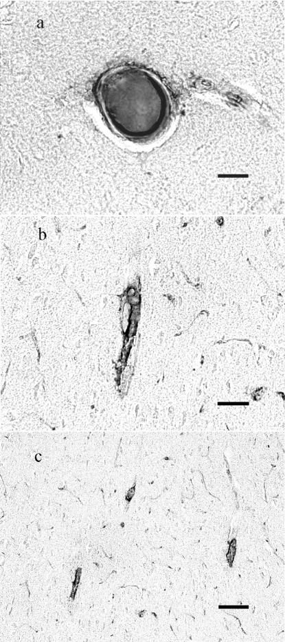
Vascular damage usually accompanies traumatic brain injury (TBI) and contributes to the resulting cerebral pathology through mechanisms such as hemorrhage and traumatic vasospasm. Blood also reacts with tissue factor (TF), which is present in high concentrations on the adventitial surface of cerebral vessels. This results in the intravascular clotting and formation of microthrombi, which occlude cerebral arterioles in the days following injury (Fig. 1). This in turn results in cerebral ischemia, that likely aggravates the impact of initial trauma (Lu et al., 2004; Stein S. C. et al., 2002; Stein S. C. et al., 2004). In theory, lysing these thrombi might alleviate secondary damage and improve the outcome of TBI, if it could be done without impairing normal hemostasis and inflicting other adverse effects in the CNS.
Fig. 1.
Microclots 48 hours following TBI are found in large (a), medium (b) and small (c – arrows) arterioles. Stain for clots uses an antibody to thrombin-antithrombin complexes and DAB counterstain (see text for details). Bar in each image = 40μ.
DAB: diaminobenzidine
Tissue type plasminogen activator (tPA) is a potent thrombolytic agent, used with success for emergency treatment of ischemic stroke. Although its use might ameliorate damage done by clots, there are several drawbacks to tPA administration. An obvious problem is the likelihood that tPA indiscriminately dissolves both pathological intravascular microclots and mural hemostatic plugs, thereby worsening post-traumatic hemorrhage. Furthermore, the pharmacokinetics of tPA seem unsuitable for prevention of secondary post-traumatic clots. Intravascular microclot formation, development and dissolution occur over days(Lu et al., 2004). Unfortunately, exogenous tPA is cleared from the circulation within minutes(Narita et al., 1995), thus limiting its therapeutic utility in this setting. In addition, the molecule has been reported to have toxic effects in the CNS, both on the blood-brain barrier (BBB) (Polavarapu et al., 2007; Yepes et al., 2003), on neurons themselves and on other elements of the neuropil(Armstead et al., 2006; Yepes and Lawrence, 2004).
To be of use in thromboprophylaxis, a fibrinolytic agent should selectively lyse potentially occlusive clots during their formation, without affecting hemostatic clots or exerting extravascular toxicity. However, all existing fibrinolytics are short-lived (<30 min) and small (<10 nm diameter) agents, capable of diffusion into hemostatic clots. None can be used safely, even in patients diagnosed with high risk of imminent or recurrent thrombosis. However, prior studies showed that coupling tPA to red blood cells (RBC) produces long-circulating enzymatically active complexes, RBC/tPA that neither lyse hemostatic clots nor enter the CNS. Therefore, coupling tPA to RBC converts tPA into an effective and safe thromboprophylactic agent. Injection of either preformed RBC/tPA(Ganguly et al., 2006; Ganguly et al., 2007; Murciano et al., 2003) or tPA derivatives targeted to circulating RBC's(Zaitsev et al., 2006) prevents subsequent formation of occlusive venous and arterial clots, with no overt harmful effects on the carrier RBC(Ganguly et al., 2006; Ganguly et al., 2007; Murciano et al., 2003), activation of coagulation(Murciano et al., 2003) or impaired post-surgical hemostasis(Zaitsev et al., 2006). Furthermore, pretreatment with RBC/tPA provides fast dissolution of cerebrovascular thromboemboli in a mouse model, thereby providing fast and stable reperfusion, alleviating ischemic brain injury and eliminating lethality without adverse side effects associated with injection of tPA (Danielyan et al., 2008).
Therefore, RBC/tPA has several key advantages over free tPA in the context of preventing thrombotic brain ischemia (Murciano et al., 2003), which makes it a good candidate for testing in TBI models. The purpose of this study was to evaluate and compare the effects of different forms of tPA after moderate TBI in the rat.
Materials and Methods
Treatment groups
In an effort to separate the various actions of tPA, we used two experimental treatment groups: RBC-bound wild-type tPA (RBC/tPA) and free (unbound) tPA. A third (control) group received vehicle, phosphate-buffered saline with bovine serum albumin (PBS-BSA) injection only.
The day prior to injury, 1.5 ml of blood was removed from each rat. All rats were assigned to one of the three groups and received the same volume of red blood cell (RBC) transfusions, to maintain hematocrits within the normal range. Preparations were readied, coded and labelled by one of us (KG); all others were blinded with respect to the groups’ compositions for the duration of the experiments. All animals were given a single 1.5 ml femoral vein injection of tPA or vehicle in autologous blood 15 minutes after injury.
Preparation of Treatments
Human recombinant tPA (Alteplase) was obtained from Genentech (South San Francisco, CA). RBCs were isolated by centrifugation from fresh anti-coagulated rat blood. Approximately 6×10⁴ molecules of tPA were attached per RBC as previously described(Ganguly et al., 2006; Ganguly et al., 2005; Murciano et al., 2003; Muzykantov and Murciano, 1996). The conjugation process does not affect enzymatic activity of tPA or RBC biocompatibility(Ganguly et al., 2007. The RBC/tPA conjugate was suspended in PBS-BSA containing 2 mg/ml BSA and intact washed RBC (final hematocrit 50%) added to all formulations in order to compensate for potential effect of hematocrit change in animals.
Parasagittal fluid percussion injury
The procedure for the fluid percussion model of brain injury has been previously described in detail(McIntosh et al., 1989). Briefly, 66 male Sprague–Dawley rats (325−375 g) were anesthetized with sodium pentobarbital (60 mg/kg, i.p.) and a 5.0-mm craniotomy was performed over the left cortex midway between lambda and bregma. With the dura intact, a female Leur–Lok fitting was secured in the craniotomy site. Animals were placed on heating pads until the time of injury. Ninety minutes after anesthesia, animals were attached to the fluid percussion device via the Leur–Lok fitting and injured at a moderate severity level (2.4−2.6 atm).
All animals were pair-housed on a 12-h light/dark cycle with access to food and water ad libitum, and food intake and animal weight were measured every two days. Twelve animals (28%) died, presumably of their injuries, prior to sacrifice. Since they were evenly distributed among treatment groups, they were excluded from further analysis. All experiments were carried out in accordance with policies set forth by the University of Pennsylvania Institutional Animal Care and Use Committee and in accordance with NIH guidelines for the humane and ethical treatment of animals.
Histological preparation
Sacrifice was at two (intravascular microthrombosis, blood-brain barrier integrity) or at seven days (lesion volume, hippocampal cell loss). All animals were given an overdose of sodium pentobarbital (200 mg/kg) and transcardially perfused with heparinized saline followed by 4% paraformaldehyde. The brains were removed, post-fixed overnight in 4% paraformaldehyde, placed in PBS buffer for three days and immersed in 30% sucrose until saturated (4−5 days). The brains were then snap frozen in isopentane at −30−40°C and sectioned coronally. Twenty micron sections (3mm behind frontal poles to cerebellum) were cut on a rotary microtome, and sections120 every microns apart were saved for analysis.
Tissue sections for IgG immunostaining were incubated in PBS, anti-rat IgG (1:200, Vector Laboratories), then incubated with antisheep IgG, 1:400 secondary antibody and 3,3'-diaminobenzidine tetrahydrochloride (Vector Laboratories, Burlingame, CA) for visualization(Hoshino et al., 1996). Tissue for intravascular microthrombi was incubated overnight with 1:300 dilution of a sheep polyclonal antibody used to recognize both free antithrombin III and antithrombin III in antithrombin III–protease complexes (American Diagnostica, Stamford, CT). Sections were then incubated with antisheep IgG, 1:400 secondary antibody and 3,3'-diaminobenzidine tetrahydrochloride (Vector Laboratories, Burlingame, CA) for visualization. Staining for evaluation of cortical tissue loss involved hematoxylin and eosin (H&E), that for evaluation of hippocampal tissue loss employed cresyl violet Nissl stain.
Evaluation of intravascular microthrombi
The total number of microclots were counted in each section of each specimen. To estimate the density of microclots, the surface area of each microscopic specimen was computed from the digital images, using MCID Imaging software (Interfocus Imaging, Cambridge, England) and the number of microclots per mm2 (microclot burden) calculated for each hemisphere of each section(Stein S. C. et al., 2002). Treatment groups were compared with respect to microclot burdens.
Evaluation of Blood-brain barrier integrity
Endogenous IgG is not normally seen in the rat brain in any quantity; its presence is an indication of extravasation secondary to BBB breakdown. All sections were examined by light microscopy. Areas containing IgG staining were outlined and measured, and the total volume of extravasation calculated using the same formula as for cortical tissue loss below.
Evaluation of cortical and hippocampal tissue loss
For cortical tissue, the area of each lesion was outlined. Where tissue had been lost at the cortical surface, the surface line was reconstructed by referring to the opposite hemisphere. Areas of neuronal necrosis, cavitation and hemorrhage were included within the lesion outline. The area of cortical tissue loss in each section was measured using NIH Image Software (v1.62). All sections containing identifiable tissue loss were included. The total volume of tissue loss was obtained using the formula:
For each animal, five nonadjacent sections were chosen from − 3.3 to − 3.8 and four nonadjacent sections from −5.3 to −5.6 bregma for evaluation of hippocampal tissue loss. The CA3 subfield of the hippocampus was centered on the image field and a precalibrated length of 1 mm along the region previously characterized as the site of maximal damage in the ipsilateral pyramidal layer was chosen for counting neurons. “Viable” neurons, which had well-defined cell bodies and nuclei, were counted in the selected area. For each slide, the corresponding cell count on the uninjured side was recorded. The percent cell loss in each section was obtained using the formula: 100% * (Ncontrol – Ninjured)/Ncontrol. A total count of neuron loss for each animal was obtained by averaging the counts from the individual sections(Leoni et al., 2000).
Statistical Analysis
Comparisons among different groups were performed using a one-way analysis of variance (ANOVA), followed by post-hoc Bonferroni tests for individual comparisons (Stata 9, StataCorp, College Station, TX). We considered differences for which the probability was < 0.05 to be significant.
Results
The means and standard deviations of microclot burden calculations (# of microthrombi/tissue area) are shown in the Table. The burden was calculated for the perilesional area of the left hemisphere, the entire left hemisphere and the entire brain. No differences were found among the three treatment groups in any of these calculations (p = .775). The mean volume of IgG staining was 27.6 (± s.d. 11.6) mm3 in the vehicle group (n =4), 33.8 (± 23.4) in the free tPA group (n = 5) and 30.5 (± 15.2) with RBC/tPA (n = 4), suggesting no significant difference with regards to BBB breakdown (p = .609).
Table.
Microclot Density – mean # per mm2(± s.d.)
| Group | Vehicle | RBC/tPA | Free tPA |
|---|---|---|---|
| n per group | 5 | 4 | 4 |
| Peri-lesional | 0.0304 (±.0313 ) | 0.0349 (±.0537) | 0.0504 (±.0306) |
| Whole Hemisphere | |||
| Left Hemis. | 0.0248 (±.0227) | 0.0337 (±.0474 ) | 0.0465 (±.0318 ) |
| Right Hemis. | 0.0193 (±.0156) | 0.0346 (±.0411 ) | 0.0672 (±.0835 ) |
| Whole Brain | 0.0221 (±.0156) | 0.0346 (±.0411 ) | 0.0566 (±.0567 ) |
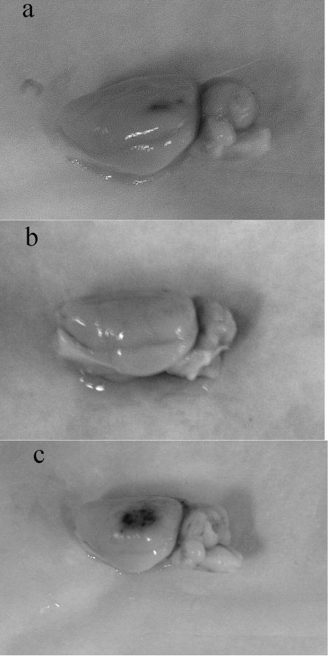
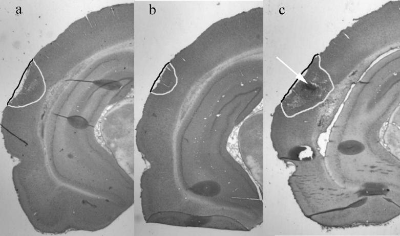
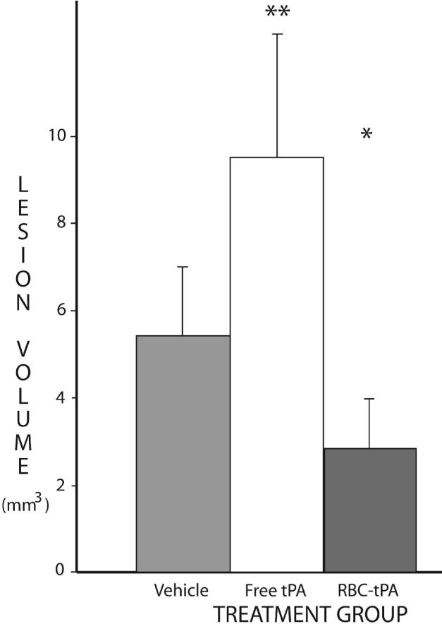
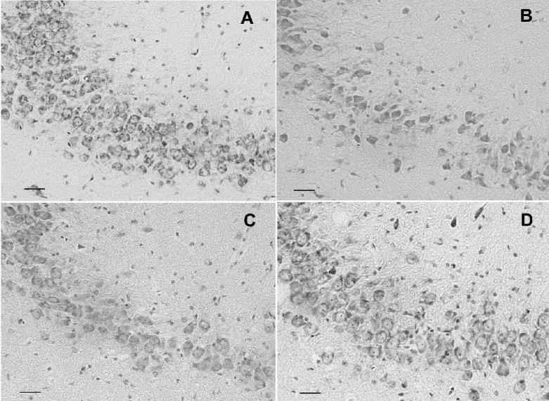
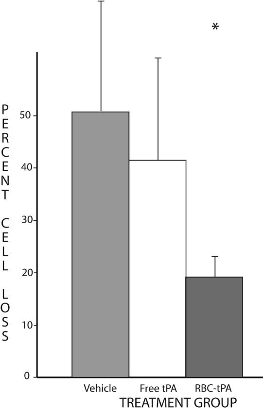
Both gross and microscopic inspection suggested that, compared to vehicle, rats treated with free tPA had more cortical hemorrhage, whereas RBC/tPA-treated rats appeared to have less (Fig. 2). At seven days after TBI, significant differences in outcome were noted among the treatment groups. Thus, mean lesion volumes were measured as 5.44 mm3 (±1.55) in the 9 control rats administered vehicle. Mean lesion volumes for the rats treated with RBC/tPA (n=6), free (n=9) tPA were 3.03 (± 1.24) and 9.56 (± 2.44), respectively. These differences, illustrated in Figures 3 and 4, are significant, both for ANOVA (p = .006) and in between-group comparisons. Lesion volumes in rats treated with RBC/tPA were significantly smaller (p < .01) than all other treatments, whereas they were significantly larger (P < .05) with free wild type tPA. In addition, hippocampal cell loss was 50.9 (± 20.9) % in the 8 vehicle-treated rats, 43.2 (± 15.7) % in the 8 treated with free tPA and 17.9 (± 8.3) in the 4 RBC/tPA treated rats (Figs. 5,6). These differences are significant (p = .007) for one-way ANOVA. The RBC/tPA group showed significantly less (p < .01) cell loss than the other two groups; between-group comparisons were not significant for the free tPA or control groups.
Fig. 2.
Amount of post-traumatic hemorrhage. Photographs of the impact site taken at 48 hours after TBI in representative animals. Treatment groups: a = vehicle, b = RBC-bound tPA, c = free tPA.
tPA: tissue plasminogen activator
Fig. 3.
Cortical tissue loss. Representative H&E-stained slices of injured brain. Tissue loss (cavitation, hemorrhage, neuronal necrosis) is outlined. Treatment groups: a = vehicle, b = RBC-bound tPA, c = free tPA (arrow points to area of hemorrhage).
tPA: tissue plasminogen activator
Fig. 4.
Volume of cortical tissue loss: The mean total volume loss (mm3) for each of the four treatment groups is shown. The vertical bars represent standard deviations. Tissue loss is significantly lower with RBC-bound tPA(*) and significantly higher with free tPA(**) than with vehicle.
tPA: tissue plasminogen activator
Fig. 5.
Hippocampal CA3 cell density. Representative Nissl-stained coronal sections of treatment groups. A. Sham-injured animal: many healthy neurons, with pale, plump nuclei and prominent nucleoli. B. Vehicle-treated: striking neuronal loss. C. Free tPA treated: intermediate neuronal loss. D. RBC/tPA treated: modest neuronal loss. Original magnification 20x; bar = 50 μm.
tPA: tissue plasminogen activator
Fig. 6.
Percent hippocampal cell loss. Cell counts in defined areas of CA3 were compared on the injured (left) and uninjured (right) side of each slice studied (see text). The mean percent loss for each of the three treatment groups is shown. The vertical bars represent standard deviations. Cell loss is significantly lower in the RBC-bound tPA group (*).
tPA: tissue plasminogen activator
Discussion
The present results reveal a neuroprotective effect of RBC/tPA in rats injured by lateral fluid percussion. There is significantly less cortical and hippocampal damage compared to animals treated with vehicle or with free tPA. No hemorrhagic complications have been observed in rats treated with RBC/tPA, which confirms its unprecedented safety and potential utility in the cerebral vasculature, even shortly after traumatic injury.
The therapeutic potency of RBC/tPA may be related to its long biological half-life, which contrasts to the short-lived yet relatively undiscriminating lytic activity of free tPA. This latter behavior is reflected in the greater apparent cortical hemorrhage associated with free tPA. In contrast to free tPA, RBC/tPA is confined to the bloodstream, does not attack hemostatic mural plugs and, due to partial masking effect of the RBC glycocalyx on tPA(Ganguly et al., 2007), is more strictly dependent on positive regulation by fibrin in activation of plasminogen(Ganguly et al., 2006).
We were unable to quantify the amount of bleeding in this study. Traumatic hemorrhage takes many forms (microscopic, contusional, subdural and parenchymal hematomas). Discrete hematomas have a tendency to fragment and separate from tissue during processing. Hence we can do no more than suggest a difference in bleeding tendency among the three groups.
We were also unable to definitively address the mechanism(s) by which RBC/tPA exerted its protective effect. Possible mechanisms and sites of RBC/tPA action include dissolution of secondary clots and initiation of signaling pathways within the cerebral vasculature. In contrast, free tPA can exert plasmin activating and signaling functions both in the vascular lumen and in the perivascular and parenchymal compartments in the CNS. Additional experiments are needed to determine whether the benefit of RBC/tPA is due to its thrombolytic activity, or is related to some other proteolytic or non-proteolytic RBC/tPA effect(s).
Acknowledgements
This study was supported in part by U.S. Public Health Service grants from the National Institutes of Health (RO1s-HL077760-01A2, HL090697, HL76406, CA83121, HL76206, HL07971, HL81864, and NS053410) and the University of Pennsylvania Institute for Translational Medicine and Therapeutics (DBC).. None of the authors has any conflicts or competing financial interests.
Footnotes
Presented in part before the International Neurotrauma Society, Rotterdam, The Netherlands, 2006.
References
- Armstead WM, Nassar T, Akkawi S, Smith DH, Chen XH, Cines DB, Higazi AA. Neutralizing the Neurotoxic Effects of Exogenous and Endogenous tPA. Nat Neurosci. 2006;9:1150–1155. doi: 10.1038/nn1757. [DOI] [PubMed] [Google Scholar]
- Danielyan K, Ganguly K, Ding B, Zaitsev S, Murciano JC, Kasner S, Cines DB, Muzykantov VR. Cerebrovascular thromboprophylaxis by tPA coupled to erythrocytes. Circulation. 2008 doi: 10.1161/CIRCULATIONAHA.107.750257. In Press. [DOI] [PMC free article] [PubMed] [Google Scholar]
- Ganguly K, Murciano JC, Westrick R, Leferovich J, Cines DB, Muzykantov VR. The Glycocalyx Protects Erythrocyte-Bound Tissue-Type Plasminogen Activator from Enzymatic Inhibition. J Pharmacol Exp Ther. 2007;321:158–164. doi: 10.1124/jpet.106.114405. [DOI] [PubMed] [Google Scholar]
- Ganguly K, Krasik T, Medinilla S, Bdeir K, Cines DB, Muzykantov VR, Murciano JC. Blood Clearance and Activity of Erythrocyte-Coupled Fibrinolytics. J Pharmacol Exp Ther. 2005;312:1106–1113. doi: 10.1124/jpet.104.075770. [DOI] [PubMed] [Google Scholar]
- Ganguly K, Goel MS, Krasik T, Bdeir K, Diamond SL, Cines DB, Muzykantov VR, Murciano JC. Fibrin Affinity of Erythrocyte-Coupled Tissue-Type Plasminogen Activators Endures Hemodynamic Forces and Enhances Fibrinolysis in Vivo. J Pharmacol Exp Ther. 2006;316:1130–1136. doi: 10.1124/jpet.105.093450. [DOI] [PubMed] [Google Scholar]
- Hoshino S, Kobayashi S, Nakazawa S. Prolonged and Extensive IgG Immunoreactivity after Severe Fluid-Percussion Injury in Rat Brain. Brain Res. 1996;711:73–83. doi: 10.1016/0006-8993(95)01329-6. [DOI] [PubMed] [Google Scholar]
- Leoni MJ, Chen XH, Mueller AL, Cheney J, McIntosh TK, Smith DH. Nps 1506 Attenuates Cognitive Dysfunction and Hippocampal Neuron Death Following Brain Trauma in the Rat. Exp Neurol. 2000;166:442–449. doi: 10.1006/exnr.2000.7513. [DOI] [PubMed] [Google Scholar]
- Lu D, Mahmood A, Goussev A, Schallert T, Qu C, Zhang ZG, Li Y, Lu M, Chopp M. Atorvastatin Reduction of Intravascular Thrombosis, Increase in Cerebral Microvascular Patency and Integrity, and Enhancement of Spatial Learning in Rats Subjected to Traumatic Brain Injury. J Neurosurg. 2004;101:813–821. doi: 10.3171/jns.2004.101.5.0813. [DOI] [PubMed] [Google Scholar]
- McIntosh TK, Vink R, Noble L, Yamakami I, Fernyak S, Soares H, Faden AL. Traumatic Brain Injury in the Rat: Characterization of a Lateral Fluid- Percussion Model. Neuroscience. 1989;28:233–244. doi: 10.1016/0306-4522(89)90247-9. [DOI] [PubMed] [Google Scholar]
- Murciano JC, Medinilla S, Eslin D, Atochina E, Cines DB, Muzykantov VR. Prophylactic Fibrinolysis through Selective Dissolution of Nascent Clots by Tpa-Carrying Erythrocytes. Nat Biotechnol. 2003;21:891–896. doi: 10.1038/nbt846. [DOI] [PubMed] [Google Scholar]
- Muzykantov VR, Murciano JC. Attachment of Antibody to Biotinylated Red Blood Cells: Immuno-Red Blood Cells Display High Affinity to Immobilized Antigen and Normal Biodistribution in Rats. Biotechnol Appl Biochem. 1996;24(Pt 1):41–45. [PubMed] [Google Scholar]
- Narita M, Bu G, Herz J, Schwartz AL. Two Receptor Systems Are Involved in the Plasma Clearance of Tissue-Type Plasminogen Activator (t-PA) in Vivo. J Clin Invest. 1995;96:1164–1168. doi: 10.1172/JCI118105. [DOI] [PMC free article] [PubMed] [Google Scholar]
- Polavarapu R, Gongora MC, Yi H, Ranganthan S, Lawrence DA, Strickland D, Yepes M. Tissue-Type Plasminogen Activator-Mediated Shedding of Astrocytic Low-Density Lipoprotein Receptor-Related Protein Increases the Permeability of the Neurovascular Unit. Blood. 2007;109:3270–3278. doi: 10.1182/blood-2006-08-043125. [DOI] [PMC free article] [PubMed] [Google Scholar]
- Stein SC, Smith DH. Coagulopathy in Traumatic Brain Injury. Neurocritical Care. 2004;1:479–488. doi: 10.1385/NCC:1:4:479. [DOI] [PubMed] [Google Scholar]
- Stein SC, Chen X-H, Sinson GP, Smith DH. Intravascular Coagulation: A Major Secondary Insult in Nonfatal Traumatic Brain Injury. J Neurosurg. 2002;97:1373–1377. doi: 10.3171/jns.2002.97.6.1373. [DOI] [PubMed] [Google Scholar]
- Stein SC, Graham DI, Chen XH, Smith DH. Association between Intravascular Microthrombosis and Cerebral Ischemia in Traumatic Brain Injury. Neurosurgery. 2004;54:687–691. doi: 10.1227/01.neu.0000108641.98845.88. discussion 691. [DOI] [PubMed] [Google Scholar]
- Yepes M, Lawrence DA. New Functions for an Old Enzyme: Nonhemostatic Roles for Tissue-Type Plasminogen Activator in the Central Nervous System. Exp Biol Med (Maywood) 2004;229:1097–1104. doi: 10.1177/153537020422901103. [DOI] [PubMed] [Google Scholar]
- Yepes M, Sandkvist M, Moore EG, Bugge TH, Strickland DK, Lawrence DA. Tissue-Type Plasminogen Activator Induces Opening of the Blood-Brain Barrier Via the LDL Receptor-Related Protein. J Clin Invest. 2003;112:1533–1540. doi: 10.1172/JCI19212. [DOI] [PMC free article] [PubMed] [Google Scholar]
- Zaitsev S, Danielyan K, Murciano JC, Ganguly K, Krasik T, Taylor RP, Pincus S, Jones S, Cines DB, Muzykantov VR. Human Complement Receptor Type 1-Directed Loading of Tissue Plasminogen Activator on Circulating Erythrocytes for Prophylactic Fibrinolysis. Blood. 2006;108:1895–1902. doi: 10.1182/blood-2005-11-012336. [DOI] [PMC free article] [PubMed] [Google Scholar]


