Abstract
On bipolar cells are connected to photoreceptors via a sign-inverting synapse. At this synapse, glutamate binds to a metabotropic receptor which couples to the closure of a cation-selective transduction channel. The molecular identity of both the receptor and the G protein are known, but the identity of the transduction channel has remained elusive. Here we show that the transduction channel in mouse rod bipolar cells, a subtype of On bipolar cell, is likely to be a member of the TRP family of channels. To evoke a transduction current, the metabotropic receptor antagonist LY341495 was applied to the dendrites of cells that were bathed in a solution containing the mGluR6 agonists L-AP4 or glutamate. The transduction current was suppressed by ruthenium red and the TRPV1 antagonists capsazepine and SB-366791. Furthermore, focal application of the TRPV1 agonists capsaicin and anandamide evoked a transduction-like current. The capsaicin-evoked and endogenous transduction current displayed prominent outward rectification, a property of the TRPV1 channel. To test the possibility that the transduction channel is TRPV1, we measured rod bipolar cell function in the TRPV1-/-mouse. The ERG b-wave, a measure of On bipolar cell function, as well as the transduction current and the response to TRPV1 agonists were normal, arguing against a role for TRPV1. However, ERG measurements from mice lacking TRPM1 receptors, another TRP channel implicated in retinal function, revealed the absence of a b-wave. Our results suggest that a TRP-like channel, possibly TRPM1, is essential for synaptic function in On bipolar cells.
Keywords: TRPV, Retina, metabotropic glutamate receptor, Knockout, Capsaicin, Patch Clamp
Introduction
Glutamate hyperpolarizes On bipolar cells by closing a cation-selective channel (Shiells et al., 1981; Slaughter and Miller, 1981). The glutamate receptor (Nakajima et al., 1993; Nomura et al., 1994) and the G protein (Vardi et al., 1993; Nawy, 1999; Dhingra et al., 2000) that mediate this response have been identified, but the cation channel has not. Two major families of cation selective channels are the cyclic nucleotide gated channels (CNG) (Craven and Zagotta, 2006) and the transient receptor potential (TRP) channels (Ramsey et al., 2006). Previous studies of On bipolar transduction suggested that the cation channel may be a member of the CNG family of channels, based on the observation that cGMP strongly potentiates the current (Nawy and Jahr, 1990; Shiells and Falk, 1990). However, it was later shown that the channel is unlikely to be gated directly by cGMP, but rather, that cGMP has a modulatory role (Nawy, 1999; Snellman and Nawy, 2004).
In the vertebrate retina, pharmacological evidence suggests that a member of the TRP channel family is likely expressed in light-sensitive ganglion cells (Warren et al., 2006; Hartwick et al., 2007; Sekaran et al., 2007). In On bipolar cells, two types of TRP channels have emerged as candidates for the transduction channel. One candidate is TRPV1, which is expressed predominantly in the peripheral nervous system and mediates heat sensation. Both TRPV1 and the On bipolar cell transduction channel are moderately permeable to Ca2+ with a Ca2+/Na+ permeability ratio of 9.6 in TRPV1 channels expressed in oocytes (Caterina et al., 1997), and 4.9 in salamander On bipolar cells (Nawy, 2000). The entry of Ca2+ activates a negative feedback pathway leading to desensitization of both the On bipolar cell transduction current (Shiells and Falk, 1999; Nawy, 2000; Berntson et al., 2004; Nawy, 2004), and the response to heat and the capsaicin mediated by TPRV1 (Liu and Simon, 1996; Caterina et al., 1997; Koplas et al., 1997; Piper et al., 1999). Here, we present evidence that the transduction channel can be activated by both capsaicin and anandamide, compounds that are thought to be specific agonists for TRPV1. Another candidate channel is the founding member of the family of melastatin-related TRP channels (TRPM1). Recent studies of Appaloosa horses have demonstrated that a dramatic reduction in the expression of mRNA encoding TRPM1, is a possible cause of night blindness and a reduced b-wave in the ERGs (Sandmeyer et al., 2007; Bellone et al., 2008). Both are indicative of a disruption of On bipolar cell function, implying that TRPM1 may play a role in mGluR6 signal transduction. We therefore set out to characterize the functional properties of the transduction channel and to further evaluate the possibility that it is composed of TRPV1 or TRPM1 subunits.
Materials and Methods
Preparation of slices
Retinal slices from 4-6 week old C57BL/6 mice (Charles River) and TRPV1 knockout mice (Trpv1tm1Jul, The Jackson Laboratory) were prepared as described elsewhere (Snellman and Nawy, 2004). Briefly, after sacrifice, whole retinas were isolated and placed on a 0.65 μm cellulose acetate/nitrate membrane filter (Millipore), secured with vacuum grease to a glass slide adjacent to the recording chamber. Slices were cut to a thickness of 100 μm using a tissue slicer (Stoelting), transferred to the recording chamber while remaining submerged, and viewed with a Nikon E600FN upright microscope equipped with a water-immersion 40X objective and DIC optics.
Solutions and drug application
Slices were continuously perfused with Ames media bubbled with 95% O2/5% CO2. Picrotoxin (100 μM), strychnine (10 μM) and 1,2,5,6-Tetrahydropyridin-4-yl) methylphosphinic acid (TPMPA, 50 μM) were included in all experiments to block inhibitory conductances. Patch pipettes of resistance 7-9 MΩ were fabricated from borosilicate glass (WPI, Sarasota, Fl) using a two-stage vertical puller (Narishige), and filled with a K+gluconate-based solution that also contained 0.5 mM EGTA, 10 mM HEPES, 4 mM ATP and 1 mM GTP (pH 7.4 by CsOH) and 14 μg/ml Alexa 488 (Invitrogen). In some experiments, the metabotropic receptor antagonist LY341495, or TRP channel reagents were delivered to the retina from a pipette using positive pressure (2-4 PSI) with a computer-controlled solenoid valve (Picospritzer, General Valve Corp), and the mGluR6 agonist L-AP4 (4 μM) was added to the bath. In other experiments, drugs were applied via a fast flow apparatus (Snellman and Nawy, 2004) and glutamate was used as an mGluR6 agonist. Drugs and chemicals were purchased from Sigma, with the exceptions of L-AP4 and LY341495 (Tocris Bioscience), and AM251 (Caymen Chemical).
Recording and analysis
Whole-cell recordings were obtained with an Axopatch 1D amplifier (Molecular Devices). Currents were acquired at a sampling rate of 2 kHz with Axograph X software and an Apple G5 computer, low pass filtered at 50 Hz (Frequency Devices) and digitized with an ITC-18 interface (Heka). Holding potentials were corrected for the liquid junction potential, which was measured to be 10 mV with the standard K+ gluconate pipette solution. Recordings were discarded if the series resistance exceeded 20 MΩ. Data were analyzed offline with Axograph X and Kaleidagraph (Synergy Software). Plots of normalized conductance of the transduction channel vs. voltage were fit with a Boltzmann relation of the form: g = (gmax-gmin)/(1 + exp((Vm - V1/2)/-k))+gmin, where gmax is the maximum conductance, gmin is the minimum conductance,V1/2 is the voltage at which the conductance is half of maximum, and k is the slope factor RT/zF, where z is the valance of the gating charge. Holding potential for all cells was +40 mV unless indicated otherwise.
ERGs were recorded to flash stimuli presented to the dark-adapted eye from TRPV1-/- mice using a previously described procedure (Gregg et al., 2007) and from TRPM1-/- mice using a procedure that was generally similar, but used a different anesthetic (urethane, 2 g/kg), maximum stimulus duration (5 ms), and sampling rate (10 KHz). The a-wave was measured at 8 ms from the pre-stimulus baseline while the b-wave was measured from the a-wave trough to the positive peak.
TRPM1-/- mice were generated by Lexicon Genetics (Trpm1tm1Lex) and obtained from the European Mouse Mutant Archive. Molecular details of the targeted allele are available at http://www.emmanet.org/. The targeted allele deletes 212 bp from exon 3 and all of exons 4 and 5 (Accession # AY180104). This will produce a frameshift mutation in the transcript terminating translation at amino acid 79. While there are several splice variants listed on ENSEMBLE (www.ensemble.org), this deletion would truncate all the splice variants. The genotype of the mice was confirmed by PCR using 1μM of each primer (LexKo-428-31, GCATAGTCCATGGACCTAGC; Neo3a, GCAGCGCATCGCCTTCTATC; trp-82, TGCAGCTTTGATTCACATCAT) and Accuprime Taq polymerase in Buffer II as described by the manufacturer (Invitrogen, Inc), yielding fragments of 319 bp for WT and 280bp for the mutant allele.
Results
The rod bipolar cell transduction current is inhibited by TRP antagonists
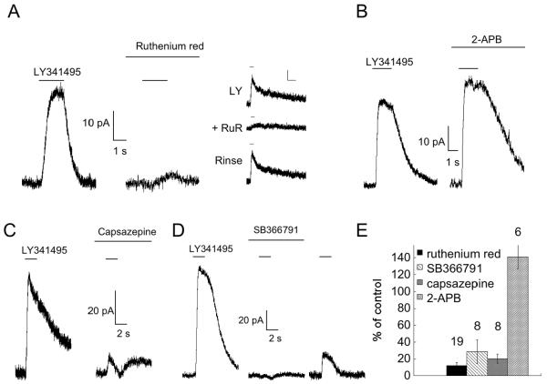
To test the hypothesis that the transduction channel is a member of the TRP family, we first examined the effects of compounds known to antagonize TRP channels on the rod bipolar cell transduction current. To evoke a transduction current, rod bipolar cells were bathed in either 1 mM glutamate or 4 μM L-AP4, and then exposed to brief applications of the mGluR antagonist LY341495 (100 μM). Blockade of mGluR6 resulted in the opening of the transduction channel which, at positive voltages, generated an outward current (fig 1A, mean amplitude 30.4 ± 3.0 pA, n=58). Using this approach, we were able to measure the transduction current for extended periods of time without any significant rundown, as reported previously (Snellman and Nawy, 2004). Application of 10 μM ruthenium red, a noncompetitive antagonist of most TRPV and TRPC channels (Clapham, 2007) using either a fast flow apparatus (see methods; fig. 1A, left, center panels), or a puffer pipet (fig. 1A, right panel), reduced the transduction current to an average of 15.3 ± 2.5% of control (fig. 1E). When applied via puffer pipet, the effects of ruthenium red were readily reversible. Conversely, application of 2-aminoethoxydiphenyl borate (2-APB), which is an antagonist at many TRPC channels, but an agonist at TRPV channels (Clapham, 2007), potentiated the transduction current (fig. 1B) to 141 ± 14.1% of control (fig. 1E).
Fig. 1. The rod bipolar cell transduction current is blocked by antagonists of TRPV1.
A. Response to 100 μM LY341495 (delivered via fast flow apparatus) before (left panel) and after a 5 minute application of 10 μM ruthenium red (center panel). Right panel, response of another cell to a 1 second puff of LY341495 delivered through a puffer pipet alone (top), or during simultaneous application of ruthenium red from a second puffer pipet (middle). Ruthenium red was applied alone for 10 seconds prior to obtaining the middle trace. The inhibition of ruthenium red was readily reversed using this approach (bottom). Scale bars: 10 pA and 2 seconds. Right panel is from a different cell. B. Response to LY341495 before and after a 5 minute application of 100 μM 2-APB. C,D. Responses to 1 second puffs of LY341495 (100 μM) before and after 5 minute bath application of 20 μM capsazepine (C) and 20 μM SB366791 (D). Responses to LY341495 typically showed partial recovery after removal of antagonists, as shown in the right panel of (D). Traces in (C) and (D) are from different cells. E. Summary of results. The number of cells for each experiment is indicated above each bar.
This pharmacological profile is consistent with a TRPV-like channel. In an attempt to further narrow this profile, we examined the effects of capsazepine and SB366791, which have been reported to be specific antagonists for TRPV1 (Caterina et al., 1997). Both compounds dramatically reduced the size of the transduction current (fig. 1C,D), SB366791 to 28.7 ± 14.1% of control, and capsazepine to 20.4 ± 5.6% of control (fig. 1E).
TRPV1 agonists evoked a current with properties that are similar to the transduction current
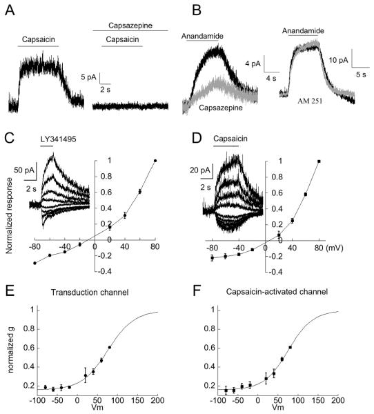
Our results suggest that TRPV1 antagonists are capable of blocking the gating of the transduction channel by the endogenous activator of the channel. We therefore tested the possibility that TRPV1 agonists can activate the rod bipolar cell transduction current. Application of 10 μM capsaicin, the prototypical TRPV1 agonist (Caterina et al., 1997), elicited a response in every rod bipolar cell that we examined (fig. 2A, mean amplitude 14.8 ± 1.4 pA, n=41). To examine the specificity of capsaicin, we applied it to Off bipolar cells, which were identified morphologically by dye filling, and physiologically by their lack of response to LY341495. Application of capsaicin to Off bipolar cells produced no detectible response (n=4, data not shown). We also recorded from rod bipolar cells in mice that were 8-9 days old. At this age, there was no detectable response to LY341495 or capsaicin (n=4, data not shown), suggesting that the transduction cascade was not yet functionally developed. Finally, the response to capsaicin was completely blocked by capsazepine (n=2, fig. 2A).
Fig. 2. Rod bipolar cells respond to agonists of TRPV1.
A. Response to a puff of 10 μM capsaicin in normal solution (left panel) and in solution containing 20 μM capsazepine (right panel). B. Response to a puff of 50 μM anandamide in normal bath solution (black trace in left and right panels) and solution containing 20 μM capsazepine (gray trace, left panel) or 5 μM AM251, a CB1 receptor antagonist (gray trace, right panel). Left and right panels are from different cells. C, D. Summary I-V relations for LY341495 (n=7 cells) and capsaicin (n=5 cells). Peak currents were normalized to the response at +80 mV for each cell, and the results were pooled. Inset: Responses to LY341495 or capsaicin obtained from representative cells. Voltage steps were from -80 mV to +80 mV in 20 mV increments. E, F. Plots of normalized conductance for the cells of the transduction channel and the capsaicin-gated channel. Lines are the fits to a Boltzmann function (see methods). Plots were obtained from the same sets of cells whose I-V relations are summarized in (C,D), using the equation g=I/(Vm-Vrev), where Vrev = 0 mV, to obtain the conductance for each cell.
The endocannabinoid anandamide, another agonist of TRPV1 receptors (Caterina et al., 1997; Jordt and Julius, 2002; van der Stelt et al., 2005), also elicited a response in rod bipolar cells (fig 2B, mean amplitude 9.6 ± 1.7 pA, n=8). The response to anandamide was inhibited by capsazepine (34.8% of control, n=2), but was unaffected by the cannabinoid-1 receptor antagonist AM251 (105.4% of control, n=3), indicating that it is not due to activation of cannabinoid receptors.
To more closely compare the transduction current and the current elicited by capsaicin, we measured the relationship between current and voltage by varying the holding potential from -80 mV to +80 mV in 20 mV increments while applying either capsaicin or LY341495. An example of each is shown in the insets of fig. 2C and fig. 2D. At negative, but not positive voltages, the transduction current often displayed a prominent peak followed by a decay to a plateau, which has been previously shown to be Ca2+ dependent (Berntson et al., 2004; Nawy, 2004). To minimize the influence of Ca2+ on the I-V relation, we measured the peak current, rather than the steady-state. For each cell, currents were normalized to the amplitude of the current at +80 mV, and the results were pooled (fig. 2C,D). The I-V relations for both the native transduction current and the current evoked by capsaicin exhibited strong outward rectification. Furthermore, the mean reversal potential (Erev) for each group were not significantly different (LY341495, Erev= -6.4 ± 3.7 mV, n=7; capsaicin, Erev= -0.6 ± 1.0 mV, n=5; p>0.15, unpaired Student t test).
By fitting the conductance of the transduction channel (fig. 2E) with a Boltzmann function, we obtained a charge valance, z, of 0.80, and a V1/2 of +76.4 mV. The charge valence is much less than for voltage gated channels such as the Shaker K+ channel (Zagotta et al., 1994; Islas and Sigworth, 1999), but very similar to values obtained in TRP channels (Nilius et al., 2005). Fitting the capsaicin-activated conductance yielded similar values, with a z of 0.87 and a V1/2 = +75.9 mV (fig. 2F). Thus, both the voltage dependence and the pharmacology of the transduction channel in mouse rod bipolar cells are consistent with the properties of a TRP channel.
Mutual occlusion of capsaicin and mGluR6-generated currents
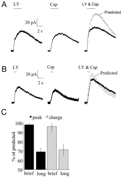
If the same population of channels is targeted by application of LY341495 and capsaicin, then one compound might be expected to occlude the actions of the other. To test this possibility, we applied LY341495 and capsaicin separately and measured the amplitude of the response to each. Next we applied the two compounds simultaneously and again measured the response. When the duration of positive pressure application was sufficient to produce a maximal response to each drug, the response to the combination of both drugs was significantly less than predicted based on linear summation of the responses obtained separately (fig. 3A,C). The same result was obtained regardless of whether we measured the peak response (p<0.01, n=7) or total charge transfer (p<0.01, n=7) during drug application. This finding is consistent with the idea that both drugs compete for a limited number of channels. If this is the case, then lowering the concentration of each compound should reduce competition for the channel. To test this possibility, we reduced the amount of drug delivered to the rod bipolar cell by shortening the duration of the application (fig. 3B). Under these conditions, the response to simultaneous delivery of both LY341495 and capsaicin coincided closely with a simple summation model (fig. 3C). These findings support the idea that mGluR6 and capsaicin operate on the same population of channels.
Fig. 3. Responses to LY31495 and capsaicin occlude each other.
A. Responses to 3 second applications of LY341495 and capsaicin, first separately and then together. Also shown in the right panel is the predicted response to simultaneous application based on linear summation. B. Similar to (A) except that the duration of the application was 1 second. Note that for subsaturating application of drugs, the predicted response more closely approximates the response to simultaneous application of LY341495 and capsaicin. Same cell as in (A). C. Comparison of evoked and predicted responses to simultaneous application of LY31495 and capsaicin as a function of puff duration. Long applications were for a duration of 3 seconds, while brief applications were either 1 second or 0.5 seconds. Charge was obtained by integration of current during the period of drug application. For both the peak response and the total charge transfer, there was no significant difference between the size of the predicted and actual response to short puffs (p>0.5, n=7), but the response to long puffs was significantly smaller than predicted based on summation (p<0.01, n=7).
TRPM1, but not TRPV1, may play a role in mGluR6 transduction
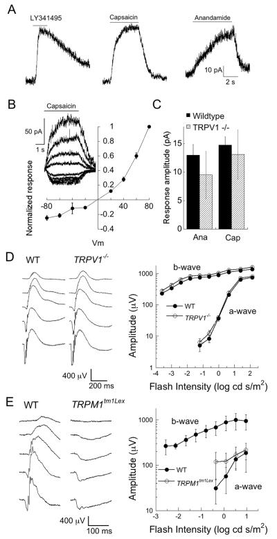
Two candidate channels for playing a role in the mGluR6 transduction pathway are TRPV1 and TRPM1. TRPV1 displays a similar pharmacology to the transduction channel as described above. On the other hand, TRPM1 has recently been implicated in transduction in the Appaloosa horse (Sandmeyer et al., 2007; Bellone et al., 2008). To address the possibility that one or both channels are a component of the transduction cascade, we recorded from two types of transgenic mice, one with a targeted deletion of TRPV1 (Caterina et al., 2000), and the other with a deletion of TRPM1 (Trpm1tm1Lex). The mGluR6 pathway appeared to be unperturbed in the TRPV1-/- mouse, as responses to LY341495, capsaicin and anandamide were all present (fig 4A). Further analysis of both the I-V relation and average amplitudes of the TRPV1 agonist responses failed to reveal any differences compared with wildtype animals (fig. 4B,C). Similarly, the amplitude and kinetics of the b-wave of the ERG were virtually identical in wildtype and Trpv1-/- mice (fig. 4D). Summary amplitudes of the b-wave, which is generated by On bipolar cell activity, and the a-wave which is generated by the activity of photoreceptors, are plotted as a function of light intensity in the right-hand panel of fig. 4D. In contrast, measurements of ERG in the TRPM1-/- mouse revealed a complete lack of a b-wave, but a normal a-wave, an indication that On bipolar cell function was completely disrupted (fig 4E).
Fig. 4. TRPV1-/- mice retain the transduction current, TRPV agonist activated currents and ERG b-waves; TRPM-/- mice lack the ERG b-wave.
A. Examples of the responses to LY341495, capsazepine and anandamide, indicating that both endogenous and exogenous gating of the transduction channel in the TRPV1-/-are preserved. B. Summary I-V relation for capsaicin in TRPV1-/- mice (n=6). Inset: responses to voltage steps from -80 mV to +80 mV in 20 mV increments. C. Responses to both anandamide and capsaicin were not significantly different in amplitude between wildtype and TRPV1-/- mice. Ana: anandamide (wt; n=13, TRPV1-/-; n=4, p>0.25). Cap: capsaicin (wt; n=41, TRPV1-/-; n=15, p>0.50). D. Left, dark-adapted ERGs from a wildtype and a TRPV1-/- mouse. Flash intensities are (in log cd s/m2) -3.6, -2.4, -1.2, -0.0, 1.4. Right, summary of the b-wave and a-wave amplitudes as a function of light intensity in wildtype (n=8) and TRPV1-/- (n=6) mice. E. Left, dark-adapted ERGs from a wildtype and a TRPM1-/- mouse. Flash intensities are (in log cd s/m2) -2.6, -1.7, -0.8, 0.1 and 1.0. Right, summary of the b-wave and a-wave amplitudes as a function of light intensity in wildtype (n=3) and TRPM1-/- (n=4) mice. For TRPM1-/-, only the a-wave data are plotted, as there was no measurable positive-going response.
Discussion
The identity of the postsynaptic channel that mediates synaptic transmission from photoreceptor to On bipolar cells is currently unknown. Here we present evidence that the channel is likely to be a member of the family of TRP channels, perhaps TRPM1. The synaptic current is reduced by antagonists of TRP channels, and mimicked by TRP channel agonists. Furthermore, the b-wave, which is thought to be generated by the opening of On bipolar cell synaptic channels, is normal in a TRPV1 knockout mouse, but completely eliminated in a mouse lacking functional TRPM1 channels. Our results are consistent with a recent study showing expression of TRPM1 RNA in mouse On bipolar cells (Kim et al., 2008). To date, a physiological characterization of TRPM1 has yet to be reported, and so it is unclear if this TRP channel can be gated by endovanilloids, or whether TRPM1 currents rectify as do those of many other TRP channels. Although it is tempting to speculate that the current evoked by endovanilloids in rod bipolar cells is due to the opening of TRPM1 channels, confirmation of this hypothesis will require further investigation of the functional properties of TRPM1.
Our findings are consistent with the findings of several recent studies of bipolar cell function in Appaloosa horses. In these horses, there is a link between a specific pattern of coat coloration and CSNB (Sandmeyer et al., 2007). Animals with this coloration lack the ERG b wave, indicating a loss of function of On bipolar cells, although the structure of the retina appears normal (Witzel et al., 1978). Genetic analysis of this phenotype revealed decreased expression of mRNA encoding TRP channel TRPM1 (Bellone et al., 2008). Of course the loss of On bipolar cell function could potentially result from a number of underlying etiologies other than a mutation in the transduction channel (McCall and Gregg, 2008). Never-the-less, an intriguing possibility, based on the results presented here and previous work on the Appaloosa horse, is that the transduction channel in the dendrites of rod bipolar cells is composed of TRPM1, either as a homomer, or in association with other TRP channels.
Acknowledgements
TRPM1-/- ERGs were done in the lab of Dr. Christiaan N. Levelt by AH with initial help of Jochem Cornelis and discussions with Dr. Reed Carroll. This work was supported by the NEI EY010254 (SN), EY12354 (RGG), Foundation Fighting Blindness, Research to Prevent Blindness, VA Medical Research Service (NSP), grant BSIK 03053 from SenterNovem (AH) and the Welcome Trust for providing funds for distribution of the TRPM1-/- mice.
References
- Bellone RR, Brooks SA, Sandmeyer L, Murphy BA, Forsyth G, Archer S, Bailey E, Grahn B. Differential Gene Expression of TRPM1, the Potential Cause of Congenital Stationary Night Blindness and Coat Spotting Patterns (LP) in the Appaloosa Horse (Equus caballus) Genetics. 2008;179:1861–1870. doi: 10.1534/genetics.108.088807. [DOI] [PMC free article] [PubMed] [Google Scholar]
- Berntson A, Smith RG, Taylor WR. Postsynaptic calcium feedback between rods and rod bipolar cells in the mouse retina. Vis Neurosci. 2004;21:913–924. doi: 10.1017/S095252380421611X. [DOI] [PubMed] [Google Scholar]
- Caterina MJ, Schumacher MA, Tominaga M, Rosen TA, Levine JD, Julius D. The capsaicin receptor: a heat-activated ion channel in the pain pathway. Nature. 1997;389:816–824. doi: 10.1038/39807. [DOI] [PubMed] [Google Scholar]
- Caterina MJ, Leffler A, Malmberg AB, Martin WJ, Trafton J, Petersen-Zeitz KR, Koltzenburg M, Basbaum AI, Julius D. Impaired Nociception and Pain Sensation in Mice Lacking the Capsaicin Receptor. Science. 2000;288:306–313. doi: 10.1126/science.288.5464.306. [DOI] [PubMed] [Google Scholar]
- Clapham DE. SnapShot: Mammalian TRP Channels. Cell. 2007;129:220.e221–220.e222. doi: 10.1016/j.cell.2007.03.034. [DOI] [PubMed] [Google Scholar]
- Craven KB, Zagotta WN. CNG AND HCN CHANNELS: Two Peas, One Pod. Annual Review of Physiology. 2006;68:375–401. doi: 10.1146/annurev.physiol.68.040104.134728. [DOI] [PubMed] [Google Scholar]
- Dhingra A, Lyubarsky A, Jiang M, Pugh EN, Birnbaumer L, Sterling P, Vardi N. The light response of ON bipolar neurons requires G[alpha]o. J Neurosci. 2000;20:9053–9058. doi: 10.1523/JNEUROSCI.20-24-09053.2000. [DOI] [PMC free article] [PubMed] [Google Scholar]
- Gregg RG, Kamermans M, Klooster J, Lukasiewicz PD, Peachey NS, Vessey KA, McCall MA. Nyctalopin expression in retinal bipolar cells restores visual function in a mouse model of complete X-linked congenital stationary night blindness. J Neurophysiol. 2007;98:3023–3033. doi: 10.1152/jn.00608.2007. [DOI] [PMC free article] [PubMed] [Google Scholar]
- Hartwick ATE, Bramley JR, Yu J, Stevens KT, Allen CN, Baldridge WH, Sollars PJ, Pickard GE. Light-Evoked Calcium Responses of Isolated Melanopsin-Expressing Retinal Ganglion Cells. J Neurosci. 2007;27:13468–13480. doi: 10.1523/JNEUROSCI.3626-07.2007. [DOI] [PMC free article] [PubMed] [Google Scholar]
- Islas LD, Sigworth FJ. Voltage Sensitivity and Gating Charge in Shaker and Shab Family Potassium Channels. J Gen Physiol. 1999;114:723–742. doi: 10.1085/jgp.114.5.723. [DOI] [PMC free article] [PubMed] [Google Scholar]
- Jordt SE, Julius D. Molecular basis for species-specific sensitivity to “hot” chili peppers. Cell. 2002;108:421–430. doi: 10.1016/s0092-8674(02)00637-2. [DOI] [PubMed] [Google Scholar]
- Kim DS, Ross SE, Trimarchi JM, Aach J, Greenberg ME, Cepko CL. Identification of molecular markers of bipolar cells in the murine retina. J Comp Neurol. 2008;507:1795–1810. doi: 10.1002/cne.21639. [DOI] [PMC free article] [PubMed] [Google Scholar]
- Koplas PA, Rosenberg RL, Oxford GS. The Role of Calcium in the Desensitization of Capsaicin Responses in Rat Dorsal Root Ganglion Neurons. J Neurosci. 1997;17:3525–3537. doi: 10.1523/JNEUROSCI.17-10-03525.1997. [DOI] [PMC free article] [PubMed] [Google Scholar]
- Liu L, Simon SA. Capsaicin-induced currents with distinct desensitization and Ca2+ dependence in rat trigeminal ganglion cells. J Neurophysiol. 1996;75:1503–1514. doi: 10.1152/jn.1996.75.4.1503. [DOI] [PubMed] [Google Scholar]
- McCall MA, Gregg RG. Comparisons of structural and functional abnormalities in mouse b-wave mutants. J Physiol. 2008;586:4385–4392. doi: 10.1113/jphysiol.2008.159327. [DOI] [PMC free article] [PubMed] [Google Scholar]
- Nakajima Y, Iwakabe H, Akazawa C, Nawa H, Shigemoto R, Mizuno N, Nakanishi S. Molecular characterization of a novel retinal metabotropic glutamate receptor mGluR6 with a high agonist selectivity for L-2-amino-4-phosphonobutyrate. Journal of Biological Chemistry. 1993;268:11868–11873. [PubMed] [Google Scholar]
- Nawy S. The Metabotropic Receptor mGluR6 May Signal Through Go, but not Phosphodiesterase, in Retinal Bipolar Cells. J Neurosci. 1999;19:2938–2944. doi: 10.1523/JNEUROSCI.19-08-02938.1999. [DOI] [PMC free article] [PubMed] [Google Scholar]
- Nawy S. Regulation of the On bipolar cell mGluR6 pathway by Ca2+ J Neurosci. 2000;20:4471–4479. doi: 10.1523/JNEUROSCI.20-12-04471.2000. [DOI] [PMC free article] [PubMed] [Google Scholar]
- Nawy S. Desensitization of the mGluR6 transduction current in tiger salamander On bipolar cells. J Physiol. 2004;558:137–146. doi: 10.1113/jphysiol.2004.064980. [DOI] [PMC free article] [PubMed] [Google Scholar]
- Nawy S, Jahr CE. Suppression by glutamate of cGMP-activated conductance in retinal bipolar cells. Nature. 1990;346:269–271. doi: 10.1038/346269a0. [DOI] [PubMed] [Google Scholar]
- Nilius B, Talavera K, Owsianik G, Prenen J, Droogmans G, Voets T. Gating of TRP channels: a voltage connection? J Physiol. 2005;567:35–44. doi: 10.1113/jphysiol.2005.088377. [DOI] [PMC free article] [PubMed] [Google Scholar]
- Nomura A, Shigemoto R, Nakamura Y, Okamoto N, Mizuno N, Nakanishi S. Developmentally regulated postsynaptic localization of a metabotropic glutamate receptor in rat rod bipolar cells. Cell. 1994;77:361–369. doi: 10.1016/0092-8674(94)90151-1. [DOI] [PubMed] [Google Scholar]
- Piper AS, Yeats JC, Bevan S, Docherty RJ. A study of the voltage dependence of capsaicin-activated membrane currents in rat sensory neurones before and after acute desensitization. J Physiol. 1999;518(Pt 3):721–733. doi: 10.1111/j.1469-7793.1999.0721p.x. [DOI] [PMC free article] [PubMed] [Google Scholar]
- Ramsey IS, Delling M, Clapham DE. AN INTRODUCTION TO TRP CHANNELS. Annual Review of Physiology. 2006;68:619–647. doi: 10.1146/annurev.physiol.68.040204.100431. [DOI] [PubMed] [Google Scholar]
- Sandmeyer LS, Breaux CB, Archer S, Grahn BH. Clinical and electroretinographic characteristics of congenital stationary night blindness in the Appaloosa and the association with the leopard complex. Veterinary Ophthalmology. 2007;10:368–375. doi: 10.1111/j.1463-5224.2007.00572.x. [DOI] [PubMed] [Google Scholar]
- Sekaran S, Lall GS, Ralphs KL, Wolstenholme AJ, Lucas RJ, Foster RG, Hankins MW. 2-Aminoethoxydiphenylborane is an acute inhibitor of directly photosensitive retinal ganglion cell activity in vitro and in vivo. J Neurosci. 2007;27:3981–3986. doi: 10.1523/JNEUROSCI.4716-06.2007. [DOI] [PMC free article] [PubMed] [Google Scholar]
- Shiells RA, Falk G. Glutamate receptors of rod bipolar cells are linked to a cyclic GMP cascade via a G-protein. Proceedings of the Royal Society of London - Series B: Biological Sciences; 1990. pp. 91–94. [DOI] [PubMed] [Google Scholar]
- Shiells RA, Falk G. A rise in intracellular Ca2+ underlies light adaptation in dogfish retinal `on' bipolar cells. J Physiol. 1999;514(Pt 2):343–350. doi: 10.1111/j.1469-7793.1999.343ae.x. [DOI] [PMC free article] [PubMed] [Google Scholar]
- Shiells RA, Falk G, Naghshineh S. Action of glutamate and aspartate analogues on rod horizontal and bipolar cells. Nature. 1981;294:592–594. doi: 10.1038/294592a0. [DOI] [PubMed] [Google Scholar]
- Slaughter MM, Miller RF. 2-amino-4-phosphonobutyric acid: a new pharmacological tool for retina research. Science. 1981;211:182–185. doi: 10.1126/science.6255566. [DOI] [PubMed] [Google Scholar]
- Snellman J, Nawy S. cGMP-dependent kinase regulates response sensitivity of the mouse on bipolar cell. J Neurosci. 2004;24:6621–6628. doi: 10.1523/JNEUROSCI.1474-04.2004. [DOI] [PMC free article] [PubMed] [Google Scholar]
- van der Stelt M, Trevisani M, Vellani V, De Petrocellis L, Schiano Moriello A, Campi B, McNaughton P, Geppetti P, Di Marzo V. Anandamide acts as an intracellular messenger amplifying Ca2+ influx via TRPV1 channels. Embo J. 2005;24:3026–3037. doi: 10.1038/sj.emboj.7600784. [DOI] [PMC free article] [PubMed] [Google Scholar]
- Vardi N, Matesic DF, Manning DR, Liebman PA, Sterling P. Identification of a G-protein in depolarizing rod bipolar cells. Visual Neuroscience. 1993;10:473–478. doi: 10.1017/s0952523800004697. [DOI] [PubMed] [Google Scholar]
- Warren EJ, Allen CN, Brown RL, Robinson DW. The light-activated signaling pathway in SCN-projecting rat retinal ganglion cells. European Journal of Neuroscience. 2006;23:2477–2487. doi: 10.1111/j.1460-9568.2006.04777.x. [DOI] [PMC free article] [PubMed] [Google Scholar]
- Witzel DA, Smith EL, Wilson RD, Aguirre GD. Congenital stationary night blindness: an animal model. Invest Ophthalmol Vis Sci. 1978;17:788–795. [PubMed] [Google Scholar]
- Zagotta WN, Hoshi T, Dittman J, Aldrich RW. Shaker potassium channel gating. II: Transitions in the activation pathway. J Gen Physiol. 1994;103:279–319. doi: 10.1085/jgp.103.2.279. [DOI] [PMC free article] [PubMed] [Google Scholar]


