
Fig. 5. Neural crest does not contribute to taste placodes or adult taste buds.
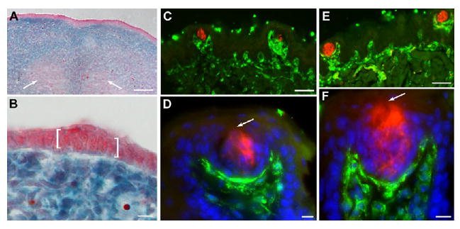
(A,B) Sections of E13.5 Wnt1cre;R26LacZ tongues stained with X-gal and counterstained with Neutral Red confirm the absence of neural crest cells in lingual epithelium. White arrows in A indicate X-gal-negative tongue muscle. In B, an X-gal-negative taste placode is bracketed. (C,D) Sections of a 6-week-old Wnt-1cre;R26LacZ tongue stained for anti-β-gal (green) and anti-PLC β2 (red) reveal neural crest cells in taste papilla mesenchyme. (E,F) In 4-month-old Wnt-1cre;R26LacZ tongue sections immunostained for a cocktail of type I, II and III taste cell markers (red) and β-gal (green), neural crest cells are found only in lingual mesenchyme. (D,F) Blue is Hoechst nuclear counterstain. Arrows indicate the taste pore. Scale bars: 50 μm in A,C,E; 10 μm in B,D,F.