Abstract
HIV-1 infection expands large populations of late-stage differentiated CD8 T cells that may persist long after viral escape from TCR recognition. Here, we investigated if such CD8 T cell populations can perform unconventional innate-like anti-viral effector functions. Chronic untreated HIV-1 infection was associated with elevated numbers of CD45RA+CD57+ terminal effector CD8 T cells expressing FcγRIIIA (CD16). The FcγRIIIA+ CD8 T cells displayed a distinctive transcriptional profile in between conventional CD8 T cells and NK cells, characterized by high levels of IKZF2 and low expression of IL7R. This transcriptional profile translated into a distinct NKp80+ IL-7Rα-surface phenotype with high expression of the Helios transcription factor. Interestingly, the FcγRIIIA+ CD8 T cells mediated HIV-specific ADCC activity at levels comparable to NK cells on a per cell basis. The FcγRIIIA+ CD8 T cells were highly activated in a manner that correlated positively with expansion of the CD8 T cell compartment, and with plasma levels of soluble mediators of antiviral immunity and inflammation such as IP-10, TNF, IL-6, and TNFRII. The frequency of FcγRIIIA+ CD8 T cells persisted as patients initiated suppressive antiretroviral therapy (ART), although their activation levels declined. These data indicate that terminally differentiated effector CD8 T cells acquire enhanced innate cell-like characteristics during chronic viral infection, and suggest that HIV-specific ADCC is a function CD8 T cells use to target HIV-infected cells. Furthermore, as the FcγRIIIA+ CD8 T cells persist on treatment they contribute significantly to the ADCC-capable effector cell pool in patients on ART.
Keywords: Cells-T Cells, Cytotoxic; Infections-AIDS; Molecules-Fc Receptors; Processes-Cell Activation; Cells-Natural Killer Cells
Introduction
The exquisite sensitivity and specificity of TCR-mediated sensing of infection is central to the function of T cells but can also, in some situations, limit their ability to provide effective immunity. This is evident in the context of HIV-1 infection in which the appearance of HIV-1-specific T cells coincides with initial viral decline; however, the response fails to completely suppress or clear infection (1–4). Since the initial characterization of HIV-specific cytotoxic CD8 T cells in the late 1980s (5–9), the limitations in their ability to control viral replication and clear infection are evident (10, 11). HIV-1 high mutation rates contribute to the ability of the virus to escape adaptive T cell responses (3, 12–14). Also, HIV-specific T cells become functionally impaired during chronic infection, additionally limiting their ability to control viral replication (15–17). Indeed, polyfunctional HIV-specific T cell responses are associated with better disease outcomes compared to those with a more narrow functional breadth (18–20). In chronic HIV-1 infection the replicating viral quasispecies have to a large extent mutated away from the originally transmitted viral sequence under T cell selection pressure, and this probably contributes to the accumulation of late-stage effector CD8 T cells with a skewed maturational phenotype (21, 22).
Persistent pathogen replication in chronic infections, such as untreated HIV-1 infection, engages T cell-mediated immune responses continuously with sustained antigenic challenge. Interestingly, some chronic infections have been associated with expansion of an unusual subset of CD8 T cells expressing CD16 (23–25). CD16 is the low affinity IgG Fc receptor and exists in two isoforms, FcγRIIIA (CD16a) and FcγRIIIB (CD16b). CD16b is expressed exclusively by neutrophils and recognizes IgG-containing immune complexes, while CD16a is best characterized for its role in mediating antibody-dependent cellular cytotoxicity (ADCC) as a function of the innate immune system (26, 27), reviewed in (28). Natural killer (NK) cells are able to mediate strong effector function in response to signaling through CD16-mediated stimulation. Whereas Fc receptors are generally not expressed by T cells, CD16 can sometimes be expressed by subsets of TCRαβ T cells (29–32). Growing evidence suggests the potential importance of ADCC in protection from HIV-1 infection (33, 34). Additionally, non-neutralizing antibodies mediate an array of effector functions through their interactions with Fc receptors that may potentiate protection from HIV-1 infection or inhibit viral replication after infection (35–40). Still, a better understanding of effector mechanisms such as ADCC involved in HIV-1 control is needed.
In this study, we hypothesized that late-stage differentiation of CD8 T cells may be associated with transcriptional changes that support innate-like effector functions in the T cell compartment. We demonstrate here that chronic, untreated HIV-1 infection is associated with the expansion of a late-stage differentiated CD8 T cell population expressing FcγRIIIA, and that this population mediates HIV-specific ADCC. Furthermore, we show that the FcγRIIIA+ CD8 T cells display a hybrid CD8 T cell and NK cell transcriptional profile characterized by high expression of NKp80 and the transcription factor Helios.
Materials and Methods
Patients and samples
Study participants aged 15–49 years were enrolled in a prospective community-based cohort to assess the prevalence and incidence of HIV-1 infection in Rakai District, Uganda, from 1998 until 2004 (Table 1) (41–43). Infected subjects were identified between 1997 and 2002 with continued annual follow up through 2008. Blood samples from 103 randomly selected HIV-1 sero-positive individuals and 40 community-matched sero-negative controls were obtained. PBMCs were then isolated and cryopreserved as described (44). None of the patients had received antiretroviral therapy. HIV-1 testing was performed as described (43). Positive samples were subjected to the Amplicor HIV-1 Monitor test, version 1.5 (Roche Diagnostics, Indianapolis, IN, USA). The HIV-1 infected study participants initiating ART were from the Couples Observation Study (COS) in Kampala Uganda as previously described (45). The index partner in each HIV-1 serodiscordant couple was followed up after the initiation of ART. Samples were collected; CD4 T cell counts determined and viral load assessments made at baseline, 6 and 12 months after initiation of ART.
Table 1.
Descriptive statistics for study population
| HIV-1 negative (n=40) | HIV-1 positive (n=103) | HIV-1 positive initiating ART (n=32) | |
|---|---|---|---|
| Age (years), median (IQR) | 30 (25 – 35) |
31 (26 – 36) |
32 (29 – 38) |
| Gender, no. (%) | |||
| Female | 20 (50) | 65(63) | 14 (44) |
| Male | 20 (50) | 38 (37) | 18 (56) |
| Viral load (log10/ml), median (IQR)a | NA | 4.5 (4.1 – 5.12) | 5.0 (4.1 – 5.3) |
| CD4 count (cells/μl), median (IQR) | NA | 513 (375 – 670) | 194 (139 – 240) |
IQR, interquartile range;
Viral load was measured by Roche Amplicor Monitor version 1.5, limit of detection 400 copies per milliliter; NA, not Applicable; whole blood from these participants was used to measure the expression of CD16 on CD8 T cells and characterize their activation profile.
Ethics statement
The study was approved by institutional review boards in the US and Uganda: The institutional Review Boards of Uganda’s National Council for Science and Technology (UNCST) and the National AIDS Research Committee, as well as Division of Human Subjects Protection at the Walter Reed Army Institute of Research. All participants gave written informed consent, or written informed consent was obtained from the parent or legal guardian of those aged 17. For samples from the Couples Observation Study (COS) in Kampala Uganda all participants gave written informed consent and ethical approvals for the study were obtained from Uganda’s National Council for Science and Technology (UNCST) and the National AIDS Research Committee and the University of Washington.
Flow cytometry and mAbs
Cryopreserved specimens were thawed and washed. Counts and viability were assessed on the Guava PCA (Guava Technologies, Hayward, CA, USA), using Guava ViaCount reagent. Standard flow cytometry phenotyping was performed as previously described (46). Commercial mAbs (clone) used in flow cytometry were; CCR5/CD195 BV421 (2D7), CCR7/CD197 FITC (150503), CD14 APC H7 (MΦP9), CD14 Alexa Fluor 700 (M5E2), CD19 Alexa Fluor 700 (HIB19), CD16 APC Cy7, PE-Cy5, Pacific Blue (PB) and BUV496 (3G8), CD161 PE-Cy5 (DX12), CD27 PerCP Cy5.5 (L128), PD-1/CD279 Alexa Flour 647 and PE (EH12.1), CD3 AmCyan, APC-H7, and PerCP-Cy5.5 (SK7), CD3 PE-CF594 (UCHT1), CD4 BV605 and APC-H7 (SK3), CD38 APC (HB7), CD45RA APC (HI100), CD56 PE-Cy7 (NCAM16.2) and (B159), CD8 PE-Cy7 and PerCP-Cy5.5 (SK1), CD8 PE and PerCP-Cy5.5 (RPA-T8), CD8b PE (2ST8.5H7), HLA-DR FITC (G46–6), IL-7R/CD127 FITC and Alexa Fluor 647 (HIL-7R-M21), KIR2DL2/DS2/DL3 PE (DX27), NKG2D/CD314 PerCP Cy.5 (ID11), TCRαβ FITC and APC (T10B9.1A-31), TRAIL/CD253 PE (RIK-2) all from (all from BD Biosciences, San Jose, CA, USA); Aqua Live/Dead viability stain, CD3 PE Texas Red (7D6), CD14 PE-Cy5 (Tuk4) and CD19 PE-Cy5 (SJ25-C1) were obtained from Invitrogen (Carlsbad, CA, USA); CD4 ECD (SFCL12T4D11), NKG2A APC (Z199), and NKp46/CD335 PE (BAB281) were all from Beckman Coulter (Brea, CA, USA); CD27 Alexa Fluor 700 (O323), NKp80 PE (5D12), CD45RA BV785 (HI100), CD57 APC, Pacific Blue, and FITC (HCD57), CD8 APC-H7 (SK1), CXCR3 FITC (G025H7), KIR3DL1 Alexa 700 (DX9), and T-bet FITC (4B10) from BioLegend (San Diego, CA, USA); Eomesodermin PE (WD1928), Helios eFluor450 (22F6), KIR2DL1/DS1 PerCP Cy5.5 (HP-MA4), and Perforin FITC (DG9) from eBioscience (San Diego, CA, USA). For assessment of transcription factors, cells were washed, permeabilized and fixed using an optimized kit (FoxP3 staining fix/perm) before intranuclear stain. Flow cytometry data were acquired with a BD LSR II instrument or a BD FACS Canto II instrument (BD Biosciences). Sorting was performed on a 4-laser BD FACS ARIA II SORP (BD Biosciences) contained in a biosafety cabinet. Clinical lymphocyte immunophenotyping was performed using the FACS MultiSET System and run on a FACSCalibur using the single platform Multi-test 4-color reagent in combination with TruCount tubes (BD Biosciences) (47).
Soluble factor analysis
A custom multiplex cytokine array was used to quantify 16 analytes from cryopreserved plasma, including IFN-γ, IL-1α, IL-1β, IL-2, IL-4, IL-5, IL-6, IL-8, IL-10, IL-12p70, IL-15, IL-17, IP-10, MCP-1, TNF, and TNFRII, according to the manufacturer’s instructions (Quansys Biosciences, Logan, UT, USA). Commercial single ELISAs were used to measure neopterin (GenWay Biotech, San Diego, CA, USA), IFNα, I-FABP, and sCD14 (R&D Systems). All samples were run in triplicate and mean values were used for data analysis.
Gene expression analysis
Targeted gene expression analysis was performed as previously described (48). Cells from seven donors were stained and four phenotypically distinct cell populations (CD8 T cells; CD45RA-CD57-, CD45RA+CD57+FcγRIIIA-, CD45RA+CD57+FcγRIIIA+, as well as CD56dimFcγRIIIA+ NK cells) (500–1,000 cells/well) were sorted into wells containing 10 μl of reaction buffer (SuperScript III Reverse Transcriptase/Platinum Taq Mix, Cells Direct 2× Reaction Mix, Invitrogen). Reverse transcription and specific transcript amplification were performed using a thermocycler (Applied Biosystems Gene Amp PCR System 9700) as follows: 50°C for 15 min, 95°C for 2 min, then 95°C for 15 sec, 60°C for 30 sec for 18 cycles. The amplified cDNA was loaded into Biomark 96.96 Dynamic Array chips using the Nanoflex IFC controller (Fluidigm). This microfluidic platform was then used to conduct qPCR in nl reaction volumes. Threshold cycle (Ct), as a measurement of relative fluorescence intensity, was extracted from the BioMark Real-Time PCR analysis software. A panel of 96 pre-selected genes related to both NK cell and CD8 T cell biology was qualified as previously described and using a script provided courtesy of Mario Roederer (49). Subsequent data analysis was performed using JMP software (version 10). Initial analyses of the transcriptome data from the Fluidigm Biomark confirmed the quality of 74 of the 96 genes, whereas data on 22 genes were discarded due to lack of amplification.
ADCC assays
Measurement of ADCC was performed using the PanToxiLux (PTL) assay (OncoImmunin, Inc., Gaithersburg, MD, USA) similar to the previously described assay (50). Recombinant HIV-1 BaL gp120 from DAIDS, NIAID catalog #4961 (obtained through the NIH AIDS Reagent Program, Division of AIDS, NIAID, NIH) were used to coat target CEM.NKRCCR5 cells. Optimal concentration used to coat target cells was determined for each gp120 through an 11-point titration starting with 20 μg/ml and serial dilution 2-fold. After coating NKR.CEMCCR5 target cells with gp120 in 0.5% FBS RPMI media, cells were labeled with TFL4 (OncoImmunin, Inc.), a fluorescent target-cell marker, for 15 min at 37°C and 5% CO2. Cells were then washed twice with 1× PBS and stained with viability dye LIVE/DEAD Fixable Aqua Dead Cell Stain (Life Technologies) for 30 min at room temperature. After washing in 0.5% FBS-RPMI media, cells were counted as above, and then re-suspended to reach a final concentration of 8.0×10⁵ cells/ml. At this point, sorted effector cell populations (NK Cells, CD45RA+CD57+ CD8 T cells, and CD45RA-CD57-CD8 T cells) were washed in 0.5% FBS-RPMI media and re-suspended to final concentration of 24×106 cells/ml for an effector to target ratio of 30:1. In a 96-well polypropylene plate, 25 μl of both target and effector cell suspensions were both added to each well, along with 75 μl of Granzyme B (GzB) substrate (OncoImmunin, Inc.). After incubation for 5 min at room temperature, 25 μl of HIV-Immune Globulin (HIV-IG™) (North American Biologicals, Inc., Miami, FL, USA) at a 0.5 mg/ml dilution was added to each well, and the plate was incubated for another 15 min at room temperature. The plate was then spun at 300g for 1 min and placed at 37°C and 5% C02 for 1 hour. Cells were washed twice with wash buffer and acquired on the LSRII (BD Bioscience) the same day. Fluorophores were detected using: a 488 nm 50 mW laser with 515/20 filters to detect GrzB substrate, a 406 nm 100 mW laser with 525/50 filters to detect Aqua L/D stain, and 640 nm 40 mW laser with 670/30 filters to detect TFL4 stain. Because of the spectral properties of the fluorescent molecules used in this panel, manual compensation of detected signals was performed to analyze the data. Data were analyzed by using FlowJo 9.7.5 (Ashland, OR, USA).
Statistical analysis
Statistical analysis was performed using Graph Pad Prism 6.0g for Macintosh version (GraphPad Software Inc., La Jolla, CA, USA) or JMP software (version 10, SAS Institute Inc., Cary, NC, USA). Direct comparisons between two groups were performed using the non-parametric Mann-Whitney U test. Associations between groups were determined by Spearman’s rank correlation. To correct for multiple comparisons, the Benjamini-Hochberg False Discovery Rate (FDR) (51) was calculated for all observations. An FDR < 0.05 was considered statistically significant. For paired observations a paired t test was used. A p value < 0.05 was considered statistically significant. Flow cytometry analysis and presentation of distributions was performed using SPICE version 5–1.2, downloaded from http://exon.niaid.nih.gov/spice (52). Comparison of distributions was performed using a Student’s T test and a partial permutation test as described (52).
Results
FcγRIIIA+ CD8 T cells expand in chronic untreated HIV-1 infection
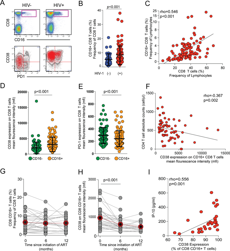
HIV-1 negative (n=40) and HIV-1 positive (n=103) individuals from a cohort in Rakai, Uganda, were chosen for the investigation of FcγRIIIA expression in CD8 T cells (Table 1). The FcγRIIIA+ CD8 T cell population was identified as positive for CD3, TCRαβ, CD8, and FcγRIIIA and negative for CD14, CD19, and CD4 (Fig. 1A and Supplemental Fig. 1). FcγRIIIA expression was detectable in T cells from healthy donors at a median (range) frequency of 3.8% (0.7 – 20.7%) of CD8 T cells (Fig. 1B). Interestingly this population was nearly doubled in HIV-1 infected donors where a median frequency of 5.9% (1.3 – 37.9%) of CD8 T cells expressed FcγRIIIA (p<0.001) (Fig. 1B). This expansion was positively associated with the overall CD8 T cell expansion in HIV-1 infected patients (p<0.001, rho=0.546) (Fig. 1C). The HIV-1 associated expansion of FcγRIIIA+ CD8 T cells was not associated with the expression levels, measured as geometric mean fluorescence intensity (MFI), of FcγRIIIA on the surface of these cells (data not shown). There was no significant difference in FcγRIIIA expression levels (MFI) on FcγRIIIA+ CD8 T cells between HIV-1 infected and uninfected participants (data not shown). Interestingly, the FcγRIIIA+ CD8 T cells were more activated than their FcγRIIIA-counterparts as assessed by CD38 expression (p<0.001) (Fig. 1D). They also expressed less of the inhibitory receptor PD-1 (p<0.001) (Fig. 1E). The CD38 expression levels were inversely associated with CD4 counts, albeit weakly (p=0.02, rho=−0.367), suggesting that the FcγRIIIA+ CD8 T cells become more activated as disease progresses (Fig. 1F).
FIGURE 1.
FcγRIIIA+ CD8 T cells expand numerically and persist in Ugandans with untreated HIV-1 infection. (A) Bivariate pseudocolor flow cytometry plots of FcγRIIIA+ CD8 T cells after gating on small lymphocytes that are Aqua Live/Dead-TCR a/b+, CD8+CD3+ T cells, in healthy donors (HIV-) (n=40) and HIV-1 infected (HIV+) individuals (n=103). Overlay plots of FcγRIIIA+ CD8 T cells (in red) and bulk CD8 T cells in grey for representative HIV- and HIV+ donors. (B) Scatter plot of the frequency of FcγRIIIA+ CD8 T cells in HIV+ versus HIV- healthy donors with lines at the mean and standard deviation (SD) shown. (C) Correlation of the FcγRIIIA+ CD8 T cell subset frequency with the overall CD8 compartment frequency. (D) CD38 MFI and (E) PD-1 MFI in FcγRIIIA+ CD8 T cells (orange) as compared to the overall CD8 compartment (green) with lines at the mean and SD. (F) Correlation between FcγRIIIA+ CD8 T cells and absolute CD4 T cell counts. Longitudinal graph of the FcγRIIIA+ CD8 T cell subset frequency (G) and the CD38 MFI on FcγRIIIA+ CD8 T cell subset (H) in patients starting ART (n=32) at baseline, 6, and 12 months after ART initiation. Grey circles and lines represent individuals while red line and outlined filled circle represent the median level. (I) Correlation between activation levels in FcγRIIIA+ CD8 T cells and TNFRII levels in plasma.
To address the stability of the FcγRIIIA+ CD8 T cell pool over time, we studied a second cohort of Ugandan HIV-1 infected subjects (n=32) located in Kampala where longitudinal samples were available from before and after initiation of antiretroviral therapy (ART) (Table 1). These patients displayed a stable population of FcγRIIIA+ CD8 T cells over 12 months of ART (Fig. 1G). However, of note, the activation levels of these FcγRIIIA+ CD8 T cells declined over the course of treatment, as measured by CD38 expression (p<0.001) (Fig. 1H). These data show that HIV-1 infected Ugandans have an expanded population of activated TCRαβ CD8 T cells expressing FcγRIIIA and that this population is stable over 12 months of ART.
Next, multiplexed assays and ELISA were used to quantify a suite of 20 soluble factors in plasma in relation to the size and activation level of the FcγRIIIA+ CD8 T cell population in HIV-1 infected individuals. Although none of the analytes measured showed a relationship to the percentage of CD8 T cells expressing FcγRIIIA, several markers were directly associated with the activation levels of FcγRIIIA+ CD8 T cells (i.e., cells co-expressing CD38) (Table 2). Statistically significant correlations between the frequency of FcγRIIIA+ CD8 T cells expressing CD38 and plasma levels of the inflammatory cytokines IL-6 (p=0.011, rho=0.446, FDR=0.040), IP-10 (p<0.001, rho=0.582, FDR=0.009), MCP-1 (p=0.016, rho=0.424, FDR=0.048), TNF (p=0.008, rho=0.459, FDR=0.036), and TNFRII (p=0.001, rho=0.556, FDR=0.009) were observed (Fig. 1I) (Table 2). Similar correlations were observed for the MFI of CD38 on FcγRIIIA+ CD8 T cells and IP-10 (p=0.001, rho=0.547, FDR=0.009), MCP-1 (p=0.009, rho=0.456, FDR=0.032), TNF (p=0.009, rho=0.458, FDR=0.032), and TNFRII (p<0.001, rho=0.569, FDR=0.009). Thus, expansion and activation of the FcγRIIIA+ CD8 T cells is associated with plasma markers of HIV-driven systemic immune activation. In contrast, neither soluble markers of an innate antiviral response such as IFNα, nor the common indices of microbial translocation sCD14 and IFABP were associated with the size of the FcγRIIIA+ CD8 T cell population or the extent of their activation.
Table 2.
Correlative analysis between plasma derived soluble factors and CD16+ CD8+ T cells in HIV+ donors.
| Cytokine | CD16+CD8+ T cell (%) | CD16+CD8+ CD38+ T cell (%) | CD16+CD8+ CD38+ T cell (CD38 mfi) | ||||||
|---|---|---|---|---|---|---|---|---|---|
| rho | p value | FDR | rho | p value | FDR | rho | p value | FDR | |
| IFNγ | −0.217 | 0.234 | 0.904 | −0.082 | 0.655 | 0.737 | 0.003 | 0.986 | 0.986 |
| IL-1a | −0.137 | 0.455 | 0.904 | −0.228 | 0.210 | 0.344 | −0.154 | 0.400 | 0.600 |
| IL-1b | −0.079 | 0.669 | 0.904 | 0.015 | 0.935 | 0.935 | 0.062 | 0.735 | 0.882 |
| IL-2 | NA | NA | NA | NA | NA | NA | NA | NA | NA |
| IL-4 | −0.213 | 0.242 | 0.904 | 0.139 | 0.448 | 0.620 | 0.201 | 0.271 | 0.443 |
| IL-5 | −0.077 | 0.673 | 0.904 | 0.118 | 0.522 | 0.671 | 0.045 | 0.807 | 0.908 |
| IL-6 | 0.149 | 0.417 | 0.904 | 0.446 | 0.011 | 0.040 | 0.378 | 0.033 | 0.099 |
| IL-8 | 0.067 | 0.714 | 0.904 | 0.032 | 0.864 | 0.915 | 0.018 | 0.921 | 0.975 |
| IL-10 | −0.095 | 0.604 | 0.904 | 0.505 | 0.003 | 0.018 | 0.465 | 0.007 | 0.032 |
| IL-12p70 | −0.034 | 0.854 | 0.904 | 0.338 | 0.059 | 0.133 | 0.290 | 0.108 | 0.216 |
| IL-15 | 0.044 | 0.812 | 0.904 | 0.373 | 0.036 | 0.093 | 0.242 | 0.182 | 0.328 |
| IL-17 | NA | NA | NA | NA | NA | NA | NA | NA | NA |
| IP-10 | −0.006 | 0.975 | 0.975 | 0.582 | 0.001 | 0.009 | 0.547 | 0.001 | 0.009 |
| MCP-1 | −0.055 | 0.765 | 0.904 | 0.424 | 0.016 | 0.048 | 0.456 | 0.009 | 0.032 |
| TNF-α | −0.176 | 0.336 | 0.904 | 0.459 | 0.008 | 0.036 | 0.458 | 0.009 | 0.032 |
| TNFR-II | −0.199 | 0.276 | 0.904 | 0.556 | 0.001 | 0.009 | 0.569 | 0.001 | 0.009 |
| IFABP | 0.041 | 0.823 | 0.904 | −0.157 | 0.390 | 0.585 | −0.136 | 0.457 | 0.633 |
| sCD14 | −0.077 | 0.674 | 0.904 | 0.307 | 0.087 | 0.157 | 0.312 | 0.082 | 0.200 |
| IFNα | −0.071 | 0.706 | 0.904 | 0.094 | 0.616 | 0.737 | 0.069 | 0.714 | 0.882 |
| Neopterin | 0.106 | 0.563 | 0.904 | 0.317 | 0.077 | 0.154 | 0.305 | 0.089 | 0.200 |
mfi, geometric mean fluorescence intensity; FDR, false discovery rate; NA, not applicable
FcγRIIIA+ CD8 T cells are late-stage effector cells and characterized by expression of Helios
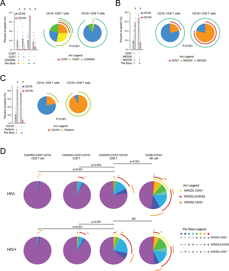
Because of the significant expansion and activation of FcγRIIIA+ CD8 T cells in HIV-1 infected individuals, we next investigated the detailed phenotype of these cells in HIV-infected subjects from the Rakai cohort. The combinatorial co-expression pattern of CCR7, CD27, and CD45RA was significantly different between CD8 T cells positive or negative for FcγRIIIA (Fig. 2A) (Supplemental Table 1) (p<0.001). Expression of CD45RA in the absence of CCR7 and CD27 was the dominant pattern among the FcγRIIIA+ CD8 T cells, consistent with a terminally differentiated status, while this phenotype was less common among CD8 T cells lacking FcγRIIIA (74% vs. 18%, respectively) (p<0.001). Next, the expression patterns of CD57, NKG2A, and NKG2D was evaluated, and frequency of the subsets defined by these receptors were different in CD8 T cells expressing FcγRIIIA compared to those that did not (Fig. 2B) (p<0.001). The majority of FcγRIIIA+ CD8 T cells expressed CD57, while maintaining NKG2D expression. In fact, all Boolean subsets containing CD57 expressing cells were higher in FcγRIIIA+ CD8 T cells compared to FcγRIIIA-CD8 T cells (all p≤0.001) (data not shown). The next panel examined CD161 and perforin, and comparison of the distribution of cell subsets expressing combinations of these two markers, again, revealed differences between the FcγRIIIA+ and FcγRIIIA-CD8 T cells (Fig. 2C) (p<0.001). The vast majority of FcγRIIIA+ CD8 T cells expressed perforin as compared to approximately 20% of FcγRIIIA-CD8 T cells. In summary, FcγRIIIA+ CD8 T cells are distinct from their FcγRIIIA-CD8 T cell counterparts by lack of CD27 expression, higher proportion of cells expressing CD57, and they are predominantly perforin positive.
FIGURE 2.
FcγRIIIA+ CD8 T cells display a late-stage effector phenotype in chronic untreated infection. A detailed phenotype of FcγRIIIA+ CD8 T cells after gating on small lymphocytes, singlets, Aqua Live/Dead negative, CD8+CD3+ T cells in HIV+ (n=15) and HIV- (n=15) individuals was examined. (A) Expression of CD27, CCR7 and CD45RA in CD8 T cell subsets having or lacking FcγRIIIA surface expression. (B) Expression of CD57, NKG2A and NKG2D in CD8 T cell subsets having or lacking FcγRIIIA surface expression. (C) Expression of CD161 and perforin in CD8 T cell subsets having or lacking FcγRIIIA surface expression. (E) Analysis of KIR surface expression patterns in CD45RA+CD57+FcγRIIIA+ CD8 T cells, CD45RA+CD57+FcγRIIIA-CD8 T cells, CD56dim NK cells, and CD45RA-CD57-CD8 T cells.
The patterns of expression of maturation markers observed in FcγRIIIA+ CD8 T cells in HIV-1-infected donors were not significantly different from HIV-1 uninfected control subjects (all p>0.05) (data not shown), suggesting that the elevated levels of FcγRIIIA+ CD8 T cells in infected individuals represent an expansion of a phenotypic cell subset retaining relatively normal characteristics. To address this question further, we investigated the expression of killer immunoglobulin-like receptors (KIRs) in CD8 T cells and NK cells expressing FcγRIIIA, as well as in late-stage differentiated CD8 T cells defined by co-expression of CD45RA and CD57, and memory CD8 T cells negative for these markers (Fig. 2D). In uninfected donors, T cell populations lacking FcγRIIIA had low levels of KIR expression, whereas NK cells had high KIR levels in diverse combinations. The FcγRIIIA+ CD8 T cells displayed a pattern intermediate between T cells and NK cells. Strikingly, this pattern was altered in HIV-1 infected subjects whose FcγRIIIA+ CD8 T cells had adopted a KIR co-expression profile very similar to that of NK cells (p<0.001 for FcγRIIIA+ CD8 T cells in HIV-1 uninfected donors compared to HIV-1 infected donors, and p=0.250 for FcγRIIIA+ CD8 T cells compared to NK cells in HIV-1 infected donors).
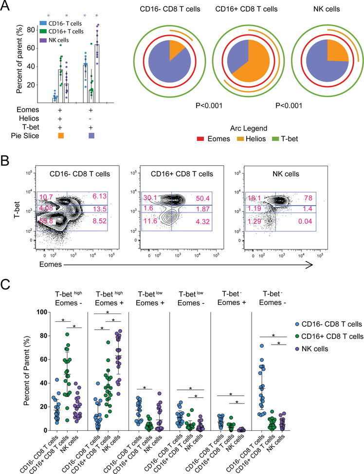
T cell differentiation and maturation are controlled by a set of transcription factors including T-bet, eomesodermin (Eomes), and Helios. PBMC from HIV-infected donors were stained intracellularly for these transcription factors, and their expression patterns were analyzed in CD8 T cells lacking or expressing FcγRIIIA, as well as in NK cells (Fig. 3A). FcγRIIIA+ CD8 T cells displayed a T-bet, Eomes, and Helios expression pattern distinct from both the general CD8 T cell population and from CD56dim NK cells, with higher levels of co-expression as compared to FcγRIIIA-CD8 T cells. Co-expression of all three transcription factors was common in FcγRIIIA+ CD8 T cells, and also relatively frequent in NK cells, but uncommon in the general CD8 T cell pool. Notably, 61% of the FcγRIIIA+ CD8 T cells expressed Helios, and this was significantly higher compared to the FcγRIIIA-CD8 T cells and NK cells (p<0.001), in which a median of 10% and 28% expressed Helios, respectively. Characterization of Tbet and Eomes can be discriminated based on a continuum of expression and varies on lymphocyte subsets (53). FcγRIIIA+ CD8 T cells were dominated by a high Tbet expression profile with variable Eomes expression, very similar to CD16+ NK cells (Figure 3B–C). HIV-1 infection status had minimal effect on Tbet and Eomes in these populations. FcγRIIIA-CD8 T cells showed a much more variable expression pattern of both transcription factors, which may reflect the different states of maturation and differentiation within this compartment.
FIGURE 3.
Transcription factors T-bet, eomesodermin (Eomes), and Helios expression in CD8 T cells with or without FcγRIIIA and NK cells. In HIV+ (n=10) and HIV- (n=10) individuals (A) expression of T-bet, Eomes and Helios, assessed by intracellular staining, is presented for CD8 T cell subsets having or lacking FcγRIIIA (CD16) surface expression, in comparison with NK cells. FcγRIIIA-CD8 T cells (blue), FcγRIIIA+ CD8 T cells (green), and NK cells (purple) are displayed on a bar graph with individual points shown. Asterics denote statistically significant differences to the FcγRIIIA+ CD8 T cells (p<0.05). Major populations expressing all three transcription factors (orange box) and positive for T-bet and Eomes in the absence of Helios (lavender) are presented which correspond to slices of the pie chart. Individual expression of each transcription factor is shown by arc (Eomes in red, Helios in gold, and T-bet in light green). P-value is presented for comparison of distribution of each pie between groups. (B) Example flow cytometry plots showing the coordinated expression of T-bet and Eomesodermin for FcγRIIIA-CD8 T cells, FcγRIIIA+ CD8 T cells, and NK cells. (C) Scatter plot for FcγRIIIA-CD8 T cells (blue), FcγRIIIA+ CD8 T cells (green), and NK cells (purple) for subsets of cells expressing high, medium, or low levels of T-bet and positive or negative for Eomes with lines at the mean and SD shown. Line with * denotes statistical significance (p<0.05) between cell populations.
Altogether, these data indicate that the FcγRIIIA+ CD8 T cell population expanded in HIV-1 infected people is characterized by Helios expression and has a late-stage differentiated effector phenotype. This population mostly retains the characteristics seen in healthy donors as it expands during HIV-1 infection, although KIR expression is significantly elevated.
The FcγRIIIA+ CD8 T cell transcriptome reveals a mixed effector CD8 T cell and NK cell character
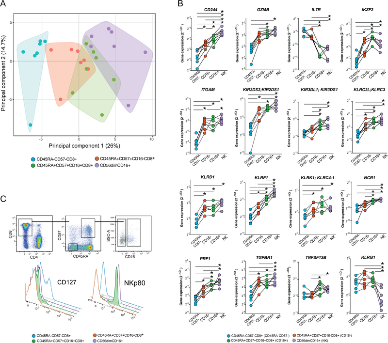
To better understand the identity of the FcγRIIIA+ CD8 T cells, we next analyzed their transcriptional profile by Fluidigm Biomark. A panel of 96 genes involved in T cell function or NK cell function was selected (Supplemental Table 2), and the expression of these genes was analyzed in cell populations purified by flow cytometry sorting. For these analyses, cells from seven HIV-1 infected donors were sorted into four populations, 500–1,000 cells per population: 1) CD45RA+CD57+ CD8 T cells expressing FcγRIIIA, 2) CD45RA+CD57+ CD8 T cells lacking expression of FcγRIIIA, 3) CD45RA-CD57-CD8 T cells not expressing FcγRIIIA, and 4) CD56dimCD16+ NK cells. The data for 74 out of the 96 genes passed quality control, and principal component analysis (PCA) was performed on the total data set of expression of these 74 genes in all four of the cell subsets (Fig. 4A). Notably, the transcriptional profile of FcγRIIIA+ CD8 T cells overlapped with both the CD45RA+CD57+ CD8 T cells lacking expression of FcγRIIIA and the CD56dimCD16+ NK cells, whereas the CD45RA-CD57-memory CD8 T cell subset was most distant. Principal component 1 contributed 26% of the variability in the data set, and component 2 contributed 14.7%. Expression of genes GZMB, LAIR1, GZMK, PRF1, and CD244 contributed most to principal component 1, and genes GZMK, IL6ST, TGFB1, CD38, and CD160 contributed most to principal component 2.
FIGURE 4.
Transcriptome analysis reveals a mixed CD8 T cell and NK cell character in the FcγRIIIA+ CD8 T cells. Supervised expression analysis of 74 genes involved in the regulation and function of innate and adaptive immune responses in 7 HIV-1 infected donors using the Fluidigm Biomark system. (A) Principal component analysis of the transcriptional data from four sorted cell populations reflecting CD45RA-CD57-(blue), CD45RA+CD57+FcγRIIIA-(red), CD45RA+CD57+FcγRIIIA+ (green), as well as CD56dimFcγRIIIA+ NK cells (purple). Polygons represent 95% confidence intervals in the data. (B) Expression of 10 selected genes in the same sorted subsets; (C) Successive flow cytometry gating strategy used for confirmation of IL7Ra and KLRF1 genes at the protein level. Offset histograms showing the relative expression of IL7Ra (CD127) and KLRF1 (NKp80) on CD8 T cells; CD45RA-CD57-(blue), CD45RA+CD57+FcγRIIIA-(red), CD45RA+CD57+FcγRIIIA+ (green/filled), as well as CD56dimFcγRIIIA+ NK cells (purple).
A subset of genes showed expression patterns that segregated the FcγRIIIA+ CD8 T cell population from the NK cells and the FcγRIIIA-CD8 T cell populations (Fig. 4B and Supplemental Fig. 2). Notably, the FcγRIIIA+ CD8 T cell displayed significantly higher IKZF2 expression than any of the three other reference populations, and lower IL7R expression than the other T cell populations and at levels similar to CD56dimCD16+ NK cells. Regarding a range of genes encoding NK cell-associated receptors, including KIR2DS2;KIR2DS1, KIR3DL1;KIR3DS1, KLRC2L;KLRC3, KLRD1, KLRF1, KLRK1;KLRC4–1 and NCR1, the FcγRIIIA+ CD8 T cells showed a pattern intermediate between FcγRIIIA-CD45RA+CD57+ CD8 T cells and the CD56dimCD16+ NK cells. In fact, KLRF1 encoding the NK cell-associated receptor NKp80, expressed at the highest levels by CD56dimCD16+ NK cells, was expressed at significantly higher levels compared to the FcγRIIIA-negative terminal effector CD8 T cells and effector memory CD8 T cells. Compared to their FcγRIIIA-counterparts, the FcγRIIIA+ CD8 T cells also expressed higher levels of genes involved in regulating T cell function including TNFSF13B. Additionally, the FcγRIIIA+ CD8 T cells had significantly lower expression of TGFBR1 than the CD56dim NK cells, but levels were above that of the other CD8 T cells populations. Altogether, the gene expression analysis indicates that FcγRIIIA+ CD8 T cells have a transcriptional profile intermediate between effector CD8 T cells and CD56dim NK cells.
Because of the distinct transcriptional signature of FcγRIIIA+ CD8 T cells, we were interested to confirm expression of the IL7R and KLRF1 genes at the protein level. We further examined 10 chronically HIV-1 infected and 10 uninfected individuals for surface expression of these receptors by flow cytometry. The majority of FcγRIIIA+ CD45RA+CD57+ CD8 T cells expressed NKp80 (median 68%) and lacked expression of the IL-7 receptor, CD127 (median 2%) (Fig. 4C). No differences were observed in FcγRIIIA+ CD45RA+CD57+ CD8 T cells expressing NKp80 or IL-7Rα between HIV-1 positive and negative individuals, and no relationship was observed between expression and markers of HIV-1 disease progression. IL-7Rα protein expression was similar between NK cells and CD45RA+CD57+ CD8 T cells, irrespective of FcγRIIIA+ expression. Interestingly, NKp80 was only found at appreciable levels in the T cells with the FcγRIIIA+ CD45RA+CD57+ phenotype. Together, the FcγRIIIA+ CD8 T cells have a distinct NKp80+ IL-7Rα-character different from other effector CD8 T cells and more akin to CD56dim NK cells.
Potent HIV-specific ADCC activity mediated by FcγRIIIA+ CD8 T cells
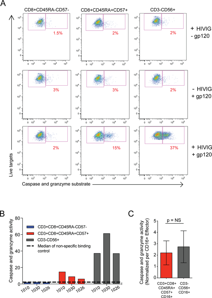
ADCC is part of the repertoire of effector functions employed by NK cells to detect and target HIV-1 infected cells. Recent data indicating that non-neutralizing antibody-mediated effects may contribute to HIV vaccine-efficacy have spurned a renewed interest in ADCC as a protective mechanism (54, 55). The present observation that HIV-1 infection drives the expansion of late-stage effector CD8 T cells with a hybrid NK cell-CD8 T cell character including FcγRIIIA and lytic protein expression suggests that CD8 T cells might actually mediate ADCC. To test this possibility, effector cell populations from HIV-1 infected donors were sorted by flow cytometry, and these cells’ ability to mediate ADCC against HIV BaL gp120-coated CEM.NKRCCR5 target cells was evaluated by the PanToxiLux granzyme B substrate cytotoxicity assay (Fig. 5A). To avoid FcγRIIIA downregulation or blocking due to staining, CD45RA+CD57+ CD8 T cells were sorted to enrich for FcγRIIIA+ cells (9–21% FcγRIIIA+), and then compared with FcγRIIIA-CD45RA-CD57-memory CD8 T cells and with NK cells sorted from the same donors. In the presence of HIV Immune Globulin (HIV-IG), the CD45RA+CD57+ cells from three HIV+ donors clearly mediated ADCC, as did the NK cells, whereas the CD45RA-CD57-CD8 T cell population did not (Fig. 5A–B). As such, bulk CD45RA+CD57+ CD8 T cells performed ADCC lower than the NK cells (Fig. 5B). However, after adjusting for the frequency of FcγRIIIA expression in these populations, 9–21% in CD45RA+CD57+ CD8 T cells and 69–96% in CD56dim NK cells, ADCC capacity of FcγRIIIA+ CD8 T cells was similar to that of FcγRIIIA+ NK cells (Fig. 5C). Interestingly, the FcγRIIIA+ MFI on FcγRIIIA+ CD8 T cells was significantly lower compared to FcγRIIIA+ MFI on CD56dim NK cells (p<0.001). FcγRIIIA+ CD8 T cell ability to mediate ADCC based on normalized FcγRIIIA+ MFI or the integrated MFI (frequency multiplied by the MFI) was as good as NK cells (data not shown). These data demonstrate that the FcγRIIIA+ CD8 T cell population expanding during chronic HIV-1 infection can mediate HIV-specific ADCC at levels comparable to NK cells.
FIGURE 5.
HIV-specific ADCC mediated by FcγRIIIA+ CD8 T cells. (A) Representative FACS plots of the cytolysis PanToxiLux assay from HIV+ (n=3) individuals. (B) HIV-1 gp120-specific ADCC mediated by HIVIG. (C) Comparison of ADCC mediated by FcγRIIIA+ CD8 T cells and NK cells on a per FcγRIIIA+ cell basis.
Discussion
CD8 T cells use a range of effector functions to combat viral infections, including cytolysis and effects mediated by cytokines and chemokines. A hallmark of these anti-viral functions is that they depend on the exquisite antigen specificity of T cell receptors and their recognition of viral antigen in a MHC-restricted manner. In this study, we demonstrate that late-stage effector CD8 T cells acquire FcγRIIIA expression in HIV-1 infected individuals and use this Fc-receptor to mediate HIV-specific ADCC in the absence of TCR recognition of antigen. Using a commercial in vitro assay, commonly used in assessing HIV-1 ADCC activity (50), we measured the effector capacity, on a per cell basis, of FcγRIIIA+ CD8 T cells to mediate antigen-specific ADCC against gp120-coated targets as efficiently as NK cells from the same donors. These findings indicate that in the context of chronic uncontrolled HIV-1 infection, a significant subset of CD8 T cells acquires innate characteristics and performs a function in the immune system normally associated with NK cells. Functional diversification of adaptive CD8 T cells may be important as therapeutic strategies evolve to include antibody mediated mechanisms to eliminate HIV-1 reservoirs (56–58).
In the Ugandan population studied here, expression of FcγRIIIA occurs on approximately 5% of CD8 T cells from healthy donors, and this frequency is doubled in patients with chronic untreated HIV-1 infection. In fact, some patients have more than 30% of their CD8 T cells expressing FcγRIIIA. The finding that the size of this population is positively associated with the global CD8 T cell expansion in these patients suggests that the FcγRIIIA+ CD8 T cells expand in response to the chronic uncontrolled viral replication. These expanded cell populations have a terminally differentiated phenotype with frequent expression of CD45RA, CD57 and perforin, but little expression of CD27 and CCR7, further supporting this notion. The phenotypic profile of these cells is similar between HIV-1 infected patients and healthy donors (data not shown and (23)). However, we found one exception to this observation in that the FcγRIIIA+ CD8 T cells adopt a KIR expression profile similar to NK cells in HIV-1 infected subjects, an observation not seen in healthy donors (Fig. 2D). The conditions in vivo during HIV-1 infection thus seem to drive not only an expansion of these cells, but also expression of surface receptors beyond FcγRIIIA normally associated with NK cells and reflective of the rise in terminally differentiated CD8 T cells in chronic viral infections (59).
These functional, and to some extent phenotypic, similarities with NK cells led us to ask how the FcγRIIIA+ CD8 T cells relate to FcγRIIIA-T cell subsets as well as FcγRIIIA+ NK cells on the transcriptional level. Based on a supervised transcriptional analysis of 74 genes in seven donors, the FcγRIIIA+ CD8 T cells appear to have a transcriptional program intermediate between late-stage effector CD8 T cells lacking FcγRIIIA and CD56dim NK cell expressing FcγRIIIA. Most interestingly, transcript and protein levels for KLRF1, encoding the activating NKp80 receptor, were expressed at high levels similar to NK cells, compared to effector memory or FcγRIIIA negative CD8 T cells. NKp80 has recently been shown to associate with the development and maturation of fully functional NK cells (60). While FcγRIIIA+ CD8 T cells show some features similar to CD56dim NK cells, PCA revealed that FcγRIIIA+ CD8 T cells, FcγRIIIA-CD8 T cells, and CD56dim NK cells were distinct from the CD45RA-CD57-memory CD8 T cell population. Consistent with this notion, when the genes differentially expressed between FcγRIIIA+ CD8 T cell and the effector memory T cell population were entered into the Reactome pathway analysis database, the DAP12 pathway, implicated in activation of NK cells, was indicated as enriched in the FcγRIIIA+ CD8 T cells (Supplemental Table 2 and Supplemental Fig. 2) (61, 62). Furthermore, we observed the upregulation of 10 genes in FcγRIIIA+ CD8 T cells compared to the effector memory CD8 T cell population that are associated with NK-like rapid effector function and the “innateness gradient” defined by Gitierrez-Arcelus et al., including GZMB, PRF1, KIR3DL1, KLRK1, KLRD1, KLRF1, NCR1, KLRC2L;KLRC3, KIR2DS2, and ITGAM (Supplemental Table 2 and Supplemental Fig. 2) (63). Although the overall pattern is that FcγRIIIA+ CD8 T cells overlap with both FcγRIIIA-T cells and CD56dim NK cells, these cells also manifest distinctive features somewhere between innate and adaptive immune cells (64). A pattern that stands out is the high expression by FcγRIIIA+ CD8 T cells of the transcription factor Helios, encoded by the IKZF2 gene, both at the protein and gene levels. These cells also have very low expression of IL-7Rα. The low IL-7Rα expression level is consistent with a model in which these cells are either maintained by non-IL-7 dependent factors or rather short-lived in vivo. Our finding that patients initiating ART largely maintain the expanded FcγRIIIA+ CD8 T cell population over 12 months suggests that these cells are not intrinsically short-lived, and thus may even be maintained by IL-7-independent mechanisms. This interpretation is supported by the recent finding of expansion of long-lived effector CD45RA+ CD8 T cells that are IL7Rlo KLRG1high in latent CMV and EBV infection, a population which phenotypically overlaps with the FcγRIIIA+ CD8 T cell identified here (65).
The expansion of FcγRIIIA+ CD8 T cells we observe here is reminiscent of the expansion of CD8 T cells with a similar phenotype in HCV-infected patients (23). While HIV-1 and HCV differ in target cell tropism and mechanisms of pathogenesis, for example, they have in common establishment of chronic infections that are very difficult for the immune system to control. This is partly due to the shared features of rapid viral replication and high mutation rates. These features lead to selection of epitope immune escape variants that allow these viruses to avoid efficient recognition by clonally expanded populations of T cells. Viral quasispecies mutate away from the originally transmitted viral sequence under T cell selection pressure and some of the early responding epitope-specific T cell populations may thus lose their efficiency in targeting infected cells. Future studies are warranted to test the hypothesis that accumulation of FcγRIIIA+ CD8 T cells may be a clonally driven process and this could be addressed by TCR repertoire analysis. The FcγRIIIA+ CD8 T cells have a phenotype that would be expected from a T cell population expanded by antigen recognition, since they are largely negative for CD27 and CCR7, but positive for CD57, perforin and CD45RA. In the Yellow Fever Virus (YFV) vaccine model, the YFV-specific CD8 T cells are CD45RO+ during the peak of the effector response and then revert back to CD45RA expression as the antigen is cleared and memory is established (66, 67). This is consistent with a model in which CD45RA may be re-expressed when the T cells have not seen their cognate epitope for some time. This allows for the possibility that the FcγRIIIA+ CD8 T cells that expand numerically after HIV-1 infection, as well as in HCV infection, may be driven by viral epitopes that later accumulate escape mutations. Interestingly, the expanded FcγRIIIA+ CD8 T cell population described here display frequent expression of inhibitory KIRs. Recent findings indicate that inhibitory KIR expression on CD8 T cells may enhance T cell survival in chronic viral infections, and may facilitate the rescuing of an activated immunodominant T cell population after chronic antigen exposure (59). This population may then be viewed as a way for the immune system to re-purpose antigen experienced T cells in defense against chronic viral infection (65). A recent study by Phaala, et. al confirms the expansion of FcγRIIIA expressing, ADCC-mediating CD8 T cells in an HIV-positive South African cohort (68). Within the same cohort, FcγRIIIA expression declined on NK cells during HIV infection, which could potentially contribute to the observed decline of their capacity to mediate ADCC. These findings further support a model where cyotoxic CD8 T cells are repurposed towards this innate-like function in the context of chronic HIV infection.
In summary, we describe a subset of late-stage differentiated CD8 T cells that acquire a distinctive hybrid NK cell and effector CD8 T cell character during untreated chronic HIV-1 infection, with expression of FcγRIIIA and potent HIV-specific ADCC activity. The development of this NK-like functionality in CD8 T cells may represent a way for the immune system to take full advantage of the cytolytic effector program of terminally differentiated cytolytic effector CD8 T cells during chronic viral infections and situations of epitope escape. In addition, the fact that expanded FcγRIIIA+ CD8 T cell populations persist after initiation of suppressive ART suggests that they may be engaged and contribute to antibody-based HIV cure strategies.
Supplementary Material
Key Points:
Chronic HIV-1 is associated with increased levels of FcγRIIIA+ CD8 T cells.
FcγRIIIA+ CD8 T cells display an innate transcriptomic profile akin to NK cells.
ADCC is mediated by FcγRIIIA+ CD8 T cells at levels comparable to NK cells.
Acknowledgements
The authors would like to thank the study participants from the RV228 MER Rakai community cohort study and the Couples Observation Study (COS) in Kampala Uganda. In addition, the authors appreciate the efforts from the Rakai District Health Science Program (RHSP), Makerere University Walter Reed Project (MUWRP), and the Infectious Diseases Institute (IDI) at Makerere University for management of the cohorts and repositories that facilitated this research. The views expressed are those of the authors and should not be construed to represent the positions of the U.S. Army, the Department of Defense, or the National Institutes of Health.
Funding: This work was supported by a cooperative agreement (W81XWH-07-2-0067) between the Henry M. Jackson Foundation for the Advancement of Military Medicine, Inc., and the U.S. Department of Defense (DOD) and with an Inter Agency Agreement (1Y-A1-26-42-07) with Division of Acquired Immunodeficiency Syndrome (DAIDS), National Institute of Allergy and Infectious Disease (NIAID), the National Institutes of Health (NIH), the Swedish Research Council, the Swedish Cancer Society, and Karolinska Institutet. Sample collection for the Couples Observational Study was supported by the Bill and Melinda Gates Foundation (grant 41185).
References
- 1.Borrow P, Lewicki H, Hahn BH, Shaw GM, and Oldstone MB. 1994. Virus-specific CD8+ cytotoxic T-lymphocyte activity associated with control of viremia in primary human immunodeficiency virus type 1 infection. J Virol 68: 6103–6110. [DOI] [PMC free article] [PubMed] [Google Scholar]
- 2.Freel SA, Picking RA, Ferrari G, Ding H, Ochsenbauer C, Kappes JC, Kirchherr JL, Soderberg KA, Weinhold KJ, Cunningham CK, Denny TN, Crump JA, Cohen MS, McMichael AJ, Haynes BF, and Tomaras GD. 2012. Initial HIV-1 antigen-specific CD8+ T cells in acute HIV-1 infection inhibit transmitted/founder virus replication. J Virol 86: 6835–6846. [DOI] [PMC free article] [PubMed] [Google Scholar]
- 3.Goonetilleke N, Liu MK, Salazar-Gonzalez JF, Ferrari G, Giorgi E, Ganusov VV, Keele BF, Learn GH, Turnbull EL, Salazar MG, Weinhold KJ, Moore S, C. C. C. B, Letvin N, Haynes BF, Cohen MS, Hraber P, Bhattacharya T, Borrow P, Perelson AS, Hahn BH, Shaw GM, Korber BT, and McMichael AJ. 2009. The first T cell response to transmitted/founder virus contributes to the control of acute viremia in HIV-1 infection. J Exp Med 206: 1253–1272. [DOI] [PMC free article] [PubMed] [Google Scholar]
- 4.Koup RA, Safrit JT, Cao Y, Andrews CA, McLeod G, Borkowsky W, Farthing C, and Ho DD. 1994. Temporal association of cellular immune responses with the initial control of viremia in primary human immunodeficiency virus type 1 syndrome. J Virol 68: 4650–4655. [DOI] [PMC free article] [PubMed] [Google Scholar]
- 5.Koenig S, Earl P, Powell D, Pantaleo G, Merli S, Moss B, and Fauci AS. 1988. Group-specific, major histocompatibility complex class I-restricted cytotoxic responses to human immunodeficiency virus 1 (HIV-1) envelope proteins by cloned peripheral blood T cells from an HIV-1-infected individual. Proc Natl Acad Sci U S A 85: 8638–8642. [DOI] [PMC free article] [PubMed] [Google Scholar]
- 6.Koup RA, Sullivan JL, Levine PH, Brettler D, Mahr A, Mazzara G, McKenzie S, and Panicali D. 1989. Detection of major histocompatibility complex class I-restricted, HIV-specific cytotoxic T lymphocytes in the blood of infected hemophiliacs. Blood 73: 1909–1914. [PubMed] [Google Scholar]
- 7.Nixon DF, Townsend AR, Elvin JG, Rizza CR, Gallwey J, and McMichael AJ. 1988. HIV-1 gag-specific cytotoxic T lymphocytes defined with recombinant vaccinia virus and synthetic peptides. Nature 336: 484–487. [DOI] [PubMed] [Google Scholar]
- 8.Plata F, Autran B, Martins LP, Wain-Hobson S, Raphael M, Mayaud C, Denis M, Guillon JM, and Debre P. 1987. AIDS virus-specific cytotoxic T lymphocytes in lung disorders. Nature 328: 348–351. [DOI] [PubMed] [Google Scholar]
- 9.Walker BD, Chakrabarti S, Moss B, Paradis TJ, Flynn T, Durno AG, Blumberg RS, Kaplan JC, Hirsch MS, and Schooley RT. 1987. HIV-specific cytotoxic T lymphocytes in seropositive individuals. Nature 328: 345–348. [DOI] [PubMed] [Google Scholar]
- 10.Eller MA, Goonetilleke N, Tassaneetrithep B, Eller LA, Costanzo MC, Johnson S, Betts MR, Krebs SJ, Slike BM, Nitayaphan S, Rono K, Tovanabutra S, Maganga L, Kibuuka H, Jagodzinski L, Peel S, Rolland M, Marovich MA, Kim JH, Michael NL, Robb ML, and Streeck H. 2016. Expansion of Inefficient HIV-Specific CD8 T Cells during Acute Infection. J Virol 90: 4005–4016. [DOI] [PMC free article] [PubMed] [Google Scholar]
- 11.Hatano H, Delwart EL, Norris PJ, Lee TH, Dunn-Williams J, Hunt PW, Hoh R, Stramer SL, Linnen JM, McCune JM, Martin JN, Busch MP, and Deeks SG. 2009. Evidence for persistent low-level viremia in individuals who control human immunodeficiency virus in the absence of antiretroviral therapy. J Virol 83: 329–335. [DOI] [PMC free article] [PubMed] [Google Scholar]
- 12.Koup RA 1994. Virus escape from CTL recognition. J Exp Med 180: 779–782. [DOI] [PMC free article] [PubMed] [Google Scholar]
- 13.Liu MK, Hawkins N, Ritchie AJ, Ganusov VV, Whale V, Brackenridge S, Li H, Pavlicek JW, Cai F, Rose-Abrahams M, Treurnicht F, Hraber P, Riou C, Gray C, Ferrari G, Tanner R, Ping LH, Anderson JA, Swanstrom R, C. C. B, Cohen M, Karim SS, Haynes B, Borrow P, Perelson AS, Shaw GM, Hahn BH, Williamson C, Korber BT, Gao F, Self S McMichael A, and Goonetilleke N. 2013. Vertical T cell immunodominance and epitope entropy determine HIV-1 escape. J Clin Invest 123: 380–393. [DOI] [PMC free article] [PubMed] [Google Scholar]
- 14.Kijak GH, Sanders-Buell E, Chenine AL, Eller MA, Goonetilleke N, Thomas R, Leviyang S, Harbolick EA, Bose M, Pham P, Oropeza C, Poltavee K, O’Sullivan AM, Billings E, Merbah M, Costanzo MC, Warren JA, Slike B, Li H, Peachman KK, Fischer W, Gao F, Cicala C, Arthos J, Eller LA, O’Connell RJ, Sinei S, Maganga L, Kibuuka H, Nitayaphan S, Rao M, Marovich MA, Krebs SJ, Rolland M, Korber BT, Shaw GM, Michael NL, Robb ML, Tovanabutra S, and Kim JH. 2017. Rare HIV-1 transmitted/founder lineages identified by deep viral sequencing contribute to rapid shifts in dominant quasispecies during acute and early infection. PLoS Pathog 13: e1006510. [DOI] [PMC free article] [PubMed] [Google Scholar]
- 15.Day CL, Kaufmann DE, Kiepiela P, Brown JA, Moodley ES, Reddy S, Mackey EW, Miller JD, Leslie AJ, DePierres C, Mncube Z, Duraiswamy J, Zhu B, Eichbaum Q, Altfeld M, Wherry EJ, Coovadia HM, Goulder PJ, Klenerman P, Ahmed R, Freeman GJ, and Walker BD. 2006. PD-1 expression on HIV-specific T cells is associated with T-cell exhaustion and disease progression. Nature 443: 350–354. [DOI] [PubMed] [Google Scholar]
- 16.Petrovas C, Casazza JP, Brenchley JM, Price DA, Gostick E, Adams WC, Precopio ML, Schacker T, Roederer M, Douek DC, and Koup RA. 2006. PD-1 is a regulator of virus-specific CD8+ T cell survival in HIV infection. J Exp Med 203: 2281–2292. [DOI] [PMC free article] [PubMed] [Google Scholar]
- 17.Trautmann L, Janbazian L, Chomont N, Said EA, Gimmig S, Bessette B, Boulassel MR, Delwart E, Sepulveda H, Balderas RS, Routy JP, Haddad EK, and Sekaly RP. 2006. Upregulation of PD-1 expression on HIV-specific CD8+ T cells leads to reversible immune dysfunction. Nat Med 12: 1198–1202. [DOI] [PubMed] [Google Scholar]
- 18.Betts MR, Nason MC, West SM, De Rosa SC, Migueles SA, Abraham J, Lederman MM, Benito JM, Goepfert PA, Connors M, Roederer M, and Koup RA. 2006. HIV nonprogressors preferentially maintain highly functional HIV-specific CD8+ T cells. Blood 107: 4781–4789. [DOI] [PMC free article] [PubMed] [Google Scholar]
- 19.Migueles SA, Laborico AC, Shupert WL, Sabbaghian MS, Rabin R, Hallahan CW, Van Baarle D, Kostense S, Miedema F, McLaughlin M, Ehler L, Metcalf J, Liu S, and Connors M. 2002. HIV-specific CD8+ T cell proliferation is coupled to perforin expression and is maintained in nonprogressors. Nat Immunol 3: 1061–1068. [DOI] [PubMed] [Google Scholar]
- 20.Migueles SA, Osborne CM, Royce C, Compton AA, Joshi RP, Weeks KA, Rood JE, Berkley AM, Sacha JB, Cogliano-Shutta NA, Lloyd M, Roby G, Kwan R, McLaughlin M, Stallings S, Rehm C, O’Shea MA, Mican J, Packard BZ, Komoriya A, Palmer S, Wiegand AP, Maldarelli F, Coffin JM, Mellors JW, Hallahan CW, Follman DA, and Connors M. 2008. Lytic granule loading of CD8+ T cells is required for HIV-infected cell elimination associated with immune control. Immunity 29: 1009–1021. [DOI] [PMC free article] [PubMed] [Google Scholar]
- 21.Champagne P, Ogg GS, King AS, Knabenhans C, Ellefsen K, Nobile M, Appay V, Rizzardi GP, Fleury S, Lipp M, Forster R, Rowland-Jones S, Sekaly RP, McMichael AJ, and Pantaleo G. 2001. Skewed maturation of memory HIV-specific CD8 T lymphocytes. Nature 410: 106–111. [DOI] [PubMed] [Google Scholar]
- 22.Seder RA, Darrah PA, and Roederer M. 2008. T-cell quality in memory and protection: implications for vaccine design. Nat Rev Immunol 8: 247–258. [DOI] [PubMed] [Google Scholar]
- 23.Bjorkstrom NK, Gonzalez VD, Malmberg KJ, Falconer K, Alaeus A, Nowak G, Jorns C, Ericzon BG, Weiland O, Sandberg JK, and Ljunggren HG. 2008. Elevated numbers of Fc gamma RIIIA+ (CD16+) effector CD8 T cells with NK cell-like function in chronic hepatitis C virus infection. J Immunol 181: 4219–4228. [DOI] [PubMed] [Google Scholar]
- 24.Clemenceau B, Vivien R, Berthome M, Robillard N, Garand R, Gallot G, Vollant S, and Vie H. 2008. Effector memory alphabeta T lymphocytes can express FcgammaRIIIa and mediate antibody-dependent cellular cytotoxicity. J Immunol 180: 5327–5334. [DOI] [PubMed] [Google Scholar]
- 25.Clemenceau B, Vivien R, Debeaupuis E, Esbelin J, Biron C, Levy Y, and Vie H. 2011. FcgammaRIIIa (CD16) induction on human T lymphocytes and CD16pos T-lymphocyte amplification. J Immunother 34: 542–549. [DOI] [PubMed] [Google Scholar]
- 26.Huizinga TW, Kerst M, Nuyens JH, Vlug A, von dem Borne AE, Roos D, and Tetteroo PA. 1989. Binding characteristics of dimeric IgG subclass complexes to human neutrophils. J Immunol 142: 2359–2364. [PubMed] [Google Scholar]
- 27.Ravetch JV, and Perussia B. 1989. Alternative membrane forms of Fc gamma RIII(CD16) on human natural killer cells and neutrophils. Cell type-specific expression of two genes that differ in single nucleotide substitutions. J Exp Med 170: 481–497. [DOI] [PMC free article] [PubMed] [Google Scholar]
- 28.Ravetch JV, and Bolland S. 2001. IgG Fc receptors. Annu Rev Immunol 19: 275–290. [DOI] [PubMed] [Google Scholar]
- 29.Sandor M, and Lynch RG. 1993. Lymphocyte Fc receptors: the special case of T cells. Immunol Today 14: 227–231. [DOI] [PubMed] [Google Scholar]
- 30.Bryceson YT, March ME, Barber DF, Ljunggren HG, and Long EO. 2005. Cytolytic granule polarization and degranulation controlled by different receptors in resting NK cells. J Exp Med 202: 1001–1012. [DOI] [PMC free article] [PubMed] [Google Scholar]
- 31.Bryceson YT, March ME, Ljunggren HG, and Long EO. 2006. Synergy among receptors on resting NK cells for the activation of natural cytotoxicity and cytokine secretion. Blood 107: 159–166. [DOI] [PMC free article] [PubMed] [Google Scholar]
- 32.Lanier LL, Le AM, Phillips JH, Warner NL, and Babcock GF. 1983. Subpopulations of human natural killer cells defined by expression of the Leu-7 (HNK-1) and Leu-11 (NK-15) antigens. J Immunol 131: 1789–1796. [PubMed] [Google Scholar]
- 33.Haynes BF, Gilbert PB, McElrath MJ, Zolla-Pazner S, Tomaras GD, Alam SM, Evans DT, Montefiori DC, Karnasuta C, Sutthent R, Liao HX, DeVico AL, Lewis GK, Williams C, Pinter A, Fong Y, Janes H, DeCamp A, Huang Y, Rao M, Billings E, Karasavvas N, Robb ML, Ngauy V, de Souza MS, Paris R, Ferrari G, Bailer RT, Soderberg KA, Andrews C, Berman PW, Frahm N, De Rosa SC, Alpert MD, Yates NL, Shen X, Koup RA, Pitisuttithum P, Kaewkungwal J, Nitayaphan S, Rerks-Ngarm S, Michael NL, and Kim JH. 2012. Immune-correlates analysis of an HIV-1 vaccine efficacy trial. N Engl J Med 366: 1275–1286. [DOI] [PMC free article] [PubMed] [Google Scholar]
- 34.Zolla-Pazner S, deCamp A, Gilbert PB, Williams C, Yates NL, Williams WT, Howington R, Fong Y, Morris DE, Soderberg KA, Irene C, Reichman C, Pinter A, Parks R, Pitisuttithum P, Kaewkungwal J, Rerks-Ngarm S, Nitayaphan S, Andrews C, O’Connell RJ, Yang ZY, Nabel GJ, Kim JH, Michael NL, Montefiori DC, Liao HX, Haynes BF, and Tomaras GD. 2014. Vaccine-induced IgG antibodies to V1V2 regions of multiple HIV-1 subtypes correlate with decreased risk of HIV-1 infection. PLoS One 9: e87572. [DOI] [PMC free article] [PubMed] [Google Scholar]
- 35.Horwitz JA, Bar-On Y, Lu CL, Fera D, Lockhart AAK, Lorenzi JCC, Nogueira L, Golijanin J, Scheid JF, Seaman MS, Gazumyan A, Zolla-Pazner S, and Nussenzweig MC. 2017. Non-neutralizing Antibodies Alter the Course of HIV-1 Infection In Vivo. Cell 170: 637–648 e610. [DOI] [PMC free article] [PubMed] [Google Scholar]
- 36.Madhavi V, Kulkarni A, Shete A, Lee WS, McLean MR, Kristensen AB, Ghate M, Wines BD, Hogarth PM, Parsons MS, Kelleher A, Cooper DA, Amin J, Emery S, Thakar M, Kent SJ, and Group ES. 2017. Effect of Combination Antiretroviral Therapy on HIV-1-specific Antibody-Dependent Cellular Cytotoxicity Responses in Subtype B- and Subtype C-Infected Cohorts. J Acquir Immune Defic Syndr 75: 345–353. [DOI] [PubMed] [Google Scholar]
- 37.Mayr LM, Decoville T, Schmidt S, Laumond G, Klingler J, Ducloy C, Bahram S, Zolla-Pazner S, and Moog C. 2017. Non-neutralizing Antibodies Targeting the V1V2 Domain of HIV Exhibit Strong Antibody-Dependent Cell-mediated Cytotoxic Activity. Sci Rep 7: 12655. [DOI] [PMC free article] [PubMed] [Google Scholar]
- 38.Wines BD, Billings H, McLean MR, Kent SJ, and Hogarth PM. 2017. Antibody Functional Assays as Measures of Fc Receptor-Mediated Immunity to HIV - New Technologies and their Impact on the HIV Vaccine Field. Curr HIV Res 15: 202–215. [DOI] [PMC free article] [PubMed] [Google Scholar]
- 39.Chung AW, Kumar MP, Arnold KB, Yu WH, Schoen MK, Dunphy LJ, Suscovich TJ, Frahm N, Linde C, Mahan AE, Hoffner M, Streeck H, Ackerman ME, McElrath MJ, Schuitemaker H, Pau MG, Baden LR, Kim JH, Michael NL, Barouch DH, Lauffenburger DA, and Alter G. 2015. Dissecting Polyclonal Vaccine-Induced Humoral Immunity against HIV Using Systems Serology. Cell 163: 988–998. [DOI] [PMC free article] [PubMed] [Google Scholar]
- 40.Sips M, Krykbaeva M, Diefenbach TJ, Ghebremichael M, Bowman BA, Dugast AS, Boesch AW, Streeck H, Kwon DS, Ackerman ME, Suscovich TJ, Brouckaert P, Schacker TW, and Alter G. 2016. Fc receptor-mediated phagocytosis in tissues as a potent mechanism for preventive and therapeutic HIV vaccine strategies. Mucosal Immunol. [DOI] [PMC free article] [PubMed] [Google Scholar]
- 41.Arroyo MA, Sateren WB, Serwadda D, Gray RH, Wawer MJ, Sewankambo NK, Kiwanuka N, Kigozi G, Wabwire-Mangen F, Eller M, Eller LA, Birx DL, Robb ML, and McCutchan FE. 2006. Higher HIV-1 incidence and genetic complexity along main roads in Rakai District, Uganda. J Acquir Immune Defic Syndr 43: 440–445. [DOI] [PubMed] [Google Scholar]
- 42.Harris ME, Serwadda D, Sewankambo N, Kim B, Kigozi G, Kiwanuka N, Phillips JB, Wabwire F, Meehen M, Lutalo T, Lane JR, Merling R, Gray R, Wawer M, Birx DL, Robb ML, and McCutchan FE. 2002. Among 46 near full length HIV type 1 genome sequences from Rakai District, Uganda, subtype D and AD recombinants predominate. AIDS Res Hum Retroviruses 18: 1281–1290. [DOI] [PubMed] [Google Scholar]
- 43.Kiwanuka N, Laeyendecker O, Robb M, Kigozi G, Arroyo M, McCutchan F, Eller LA, Eller M, Makumbi F, Birx D, Wabwire-Mangen F, Serwadda D, Sewankambo NK, Quinn TC, Wawer M, and Gray R. 2008. Effect of human immunodeficiency virus Type 1 (HIV-1) subtype on disease progression in persons from Rakai, Uganda, with incident HIV-1 infection. J Infect Dis 197: 707–713. [DOI] [PubMed] [Google Scholar]
- 44.Olemukan RE, Eller LA, Ouma BJ, Etonu B, Erima S, Naluyima P, Kyabaggu D, Cox JH, Sandberg JK, Wabwire-Mangen F, Michael NL, Robb ML, de Souza MS, and Eller MA. 2010. Quality monitoring of HIV-1-infected and uninfected peripheral blood mononuclear cell samples in a resource-limited setting. Clin Vaccine Immunol 17: 910–918. [DOI] [PMC free article] [PubMed] [Google Scholar]
- 45.Heffron R, Donnell D, Rees H, Celum C, Mugo N, Were E, de Bruyn G, Nakku-Joloba E, Ngure K, Kiarie J, Coombs RW, Baeten JM, and H. S. V. H. I. V. T. S. T. Partners in Prevention. 2012. Use of hormonal contraceptives and risk of HIV-1 transmission: a prospective cohort study. Lancet Infect Dis 12: 19–26. [DOI] [PMC free article] [PubMed] [Google Scholar]
- 46.Eller MA, Blom KG, Gonzalez VD, Eller LA, Naluyima P, Laeyendecker O, Quinn TC, Kiwanuka N, Serwadda D, Sewankambo NK, Tasseneetrithep B, Wawer MJ, Gray RH, Marovich MA, Michael NL, de Souza MS, Wabwire-Mangen F, Robb ML, Currier JR, and Sandberg JK. 2011. Innate and adaptive immune responses both contribute to pathological CD4 T cell activation in HIV-1 infected Ugandans. PLoS One 6: e18779. [DOI] [PMC free article] [PubMed] [Google Scholar]
- 47.Naluyima P, Eller LA, Ouma BJ, Kyabaggu D, Kataaha P, Guwatudde D, Kibuuka H, Wabwire-Mangen F, Robb ML, Michael NL, de Souza MS, Sandberg JK, and Eller MA. 2016. Sex and Urbanicity Contribute to Variation in Lymphocyte Distribution across Ugandan Populations. PLoS One 11: e0146196. [DOI] [PMC free article] [PubMed] [Google Scholar]
- 48.Costanzo MC, Kim D, Creegan M, Lal KG, Ake JA, Currier JR, Streeck H, Robb ML, Michael NL, Bolton DL, Steers NJ, and Eller MA. 2018. Transcriptomic signatures of NK cells suggest impaired responsiveness in HIV-1 infection and increased activity post-vaccination. Nat Commun 9: 1212. [DOI] [PMC free article] [PubMed] [Google Scholar]
- 49.Dominguez MH, Chattopadhyay PK, Ma S, Lamoreaux L, McDavid A, Finak G, Gottardo R, Koup RA, and Roederer M. 2013. Highly multiplexed quantitation of gene expression on single cells. J Immunol Methods 391: 133–145. [DOI] [PMC free article] [PubMed] [Google Scholar]
- 50.Pollara J, Hart L, Brewer F, Pickeral J, Packard BZ, Hoxie JA, Komoriya A, Ochsenbauer C, Kappes JC, Roederer M, Huang Y, Weinhold KJ, Tomaras GD, Haynes BF, Montefiori DC, and Ferrari G. 2011. High-throughput quantitative analysis of HIV-1 and SIV-specific ADCC-mediating antibody responses. Cytometry A 79: 603–612. [DOI] [PMC free article] [PubMed] [Google Scholar]
- 51.Benjamini Y, and Hochberg Y. 1995. Controlling the false discovery rate: a practical and powerful approach to multiple testing. Journal of the Royal Statistical Society, Series B 57(1): 289–300. [Google Scholar]
- 52.Roederer M, Nozzi JL, and Nason MC. 2011. SPICE: exploration and analysis of post-cytometric complex multivariate datasets. Cytometry A 79: 167–174. [DOI] [PMC free article] [PubMed] [Google Scholar]
- 53.Knox JJ, Cosma GL, Betts MR, and McLane LM. 2014. Characterization of T-bet and eomes in peripheral human immune cells. Front Immunol 5: 217. [DOI] [PMC free article] [PubMed] [Google Scholar]
- 54.Acharya P, Tolbert WD, Gohain N, Wu X, Yu L, Liu T, Huang W, Huang CC, Kwon YD, Louder RK, Luongo TS, McLellan JS, Pancera M, Yang Y, Zhang B, Flinko R, Foulke JS Jr., Sajadi MM, Kamin-Lewis R, Robinson JE, Martin L, Kwong PD, Guan Y, DeVico AL, Lewis GK, and Pazgier M. 2014. Structural definition of an antibody-dependent cellular cytotoxicity response implicated in reduced risk for HIV-1 infection. J Virol 88: 12895–12906. [DOI] [PMC free article] [PubMed] [Google Scholar]
- 55.Chung AW, Ghebremichael M, Robinson H, Brown E, Choi I, Lane S, Dugast AS, Schoen MK, Rolland M, Suscovich TJ, Mahan AE, Liao L, Streeck H, Andrews C, Rerks-Ngarm S, Nitayaphan S, de Souza MS, Kaewkungwal J, Pitisuttithum P, Francis D, Michael NL, Kim JH, Bailey-Kellogg C, Ackerman ME, and Alter G. 2014. Polyfunctional Fc-effector profiles mediated by IgG subclass selection distinguish RV144 and VAX003 vaccines. Sci Transl Med 6: 228ra238. [DOI] [PubMed] [Google Scholar]
- 56.Bruel T, Guivel-Benhassine F, Amraoui S, Malbec M, Richard L, Bourdic K, Donahue DA, Lorin V, Casartelli N, Noel N, Lambotte O, Mouquet H, and Schwartz O. 2016. Elimination of HIV-1-infected cells by broadly neutralizing antibodies. Nat Commun 7: 10844. [DOI] [PMC free article] [PubMed] [Google Scholar]
- 57.Lu CL, Murakowski DK, Bournazos S, Schoofs T, Sarkar D, Halper-Stromberg A, Horwitz JA, Nogueira L, Golijanin J, Gazumyan A, Ravetch JV, Caskey M, Chakraborty AK, and Nussenzweig MC. 2016. Enhanced clearance of HIV-1-infected cells by broadly neutralizing antibodies against HIV-1 in vivo. Science 352: 1001–1004. [DOI] [PMC free article] [PubMed] [Google Scholar]
- 58.Schoofs T, Klein F, Braunschweig M, Kreider EF, Feldmann A, Nogueira L, Oliveira T, Lorenzi JC, Parrish EH, Learn GH, West AP Jr., Bjorkman PJ, Schlesinger SJ, Seaman MS, Czartoski J, McElrath MJ, Pfeifer N, Hahn BH, Caskey M, and Nussenzweig MC. 2016. HIV-1 therapy with monoclonal antibody 3BNC117 elicits host immune responses against HIV-1. Science 352: 997–1001. [DOI] [PMC free article] [PubMed] [Google Scholar]
- 59.Boelen L, Debebe B, Silveira M, Salam A, Makinde J, Roberts CH, Wang ECY, Frater J, Gilmour J, Twigger K, Ladell K, Miners KL, Jayaraman J, Traherne JA, Price DA, Qi Y, Martin MP, Macallan DC, Thio CL, Astemborski J, Kirk G, Donfield SM, Buchbinder S, Khakoo SI, Goedert JJ, Trowsdale J, Carrington M, Kollnberger S, and Asquith B. 2018. Inhibitory killer cell immunoglobulin-like receptors strengthen CD8(+) T cell-mediated control of HIV-1, HCV, and HTLV-1. Sci Immunol 3. [DOI] [PMC free article] [PubMed] [Google Scholar]
- 60.Freud AG, Keller KA, Scoville SD, Mundy-Bosse BL, Cheng S, Youssef Y, Hughes T, Zhang X, Mo X, Porcu P, Baiocchi RA, Yu J, Carson WE 3rd, and Caligiuri MA. 2016. NKp80 Defines a Critical Step during Human Natural Killer Cell Development. Cell Rep. [DOI] [PMC free article] [PubMed] [Google Scholar]
- 61.Fabregat A, Sidiropoulos K, Viteri G, Forner O, Marin-Garcia P, Arnau V, D’Eustachio P, Stein L, and Hermjakob H. 2017. Reactome pathway analysis: a high-performance in-memory approach. BMC Bioinformatics 18: 142. [DOI] [PMC free article] [PubMed] [Google Scholar]
- 62.Lanier LL, and Bakker AB. 2000. The ITAM-bearing transmembrane adaptor DAP12 in lymphoid and myeloid cell function. Immunol Today 21: 611–614. [DOI] [PubMed] [Google Scholar]
- 63.Gutierrez-Arcelus M, Teslovich N, Mola AR, Polidoro RB, Nathan A, Kim H, Hannes S, Slowikowski K, Watts GFM, Korsunsky I, Brenner MB, Raychaudhuri S, and Brennan PJ. 2019. Lymphocyte innateness defined by transcriptional states reflects a balance between proliferation and effector functions. Nat Commun 10: 687. [DOI] [PMC free article] [PubMed] [Google Scholar]
- 64.Correia MP, Stojanovic A, Bauer K, Juraeva D, Tykocinski LO, Lorenz HM, Brors B, and Cerwenka A. 2018. Distinct human circulating NKp30(+)FcepsilonRIgamma(+)CD8(+) T cell population exhibiting high natural killer-like antitumor potential. Proc Natl Acad Sci U S A 115: E5980–E5989. [DOI] [PMC free article] [PubMed] [Google Scholar]
- 65.Remmerswaal EBM, Hombrink P, Nota B, Pircher H, Ten Berge IJM, van Lier RAW, and van Aalderen MC. 2019. Expression of IL-7Ralpha and KLRG1 defines functionally distinct CD8(+) T-cell populations in humans. Eur J Immunol. [DOI] [PMC free article] [PubMed] [Google Scholar]
- 66.Blom K, Braun M, Ivarsson MA, Gonzalez VD, Falconer K, Moll M, Ljunggren HG, Michaelsson J, and Sandberg JK. 2013. Temporal dynamics of the primary human T cell response to yellow fever virus 17D as it matures from an effector- to a memory-type response. J Immunol 190: 2150–2158. [DOI] [PubMed] [Google Scholar]
- 67.Fuertes Marraco SA, Soneson C, Cagnon L, Gannon PO, Allard M, Abed Maillard S, Montandon N, Rufer N, Waldvogel S, Delorenzi M, and Speiser DE. 2015. Long-lasting stem cell-like memory CD8+ T cells with a naive-like profile upon yellow fever vaccination. Sci Transl Med 7: 282ra248. [DOI] [PubMed] [Google Scholar]
- 68.Phaahla NG, Lassauniere R, Da Costa Dias B, Waja Z, Martinson NA, and Tiemessen CT. 2019. Chronic HIV-1 Infection Alters the Cellular Distribution of FcgammaRIIIa and the Functional Consequence of the FcgammaRIIIa-F158V Variant. Front Immunol 10: 735. [DOI] [PMC free article] [PubMed] [Google Scholar]
Associated Data
This section collects any data citations, data availability statements, or supplementary materials included in this article.


