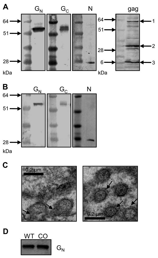
Figure 1. Characterization of RVF VLPs.
A) Western blot analysis of chimeric RVF VLPs (chimVLP): Concentrated supernatants from 293-gag cells transfected with RVFV G and N expression plasmids analyzed by Western blotting using antibodies specific for RVFV GN, GC, N and Moloney murine leukemia virus (MoMLV) gag. B) Western blot analysis of RVF VLPs: Concentrated supernatants from 293 cells transfected with RVFV G and N expression plasmids analyzed by Western blotting using antibodies specific for RVFV GN, GC and N. C) Negative staining of RVFV G and N transfected 293 cells fixed with glutaraldehyde and stained with uranyl acetate and examined by transmission electron microscopy. Scale bar represents 200nm. Left panel: arrows point to budding VLPs; right panel: arrows indicate RVFV G spikes protruding from the VLP membrane. D) Western blot analysis of RVF chimVLPs: Concentrated supernatants from 293-gag cells transfected with RVFV N and wild type (WT) or codon-optimized (CO) RVFV G sequences.

