Abstract
To improve efficiency of SCNT, it is necessary to modify differentiated donor cells to become more amendable for reprogramming by the oocyte cytoplasm. A key feature that distinguishes somatic/differentiated cells from embryonic/undifferentiated cells is cellular metabolism, with somatic cells using OXPHOS while embryonic cells utilize glycolysis. Inducing metabolic reprogramming in donor cells could improve SCNT efficiency by priming cells to become more embryonic in nature prior to SCNT.
HIF1-α, a transcription factor that allows for cell survival in low oxygen, promotes a metabolic switch from OXPHOS to glycolysis. We hypothesized that chemically stabilizing HIF1-α in donor cells by use of the hypoxia mimetic, CoCl2, would promote this metabolic switch in donor cells and subsequently improve the development of SCNT embryos. Donor cell treatment with 100 μM CoCl2 for 24 hours preceding SCNT upregulated mRNA abundance of glycolytic enzymes, improved SCNT development to the blastocyst stage and quality, and affected gene expression in the blastocysts. After transferring blastocysts created from CoCl2-treated donor cells to surrogates, healthy cloned piglets were produced. Therefore, shifting metabolism toward glycolysis in donor cells by CoCl2 treatment is a simple, economical way of improving the in vitro efficiency of SCNT and is capable of producing live animals.
Keywords: Somatic cell nuclear transfer, metabolism, hypoxia inducible factor, cellular reprogramming, porcine
Introduction
Since the birth of the first animal cloned with a somatic cell in 1996, SCNT has developed into a useful research tool (Wilmut et al, 1997). Today SCNT is used for biomedical models, including xenotransplantation, as well as agricultural models that have led to the discovery of novel treatments for human diseases, animals that are disease resistant, and have put animal-to-human organ transplant within reach (Whitworth and Prather, 2017); (Prather et al, 2013). Even with the current success of SCNT-created animals, the overall efficiency of SCNT remains low (<5%) with few live births resulting from the SCNT process (Whitworth and Prather, 2011). Due to the lack of authentic embryonic stem cells and induced pluripotent stem cell lines capable of producing live pigs, porcine SCNT is limited to the use of somatic cell types. Since somatic cells have already undergone some degree of differentiation, a possible explanation for poor SCNT efficiency is the inability to successfully remodel somatic nuclei through the SCNT process. A key feature that distinguishes embryonic/undifferentiated cells from somatic/differentiated cells is the metabolism that is used. Differentiated cells utilize mitochondrial OXPHOS, while undifferentiated cells use glycolysis. There is mounting evidence to suggest that metabolic reprogramming, or the switch from OXPHOS to glycolysis, is necessary to revert cells back to an undifferentiated state and maintain stemness (Prigione et al, 2014).
HIFs are a class of master transcription factors responsible for the cellular survival response to hypoxic conditions. HIF stabilization promotes the transcription of target genes related to glycolysis, angiogenesis, cell survival and proliferation, cell migration, apoptosis, and erythropoiesis (Hu et al, 2003). Hypoxic stress is alleviated by these downstream targets by modifying the need for oxygen for cellular mechanisms, such as energy production, or allowing for greater oxygen delivery. For example, downstream targets related to glucose metabolism, such as the glucose transporters SLC2A1 and SLC2A3, allow for energy production through glycolysis as opposed to mitochondrial OXPHOS, which can only occur in the presence of oxygen (Semenza, 2000).
Previous studies have shown that donor cell culture in hypoxia (1.25% O2) results in an upregulation of genes related to glycolysis in donor cells, as well as increased blastocyst production and in utero survivability following SCNT (Mordhorst et al, 2019). However, hypoxic cell culture can be costly and often requires specialized mixed gas tanks in order to achieve low oxygen tensions. There is also no reliable way to monitor the oxygen tension that the donor cells are being exposed to when cultured in hypoxia, as it requires culture in chambers that must remain sealed. In addition, HIF 1-α, the modulator of the hypoxic response in cells, has a high turnover rate with degradation occurring in 5–8 minutes once cells are exposed to atmospheric oxygen levels. During the SCNT process, the time between cell collection and cell-oocyte fusion/activation is typically greater than 1 hour. Therefore, the influence of HIF 1-α in these cells may be greatly diminished by the conclusion of the SCNT process.
Due to the possible instability of HIF1-α in hypoxia cultured cells, we proposed a chemical hypoxia mimetic that allows a sustained effect of HIF1-α outside of physiological hypoxia. In normoxia, HIF1-α is hydroxylated by prolyl hydroxylases that require oxygen and iron for their enzymatic activity. This hydroxylation serves as a docking site for VHL protein that marks HIF1-α for degradation by the 26S proteasome. In hypoxic conditions, the oxygen required for the prolyl hydroxylases is not available; and therefore, the cascade of events leading to HIF1-α degradation cannot be initiated. This allows HIF1-α protein to accumulate in the cytoplasm and subsequently translocate to the nucleus to dimerize with HIF1-β and direct transcription of downstream targets (Semenza, 2000). CoCl2 is a known hypoxia mimetic that inhibits the activity of prolyl hydroxylases by replacing the required iron domain of the prolyl hydroxylases with cobalt (Hirsila et al, 2005). This chemical simulation allows stabilization of the volatile HIF1-α, even in the presence of atmospheric oxygen. Once stabilized, HIF1-α can activate its downstream targets including genes that induce the reprogramming of metabolic processes to favor glycolytic metabolism over OXPHOS.
Therefore, the objective of this study was to determine if treatment of somatic donor cells with the hypoxia mimetic, CoCl2, can induce metabolic reprogramming in the donor cells and promote better nuclear reprogramming prior to SCNT to improve development of SCNT embryos.
Results
Impact of CoCl2 on cell viability
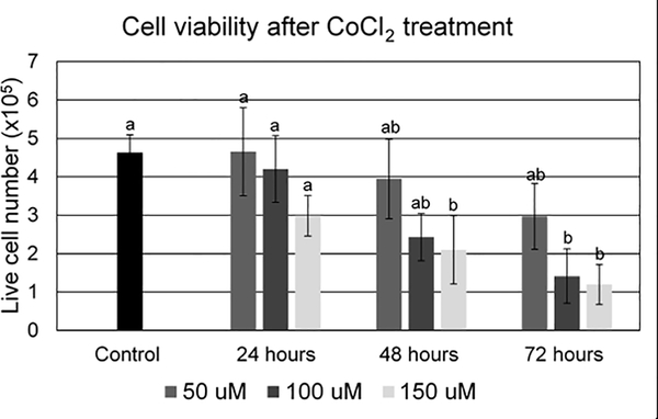
Cell number and viability was determined by Trypan blue exclusion after culture in 50, 100, or 150 μM of CoCl2 for 24, 48, or 72 hours (Figure 1). Live cell number was not different between any CoCl2 concentrations after 24 hours of culture. After 48 hours of culture, live cell number was significantly lower in the 150 μM treatment group as opposed to the 50 μM, 100 μM, or untreated cell groups. After 72 hours of culture, live cell number was negatively impacted in the 100 μM and 150 μM treatment groups as compared to the 50 μM and untreated groups.
Figure 1.
Cell viability after treatment with 0 μM, 50 μM, 100 μM, or 150 μM of CoCl2 for 24, 48 or 72 hours. Data represented as means ± SEM. Statistical differences represented by different lowercase letters (a,b).
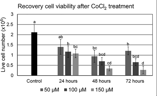
Long-term effects of CoCl2 treatment were determined by analysis of cell viability after a 3-day recovery period following CoCl2 exposure (Figure 2). Only the 24-hour 50 μM treatment of CoCl2 was capable of recovering cell viability to numbers comparable to the untreated control. The 50 μM treatment of CoCl2 did become detrimental to cell viability following 48 and 72 hours of exposure. The 100 μM CoCl2 treatment was comparable to the 50 μM treatment at all time points. The 150 μM treatment was significantly lower than the 50 μM treatment after 48 and 72 hours of CoCl2 exposure. Based on the results of these two studies, a treatment of 24-hour exposure to 100 μM CoCl2 was chosen for the remainder of the study.
Figure 2.
Cell viability following a 72 hr recovery period after treatment with 0 μM, 50 μM, 100 μM, or 150 μM of CoCl2 for 24, 48 or 72 hours. Data represented as means ± SEM. Statistical differences represented by different lowercase letters (a,b,c.d).
Gene expression in donor cells following CoCl2 exposure
Real-time quantitative PCR was used to analyze differences in message abundance between CoCl2 treated donor cells, hypoxia treated donor cells, and untreated control cells (Table 2) for HIF1-α and non HIF1-α gene targets (Liu et al, 2012). Glucose transporters, SLC2A1 and SLC2A3, as well as glycolytic enzymes HK1, HK2, GPI, ALDOC, GAPDH, PGK1, PGAM1, ENO1, PKM2, PDK1, and LDHA were upregulated in the CoCl2 group compared to the control. The same transcripts, with the exception of SLC2A1, ALDOC, GAPDH, and PGAM1 were also upregulated in the hypoxia group compared to the control. Transcript abundance of the mitophagy-associated gene BNIP3, GPI and PDK1 were differentially expressed between all treatment groups with the lowest expression present in the control cells and the highest expression in the CoCl2 cells. Non HIF1-α targets, TALDO1, EPAS1, YWHAG, LDHB, and BCL2 were not differentially expressed between the groups.
Table 2.
Normalized abundance ± SEM of gene products related to glycolysis and mitophagy. Treatments include a control (cultured at 5% O2 for 3 days), CoCl2 treatment (100 μM CoCl2 for 24 hours), and a hypoxic treatment (cultured at 1% O2 for 3 days). Superscripts represent differences between treatments with P < 0.05 considered significant.
| Gene name | Control | CoCl2 | Hypoxia |
|---|---|---|---|
| SLC2A1 † | 1.88 ± 0.38a | 3.25 ± 0.32b | 2.29 ± 0.12ab |
| SLC2A3 † | 1.61 ± 0.26a | 3.25 ± 0.39b | 3.75 ± 0.34b |
| HK1 † | 2.02 ± 0.17 a | 3.30 ± 0.25 b | 3.02 ± 0.18 b |
| HK2 † | 10.59 ± 1.92a | 22.36 ± 1.68b | 19.24 ± 1.03b |
| GAPDH † | 3.27 ± 0.34a | 6.38 ± 0.49b | 4.33 ± 0.44b |
| PGK1 † | 1.03 ± 0.10a | 2.06 ± 0.09b | 1.77 ± 0.10b |
| ENO1 † | 5.88 ± 0.44a | 10.28 ± 0.68b | 9.15 ± 0.97b |
| PKM2 † | 3.70 ± 0.25a | 6.30 ± 0.59b | 5.59 ± 0.48b |
| PDK1 † | 3.82 ± 0.48a | 7.10 ± 0.05b | 5.66 ± 0.51c |
| LDHA † | 2.16 ± 0.22a | 3.45 ± 0.28b | 3.57 ± 0.26b |
| LDHB | 0.11 ± 0.01 | 0.11 ± 0.001 | 0.12 ± 0.02 |
| BNIP3 † | 2.02 ± 0.40a | 5.54 ± 0.32b | 3.69 ± 0.31c |
| TALDO1 | 0.81 ± 0.10 | 1.03 ± 0.09 | 0.85 ± 0.09 |
| EPAS1 | 0.32 ± 0.07 | 0.55 ± 0.17 | 0.24 ± 0.04 |
| YWHAG | 0.39 ± 0.04 | 0.44 ± 0.04 | 0.39 ± 0.02 |
| BCL2 | 0.55 ± 0.03 | 0.65 ± 0.07 | 0.53 ± 0.05 |
indicates genes that are HIF targets.
SCNT embryo development and quality
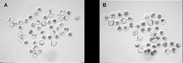
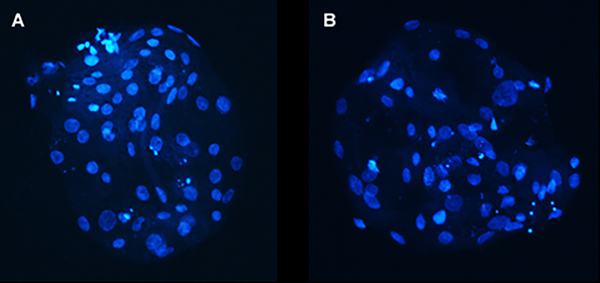
The use of CoCl2-treated donor cells for SCNT resulted in an increased rate of development to the blastocyst stage compared to untreated control donor cells (50.3±2.6% vs 32.6±1.9%, P = 0.0002) (Figure 3), as well as an increase in the total number of nuclei within the blastocyst-stage embryos (52.0±3.3 vs 39.0±3.0, P = 0.014) (Figure 4). Evaluation of DNA damage by the TUNEL assay revealed no difference in the number of apoptotic nuclei between the groups (P = 0.64) (Table 2).
Figure 3.
Representative images of blastocyst stage embryos created from (A) CoCl2 treated donor cells and (B) control donor cells.
Figure 4.
Representative images of Hoechst stained blastocyst stage embryos created from (A) CoCl2 treated donor cells and (B) control donor cells.
Gene expression in SCNT blastocyst stage embryos produced by CoCl2 donor cells
Genes that were evaluated in donor cells were also analyzed in blastocyst-stage embryos created with CoCl2 treated donor cells and blastocyst-stage embryos created from untreated control cells (Table 4). Of the genes evaluated, SLC2A1, PGAM1, and LDHA were upregulated in day 6 blastocyst-stage embryos created from CoCl2 treated donor cells compared to control donor cells (p<0.05).
Table 4.
Blastocyst stage embryo development and quality parameters on day 6 between embryos created from CoCl2 treated donor cells and control donor cells.
| Quality parameter | Control | CoCl2 |
|---|---|---|
| Blastocyst rate (%) ± SEM | 32.55 ± 1.87a | 50.29 ± 2.57b |
| Total cell number ± SEM | 38.99 ± 3.03a | 51.96 ± 3.34b |
| % TUNEL positive ± SEM | 7.04 ± 0.78 | 6.51 ± 0.72 |
Different letters represent statistical significance (P <0.05).
Different letters represent statistical significance (P <0.05).
Cloned piglet production with CoCl2 treated donor cells
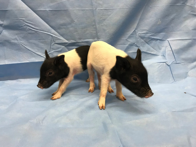
Following surgical embryo transfer to two recipient surrogates, both surrogates were confirmed pregnant by ultrasound at 25 and 38 days of gestation. At 52 days of gestation, one of the two surrogates had exhibited estrus and was no longer pregnant. At 120 days of gestation, the remaining pregnant surrogate farrowed naturally and delivered 5 piglets. Three of the five piglets were stillborn, and the surviving two piglets were healthy with no signs of abnormalities (Figure 5). No obvious defects were detected in the stillborn piglets; however, a necropsy was not performed. Birthweights ranged from 0.800 kg to 1.155 kg, with an average birthweight of 0.955 kg. Weaning weights recorded at 3 weeks were 4.720 kg and 4.120 kg, for an average weight of 4.420 kg (Table 5).
Figure 5.
Images of cloned piglets produced from SCNT embryos created from CoCl2 treated donor cells.
Table 5.
Birthweights and status of piglets born from SCNT embryos created from CoCl2 treated donor cells.
| Piglet # | Birthweight | WEANING WEIGHT |
|---|---|---|
| 1 | 0.845 kg | 4.120 kg |
| 2 | 1.155 kg | 4.720 kg |
| 3 (stillborn) | 0.980 kg | --- |
| 4 (stillborn) | 0.800 kg | --- |
| 5 (stillborn) | 0.995 kg | --- |
| Avg | 0.955 kg | 4.420 kg |
Discussion
The purpose of this study was to understand the effect of CoCl2 treatment on metabolism in SCNT donor cells and the resultant effect on SCNT efficiency in vitro with these donor cells. Analysis of HIF1-α targets related to glycolysis and cell survival in donor cells cultured in either 5% O2 (control), 1% O2 (hypoxia) or 5% O2 with CoCl2 treatment was analyzed in order to understand the effect that HIF1-α stabilization through physiological or chemical means had on gene expression (Table 1). Hypoxic culture of fibroblasts and fibroblasts cultured with CoCl2 resulted in an increase in mRNA abundance of glucose transporters SLC2A1 and SLC2A3, as well as glycolysis-related enzymes HK1 and HK2, GPI, ALDOC, GAPDH, PGK1, PGAM, ENO1, PKM2, PDK1, and LDHA, all of which are HIF downstream targets. (Figure 6).
Table 1.
RT-PCR primers
| Gene | Forward primer 5’ → 3’ | Reverse primer 5’ → 3’ | Accession # |
|---|---|---|---|
| YWHAG | TCCATCACTGAGGAAAACTGCTAA | TTTTTCCAACTCCGTGTTTCTCTA | XM_005661962.3 |
| PKM2 | ATGCAGTCTTGGATGGAGCTGACT | ATTGCAAATGGTAGATGGCGGCCT | AJ557236.1 |
| SLC2A1 | TCCACACCCACTTTGTCACACTGA | AGCCTCAACTCCCACATCACTGAA | XM_021096908.1 |
| SLC2A3 | CCCTCAGCTGCATTCTATTT | GTCTCAGGGACTTTGAAGAAG | XM_021092392.1 |
| PGK1 | CGCTTTCTGCATCTCCACTTGGCA | GCTGTGCAATGGTTCAAGGGTTCCT | NM_001099932.2 |
| PDK1 | ACCAGGACAGCCAATACAAGTGGT | ACGTGGACTTGAATAGGCGGGTAA | NM_001159608.1 |
| TALDO1 | TGAAGCGGCAGAGGATGGAGAGC | TCGTCGATGGCGTTGAAGTCGC | NM_001244935.1 |
| EPAS1 | AAGCAAAGACATGTCCACCGAGCG | GTGGCTGACTTGAGGTTGACGGTG | NM_001097420.1 |
| HK1 | TCTTGATCGACTTCACCAAGAGGG | TCGCTCTCGATCTGCGAGAGATACTT | NM_001243184.1 |
| HK2 | GAATTTGATGCGGCCGTGGATGAA | CCAGGTACATGCCGCTGATCATTT | NM_001122987.1 |
| ENO1 | TCGGAGTTCTACAGGTCGGGCAAG | TGGTCCGGTGAGATGTACCTGCTG | XM_021095280.1 |
| PGAM1 | CAGTGCTGGATGCCATTGACCAAA | GCTTGGCAGCAGTTTCTGCCTTAT | XM_003483535.4 |
| LDHA | TTCAGCCCGGTTCCGTTACCTAAT | TTCTTCAGGGAGACACCAGCAACA | NM_001172363.2 |
| LDHB | TAAGCATGGGCTTTGACTCTGGGA | ACTCCCGGCTTCTAGGTTGTAGTA | NM_001113287.1 |
| VEGFA | CAAACCTCACCAAGGCCAGCACAT | CGAGCAAGGCCCACAGGGATTTTC | NM_214084.1 |
| GPI | CCAGGAGACCATCACAAATG | TAGACAGGGCGACAAAGT | NM_214330.1 |
| ALDOC | TCTTCCATGAGACCCTCTAC | TACACCCTTGTCCACCTT | NM_001243928.1 |
| BNIP3 | GGATTACATGGAGAGGAGGA | GTGCTTGAAGAGGAGGAAC | XM_003359404.4 |
| BCL2 | ACTGAATGCCCTCCGGTACC | ATCCCCATGGCTGCAGTGAA | XM_003130557.2 |
| ACTB | TCTGGCACCACACCTTCT | TGATCTGGGTCATCTTCTCAC | DQ178122.1 |
| POU5F1 | TTTGGGAAGGTGTTCAGCCAAACG | TCGGTTCTCGATACTTGTCCGCTT | NM_001113060.1 |
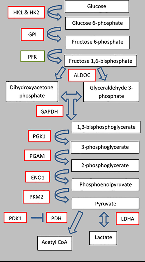
Figure 6.
Schematic of glycolysis. Red rectangles represent enzyme transcripts that were differentially expressed between CoCl2 treated fibroblasts and control fibroblasts. Green rectangles represent enzyme transcripts that were not evaluated.
Although the majority of these enzymes are basic glycolytic enzymes that could indicate that an increase in glycolytic activity is occurring, enzymes such as PKM2, PDK1, and LDHA have unique roles that are specific to less differentiated cells, such as cancer cells, that are being pushed away from oxidative metabolism. Pyruvate kinase muscle isozyme M2 is one of the four isoforms of pyruvate kinase, produced by alternative splicing, and is specifically associated with proliferating cells and cancer cells (as reviewed by Dong et al, 2016). In the analysis of mRNA abundance of glycolytic enzymes associated with the Warburg effect, it was determined that blastocyst stage-embryos exclusively expressed the fetal PKM2 as opposed to the adult PKM1 (Redel et al, 2011).
In an aerobic system, once pyruvate has been produced through glycolysis, it is subsequently converted to acetyl CoA through the mitochondrial enzyme PDH. However, in glycolytic systems, the production of the enzyme PDK1 results in phosphorylation of pyruvate dehydrogenase which inactivates the complex and directs pyruvate away from the TCA cycle, inhibiting its oxidation. PDK1 has been demonstrated by microarray and chromatin immunoprecipitation to be a direct target of HIF1-α, and is an important player in the switch from aerobic to anaerobic metabolism through its ability to block acetyl CoA production so that pyruvate can be converted to lactate (Kim et al, 2006).
Since PDK1 increases availability of pyruvate in the cell, it is then able to be converted to lactate by LDHA. The conversion of pyruvate to lactate is crucial for anaerobic glycolysis. In human pancreatic cancer cells, LDHA is upregulated by hypoxia and is directly activated by HIF1-α. Induced expression of LDHA promotes the proliferation and migration of pancreatic cancer cells, and knocked down expression inhibits cell growth and migration (Cui et al, 2017). This indicates that LDHA and its effect in hypoxic conditions is crucial for cancer cell survival.
Although the majority of gene expression changes found in this study relate to the SCNT donor cells, there were also several genes upregulated in CoCl2 treated donor cell SCNT blastocyst stage embryos (Table 3). Glucose transporter SLC2A1, and glycolytic enzymes PGAM1 and LDHA were found to be upregulated in embryos created from CoCl2 treated donor cells as compared to those created from control donor cells. Although glucose is not a component of the embryo culture media used in this study, increased glucose uptake has been shown to be associated with improved embryo viability in bovine (Renard et al, 1980), mouse (Gardner and Leese, 1987) and human (Gardner et al, 2011) systems. PGAM1 enzymatic activity has been proposed as a potential alternative glycolytic pathway in rapidly proliferating cells that do not have increased pyruvate kinase activity. Phosphorylation of PGAM1 by the phosphate donor phosphoenolpyruvate (PEP), which is typically associated with PKM2 activity, promotes increased pyruvate production and allows for a higher glycolytic flux (Vander Heiden et al, 2010). LDHA promotes lactate production, and aligning with the Warburg effect, lactate production in the presence of oxygen is associated with rapidly proliferating cells. During blastocyst formation, there is a transition from the LDHB isoform to the LDHA isoform which is associated with lactate production as opposed to pyruvate production (as reviewed by Krisher and Prather, 2012). Therefore, the upregulation of LDHA at the blastocyst stage in the embryos created from CoCl2 treated donor cells as compared to control SCNT embryos could indicate that a more natural gene expression profile in the blastocysts is promoted by metabolic reprogramming of CoCl2 treated donor cells prior to SCNT.
Table 3.
Normalized abundance ± SEM of gene products related to glycolysis and mitophagy. Treatments include day 6 blastocyst stage embryos created from control donor cells and CoCl2 treated donor cells (100 μM CoCl2 for 24 hours). Superscripts represent differences between treatments with P < 0.05 considered significant.
| Gene name | Control | CoCl2 | p-value |
|---|---|---|---|
| SLC2A1 | 5.86 ± 0.66a | 8.06 ± 0.44b | 0.0497 |
| SLC2A3 | 2.05 ± 0.47 | 2.74 ± 0.16 | 0.2370 |
| HK1 | 0.12 ± 0.01 | 0.16 ± 0.03 | 0.0978 |
| HK2 | 24.30 ± 3.32 | 30.87 ± 2.68 | 0.0989 |
| GPI | 0.66 ± 0.10 | 0.88 ± 0.04 | 0.0917 |
| ALDOC | 0.37 ± 0.12 | 0.50 ± 0.03 | 0.3755 |
| GAPDH | 2.45 ± 0.52 | 2.38 ± 0.50 | 0.4626 |
| PGK1 | 0.18 ± 0.02 | 0.25 ± 0.04 | 0.0955 |
| PGAM1 | 3.08 ± 0.10a | 3.88 ± 0.26b | 0.0446 |
| ENO1 | 1.31 ± 0.12 | 1.55 ± 0.09 | 0.0916 |
| PKM2 | 0.47 ± 0.11 | 0.66 ± 0.11 | 0.1518 |
| PDK1 | 2.26 ± 0.58 | 2.48 ± 0.56 | 0.3997 |
| LDHA | 0.08 ± 0.01a | 0.15 ± 0.02b | 0.0315 |
| BNIP3 | 4.99 ± 0.66 | 6.76 ± 0.68 | 0.1348 |
| TALDO1 | 3.90 ± 0.45 | 4.58 ± 0.74 | 0.2414 |
| YWHAG | 0.09 ± 0.01 | 0.13 ± 0.03 | 0.1302 |
| BCL2 | 4.49 ± 0.57 | 4.94 ± 0.69 | 0.6432 |
| POU5F1 | 476.97 ± 136.52 | 614.25 ± 35.49 | 0.1928 |
| VEGFA | 2.99 ± 0.23 | 3.73 ± 0.53 | 0.2678 |
CoCl2 treatment of donor cells resulted in greater (~18% increase) blastocyst stage embryo development and improved embryo quality (13 more cells per blastocyst) as compared to control embryos (Table 4). However, previous studies have shown that analysis of blastocyst stage embryo qualities alone is not indicative of the in utero survival and live birth potential of embryos (Redel et al, 2016). In order to demonstrate that CoCl2 treatment of donor cells could result in the live birth of piglets following SCNT, embryo transfer was conducted. An untreated control donor cell comparison was not conducted in this study due to the number of animals that would need to be utilized, and surgeries that would have to be performed. The purpose of the embryo transfer was to ensure that there were no lethal effects of the donor cell treatment that would prevent the in utero survival of these embryos. Of the two surrogates used for embryo transfer, one was able to maintain pregnancy to term. This surrogate delivered 5 piglets unassisted. Of the 5 piglets that were delivered, 3 were stillborn. From outward visual inspection and birth weights, the 3 piglets did not have any obvious abnormalities that would have resulted in their death and had healthy birthweights for an Ossabaw breed (Table 5). Due to the lack of outward abnormalities in these stillborn piglets, along with the birth of two live piglets that proved there was no lethal effect of the CoCl2 treatment, postmortem necropsies were not conducted. The two surviving piglets had healthy birth weights and weaning weights and have had no issues since their birth. Therefore, the birth of healthy clones from this experiment indicates that CoCl2 treatment of donor cells results in SCNT embryos that are capable of producing piglets and can be used as a viable option for future cloning studies.
Our findings indicate that the use of CoCl2 as a novel treatment for SCNT donor cells induces the same glycolytic response as culture in 1% oxygen (hypoxia) for 3 days. The use of the hypoxia mimetic allows the cells to be maintained in any oxygen tension, without the need for specialized gas tanks or chambers and eliminates the need for long term culture of donor cells in hypoxic conditions in order to establish the same effect. The upregulation of genes that are known to be downstream targets of HIF1-α in the CoCl2 treated and hypoxia treated donor cells, along with the lack of differential expression of non- HIF1-α targets suggests that the transcription factor may be activated through these treatments. Therefore, promoting metabolic reprogramming in donor cells through CoCl2 treatment improves the efficiency of the SCNT process through alterations in gene expression in donor cells and resultant SCNT blastocysts, improvement in the quality and development rate of SCNT embryos, and production of healthy, cloned piglets.
Materials and Methods
Ethics statement
Collection of ovaries from prepubertal gilts and use of live animals were in accordance with approved protocol and standard operating procedures by the Animal Care and Use Committee of the University of Missouri.
Determining optimal CoCl2 concentration
Dorsal tissue of gestational day 35 wild-type fetuses was removed and dissociated. Cells were cryopreserved in 500 μL aliquots and stored in liquid nitrogen until needed. Cells were thawed and cultured in Dulbecco’s modified Eagle’s medium (1 g/L glucose with phenol red) supplemented with 15% FBS (Corning, Corning, NY) for four days in T25 flasks (Corning). For determining the working CoCl2 concentration, cobalt chloride hexahydrate (Thermo Fisher, Waltham, Massachusetts – C8661) was mixed fresh daily for each use. In order to achieve a 10 mM concentration of CoCl2, 11.89 mg of CoCl2 was dissolved into 5 mL MilliQ H2O. The solution was then added at a 1 μL:100 μL ratio to culture media to achieve a final concentration of 100 μM. In order to evaluate the effect of increased CoCl2 concentrations on cell viability, cells were plated at equal density of 7.5 × 10⁴ cells/flask and the CoCl2 solution was added at 50 μM, 100 μM, and 150 μM. All concentrations were applied to cells for 24, 48 or 72 hours. Control cells were left untreated. Following the 72 hours, CoCl2 treated and control cells were trypsinized and Trypan blue exclusion was used to determine live and total cell number. To evaluate the recovery ability of cells after CoCl2 exposure, the beforementioned conditions were applied to cells plated at equal densities, followed by aspiration of media containing CoCl2 and replacement with fresh media. The cells were grown for 3 days subsequent to CoCl2 removal and then trypsinized and subjected to Trypan blue exclusion to determine live and total cell number.
For SCNT, fibroblast cells were thawed 4 days prior to SCNT, counted by Trypan blue exclusion, plated at a density of 7.5 × 10⁴ cells/T25 flask and cultured in a humidified incubator with an atmosphere of 5% CO2, 5% O2, and 90% N2 at 37.5° C. On day 3, 24 hours before SCNT, CoCl2 was added at a 100 μM concentration. The control cells were left untreated.
Oocyte collection and somatic cell nuclear transfer
Ovaries from a local abattoir (Smithfield, Milan, MO) were harvested and 18-gauge needles attached to disposable 10 mL syringes were used to aspirate follicles that were 3–6 mm in size and showed normal morphology. COCs in follicular fluid were washed 3 times in TL-Hepes before being placed in 100 mm polystyrene petri dishes. COC’s displaying uniform cytoplasm and at least 3 layers of cumulus cells were selected and placed in maturation medium (TCM-199 medium supplemented with 0.1% PVA, 3.05 mM D-glucose, 0.91 mM sodium pyruvate, 10 μg/mL of gentamicin, 0.57 mM cysteine, 10 ng/mL of EGF, 0.5 μg/mL of FSH, 0.5 μg/mL of LH, 40 ng/mL FGF2, 20 ng/mL LIF, and 20 ng/mL IGF1) (Yuan et al, 2017) for 42–44 hours in a humidified incubator with an atmosphere of 5% CO2 in air at 37.5° C. Cumulus cells were stripped from oocytes by gentle vortex for 3 minutes in 0.1% (w/v) hyaluronidase in TL-HEPES-buffered saline with 0.1% PVA. Metaphase II oocytes were selected based on the presence of an extruded first polar body in the perivitelline space.
Metaphase II oocytes were placed on the stage of an inverted microscope equipped with micromanipulators in drops containing manipulation medium (Lai & Prather, 2004) supplemented with 7.0 μg/mL cytochalasin B. A hand-tooled glass pipette was used to remove the polar body, and ~ 10% of the adjacent cytoplasm (presumably containing the metaphase plate). Following enucleation, a fibroblast cell was injected into the perivitelline space and pressed against the cytoplasm. Donor cells were then trypsinized and harvested for SCNT, with CoCl2 treated cells resuspended in manipulation medium containing 7.0 μg/mL cytochalasin B and 100 μM CoCl2. While injecting CoCl2 treated cells, 100 μM CoCl2 was present in the micromanipulation drops in order to sustain the treatment effect and prohibit HIF1-α degradation. Oocyte-donor cell couplets were then fused in fusion medium (0.3 M mannitol, 0.1 mM CaCl2, 0.1 mM MgCl2, 0.5 mM HEPES buffer, pH 7.2) by two direct current pulses (1‐s interval) at 1.2 kV/cm for 30 μsec by using a BTX Electro Cell Manipulator (Harvard Apparatus, Holliston, MA). At least one hour after fusion, reconstructed embryos were fully activated for 30 minutes with 200 μM TPEN (Lee et al, 2015) in TL-HEPES. Embryos were then incubated in MU-2 media with 0.5 μM of histone deacetylase inhibitor Scriptaid, for 14–16 hr in a 5% carbon dioxide (atmospheric oxygen) incubator (Whitworth, Zhao, Spate, Li, & Prather, 2011; Zhao et al., 2009). The following morning, embryos were removed from Scriptaid treatment, washed, and placed in fresh MU-2 media and cultured in an incubator with a humidified atmosphere of 5% CO2, 5% O2, and 90% N2 at 37.5° C until day 6 post-activation.
Blastocyst quality evaluation
Day 6 blastocyst-stage embryos collected in pools of 15–25 per treatment were fixed in 4% paraformaldehyde in TL-HEPES for 20 minutes, followed by permeabilization with 0.1% Triton X-100 for 30 minutes. In order to assess DNA damage, blastocyst stage embryos were incubated with TUNEL stain for 30 minutes, and then Hoechst nuclear stain (10 μg/mL) for 5 minutes. Blastocyst-stage embryos were visualized at 20× magnification on a microscope equipped with epi-fluorescence, and total cells and TUNEL positive cells were quantified. The ratio of TUNEL positive cells to total cells was calculated in order to determine a percentage of DNA damaged cells per blastocyst-stage embryo.
RNA Extraction and cDNA synthesis
In order to evaluate gene expression in donor cells, cells were subjected to either CoCl2 treatment, hypoxic treatment, or left untreated. For all treatments, cells were plated at equal densities in T25 plates. For CoCl2 treatment, cells were maintained at 5% CO2, 5% O2, and 90% N2 at 37.5° C and 100 μM CoCl2 was added on the 3rd day of culture, 24 hours prior to cell collection. For the hypoxic treatment, cells were placed in an incubator maintained at 5% CO2, 5% O2, and 90% N2 at 37.5° C for at least four hours before being transferred to a hypoxic chamber (Billups-Rothenburg, San Diego, CA) supplemented with a 100 mm petri dish of milliQ H2O. The chamber was sealed and gassed for two minutes with 1% O2 using a mixed gas LiquidGas tank (1% O2, 5% CO2). The chamber was then placed back into the incubator at 5% CO2, 5% O2, and 90% N2 at 37.5° C and were left to grow for 3 days following hypoxic exposure.
Day 6 blastocyst-stage embryos created with either CoCl2 treated donor cells or control donor cells were collected in pools of 35–50 and washed in diethyl pyrocarbonate-treated phosphate-buffered saline before being snap-frozen in liquid nitrogen for storage at −80° C. Fibroblast cells cultured in CoCl2 for 24 hours, untreated, or cultured in 1% hypoxia for 3 days were trypsinized, pelleted, and snap-frozen in liquid nitrogen for storage at −80° C. Three biological replicates were collected for each treatment. For blastocyst-stage embryos, total RNA was extracted by using an RNeasy Micro Kit (Qiagen, Germantown, MD) and eluted in 12 μL of nuclease-free water. All 12 uL of eluted RNA was used for cDNA synthesis by the SuperScript VILO cDNA Synthesis Kit (Thermo Fisher: 11754050). For fibroblast cells, total RNA was extracted by using an RNeasy Mini Kit (Qiagen, Germantown, MD) and eluted in 30 μL of nuclease-free water. RNA content was determined by using a Nanodrop 1000 Spectrophotometer (Thermo-Fischer), and an appropriate amount of eluted RNA was added accordingly for cDNA synthesis by the SuperScript VILO cDNA Synthesis Kit (Thermo Fisher: 11754050).
Relative Quantitative PCR
Relative quantitative PCR was performed with each sample from cDNA synthesis. Message evaluated included HIF1-α targets associated with glycolysis, autophagy, and pluripotency in fibroblast cells and blastocyst stage embryos (Table 1). Samples from each biological replicate were diluted to 5 ng/μL, and quantitative PCR was run in triplicate to determine differential expression of the selected transcripts with the conditions: 95°C for 3 min, and 40 cycles of 95°C for 10 s, 55°C for 10 s, and 72°C for 30 s. A dissociation curve was generated after amplification to ensure that a single product was amplified. Abundance of each mRNA transcript was calculated relative to a housekeeping gene, β actin, and a pig genome reference sample. The comparative quantification cycle method was used to determine relative mRNA expression for each treatment.
Surgical embryo transfer
For the embryo transfer experiment, donor cells used for SCNT were a wild-type Ossabaw cell line (RRID NSRRC:0008) that had been proven clonable (Mordhorst et al, 2019). Following SCNT, day 6 blastocyst-stage embryos created from CoCl2-treated donor cells were transferred into recipient surrogates. Briefly, two gilts 4 days post-observed estrus were aseptically prepared for surgery, and the infundibulum was exposed by entry though the lower abdominal wall. A Tomcat catheter containing 42 blastocyst-stage embryos was inserted into one ampullary-isthmic junction of each surrogate where the blastocysts were deposited. Pregnancy was determined by ultrasound on day 25 and monitored by biweekly ultrasounds thereafter. After farrowing, birth weights, weaning weights, and phenotypes were recorded.
Acknowledgements
The authors would like to thank Jason Dowell for his assistance with the embryo transfer surgeries, as well as the pig care workers that assisted with surrogate and piglet care. The authors would like to acknowledge funding from Food for the 21st Century and the National Institutes of Health via R01 HD080636 and NHLBI and NIAID via U42 OD011140. The authors have no conflict of interest to declare.
Grant support: Food for the 21st Century at the University of Missouri, and the National Institutes of Health via R01 HD080636 and NHLBI and NIAID via U42 OD011140.
Abbreviations
- SCNT
somatic cell nuclear transfer
- OXPHOS
oxidative phosphorylation
- HIF1-α
hypoxia inducible factor 1-α
- CoCl2
cobalt chloride
- HIFs
hypoxia inducible factors
- SLC2A1
solute carrier 2A1
- SLC2A3
solute carrier 2A3
- VHL
Von Hippel Lindeau
- COCs
cumulus-oocyte complexes
- TL-Hepes
Tyrode’s Lactate 4-(2-hydroxyethyl)-1-piperazineethanesulfonic acid
- PVA
polyvinyl alcohol
- TPEN
(N,N,N′,N′-tetrakis(2-pyridylmethyl) ethane-1,2-diamine)
- TUNEL
terminal deoxynucleotidyl transferase (TdT) dUTP nick-end labeling
- HK1
hexokinase 1
- HK2
hexokinase
- GPI
glucose-6-phosphate isomerase
- ALDOC
aldolase C
- GAPDH
glyceraldehyde 3-phosphate dehydrogenase
- PGK1
phosphoglycerate kinase 1
- PGAM1
phosphoglycerate mutase 1
- ENO1
enolase 1
- PKM2
pyruvate kinase muscle isozyme M2
- PDK1
pyruvate dehydrogenase kinase 1
- LDHA
lactate dehydrogenase A
- BNIP3
BCL2/adenovirus E1B 19 kDa protein-interacting protein 3
- TALDO1
transaldolase 1
- EPAS1
endothelial PAS domain-containing protein 1
- YWHAG
tyrosine 3-monooxygenase/tryptophan 5-monooxygenase activation protein gamma
- LDHB
lactate dehydrogenase b
- BCL2
B-cell leukemia/lymphoma 2
- PDH
pyruvate dehydrogenase
- PEP
phosphoenolpyruvate
References
- Cui X, Han Z, He S, Chen T, Shao C, Chen D, Su N, Chen Y, Wang T, Wang J, Song D, Yan W, Yang X, Liu T, Wei H, and Xiao J (2017). HIF1/2a mediates hypoxia-induced LDHA expression in human pancreatic cancer cells. Ongotarget. 8(15): 24840–24852. [DOI] [PMC free article] [PubMed] [Google Scholar]
- Dong G, Mao Q, Xia W, Xu Y, Wang J, Xu L, and Jiang F (2016). PKM2 and cancer: the function of PKM2 beyond glycolysis. Oncol. Lett 11(3): 1980–1986. [DOI] [PMC free article] [PubMed] [Google Scholar]
- Gardner DK, and Leese HJ (1987). Assessment of embryo viability prior to transfer by the noninvasive measurement of glucose uptake. J. Exp. Zool 242:103–105. [DOI] [PubMed] [Google Scholar]
- Gardner DK, Wale PL, Collins R, and Lane M (2011). Glucose consumption of single post-compaction human embryos is predictive of embryo sex and live birth outcome. Human Reproduction. 26(8): 1981–1986. [DOI] [PubMed] [Google Scholar]
- Hirsila M, Koivunen P, Leon X, Seeley T, Kivirikko KI, Myllyharju J (2005). Effect of desferrioxamine and metals on the hydroxylases in the oxygen sensing pathway. The FASEB Jrnl. 19(10): 10.1096/fj.04-3399fje. [DOI] [PubMed] [Google Scholar]
- Hu C, Wang L, Chodosh LA, Keith B, and Simon MC (2003). Differential roles of hypoxia-inducible factor 1a(HIF-1a) and HIF-2a in hypoxia gene regulation. Mol. Cell. Biol 23(24):9361–9374. [DOI] [PMC free article] [PubMed] [Google Scholar]
- Kim JW, Tchernyshyov I, Semenza GL, and Dang CV (2006). HIF-1-mediated expression of pyruvate dehydrogenase kinase: a metabolic switch required for cellular adaptation to hypoxia. Cell. Metab 3(3): 177–185. [DOI] [PubMed] [Google Scholar]
- Krisher R and Prather RS (2012). A role for the Warburg Effect in preimplantation embryo development: metabolic modification to support rapid cell proliferation. Mol. Reprod. Dev 79(5): 311–320. [DOI] [PMC free article] [PubMed] [Google Scholar]
- Lai L, and Prather RS (2004). A method for producing cloned pigs by using somatic cells as donors. Germ Cell Protocols. 245: 149–163. [DOI] [PubMed] [Google Scholar]
- Lee K, Davis A, Zhang L, Ryu J, Spate LD, Park KW, Samuel MS, Walters EM, Murphy CN, Machaty Z, and Prather RS (2015). Pig oocyte activation using a Zn2+ chelator, TPEN. Theriogenology. 84(6):1024–1032. [DOI] [PMC free article] [PubMed] [Google Scholar]
- Liu W, Shen S, Zhoa X, and Chen G (2012). Targeted genes and interacting proteins of hypoxia inducible factor-1. Int. J. Biochem. Mol. Biol 3(2): 165–178. [PMC free article] [PubMed] [Google Scholar]
- Mordhorst BR, Benne JA, Cecil RF, Whitworth KM, Samuel MS, Spate LD, Murphy CN, Wells KD, Green JA, and Prather RS (2019). Improvement of in vitro and early in utero porcine clone development after somatic donor cells are cultured under hypoxia. Mol. Reprod. Dev 86(5): 558–565. [DOI] [PMC free article] [PubMed] [Google Scholar]
- Mordhorst BR, Murphy SL, Schauflinger M, Salazar SR, Ji T, Behura SK, Wells KD, Green JA, and Prather RS (2018). Porcine fetal-derived fibroblasts alter gene expression and mitochondria to compensate for hypoxic stress during culture. Cell. Reprogram 20(4): 10.1089/cell.2018.0008. [DOI] [PMC free article] [PubMed] [Google Scholar]
- Prather RS, Lorson M, Ross JW, Whyte JJ, and Walters E (2013). Genetically engineered pig models for human diseases. Annu Rev Anim Biosci. 1:203–219. [DOI] [PMC free article] [PubMed] [Google Scholar]
- Prigione A, Rohwer N, Hoffmann S, Mlody B, Drews K, Bukowiecki R, Blumlein K, Wanker EE, Ralser M, Cramer T, and Adjaye J (2014). HIF1α modulates cell fate reprogramming through early glycolytic shift and upregulation of PDK1–3 and PKM2. Stem Cells. 32(2): 364–376. [DOI] [PMC free article] [PubMed] [Google Scholar]
- Redel BK, Brown AN, Spate LS, Whitworth KM, Green JA, and Prather RS (2011). Glycolysis in preimplantation development is partially controlled by the Warburg effect. Mol. Reprod. Dev 79(4): 262–271. [DOI] [PubMed] [Google Scholar]
- Redel BK, Spate LD, Lee K, Mao J, Whitworth KM and Prather RS (2016) Glycine supplementation in vitro enhances porcine preimplantation embryo cell number and decreases apoptosis but does not lead to live births. Mol. Reprod. Dev 83(3): 246–258. [DOI] [PMC free article] [PubMed] [Google Scholar]
- Renard JP, Philippon A, Menezo Y (1980). In-vitro uptake of glucose by bovine blastocysts. J. Reprod. Fertil 58:161–164. [DOI] [PubMed] [Google Scholar]
- Semenza GL. (2000). Expression of hypoxia‐inducible factor 1: Mechanisms and consequences. Biochem Pharmacol. 59:47–53. [DOI] [PubMed] [Google Scholar]
- Vander Heiden MG, Locasale JW, Swanson KD, Sharfi H, Heffron GJ, Amador-Noguez D, Christofk HR, Wagner G, Rabinowitz JD, Asara JM, and Cantley LC (2010). Evidence for an alternative glycolytic pathway in rapidly proliferating cells. Science. 329(5998):1492–1499. [DOI] [PMC free article] [PubMed] [Google Scholar]
- Vengellur A, and LaPres JJ (2004). The role of hypoxia inducible factor 1α in cobalt chloride induced cell death in mouse embryonic fibroblasts. Toxicological Sciences. 82(2): 638–646. [DOI] [PubMed] [Google Scholar]
- Whitworth K, and Prather RS (2010). Somatic cell nuclear transfer efficiency: how can it be improved through nuclear remodeling and reprogramming? Mol. Reprod. Dev 77(12): 1001–1015. [DOI] [PMC free article] [PubMed] [Google Scholar]
- Whitworth K, and Prather RS (2017). Gene editing as applied to prevention of reproductive porcine reproductive and respiratory syndrome. Mol Reprod Dev. 84(9): 926–933. [DOI] [PubMed] [Google Scholar]
- Whitworth KM, Zhao J, Spate LD, Li R, and Prather RS (2011). Scriptaid corrects gene expression of a few aberrantly reprogrammed transcripts in nuclear transfer pig blastocyst stage embryos. Cell Reprogram. 13(3): 191–204. [DOI] [PubMed] [Google Scholar]
- Wilmut I, Schnieke AE, McWhir J, Kind AJ, and Campbell KHS (1997). Viable offspring derived from fetal and adult mammalian cells. Nature. 385:810–813. [DOI] [PubMed] [Google Scholar]
- Zhao J, Ross JW, Hao Y, Spate LD, Walters EM, Samuel MS, Rieke A, Murphy CN, Prather RS (2009). Significant improvement in cloning efficiency of an inbred miniature pig by histone deacetylase inhibitor treatment after somatic cell nuclear transfer. Biol. Reprod 81(3):525–530. [DOI] [PMC free article] [PubMed] [Google Scholar]


