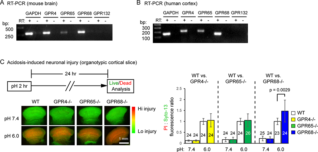
Figure 1. Functional screening identifies GPR68 as a regulator of pH 6-induced neuronal injury.
(A, B) Reverse transcription (RT)-polymerase chain reaction (PCR) result on the expression of proton-sensitive GPCRs in mouse brain (A) and human cortical tissue (B). -RT has no reverse transcriptase added into RT. Images shown were from products of 35 cycles of PCR. (C) Representative fluorescence images and quantification showing pH 6-induced neuronal injury in organotypic cortical slices from WT and corresponding GPCR knockouts. Organotypic cortical slices were treated with pH medium buffered at pH 7.4 or 6.0 for 2 hr and stained for propidium iodide (PI, red) and Syto-13 (green) 24 hrs later. Relative fluorescence intensity of PI and Syto-13 was quantified. Increased red/green ratio indicates increased neuronal injury. Dashed lines on the bar graph indicate that the WT controls for the three sets were different. For all experiments, the knockouts were compared to the WT that was cultured and treated in parallel. To better compare different experiments, injury (PI:Syto-13 ratio) in pH 6 of WT in a given experiment was normalized to 1. N on the bars indicate total number of slices quantified from 6 different experiments.

