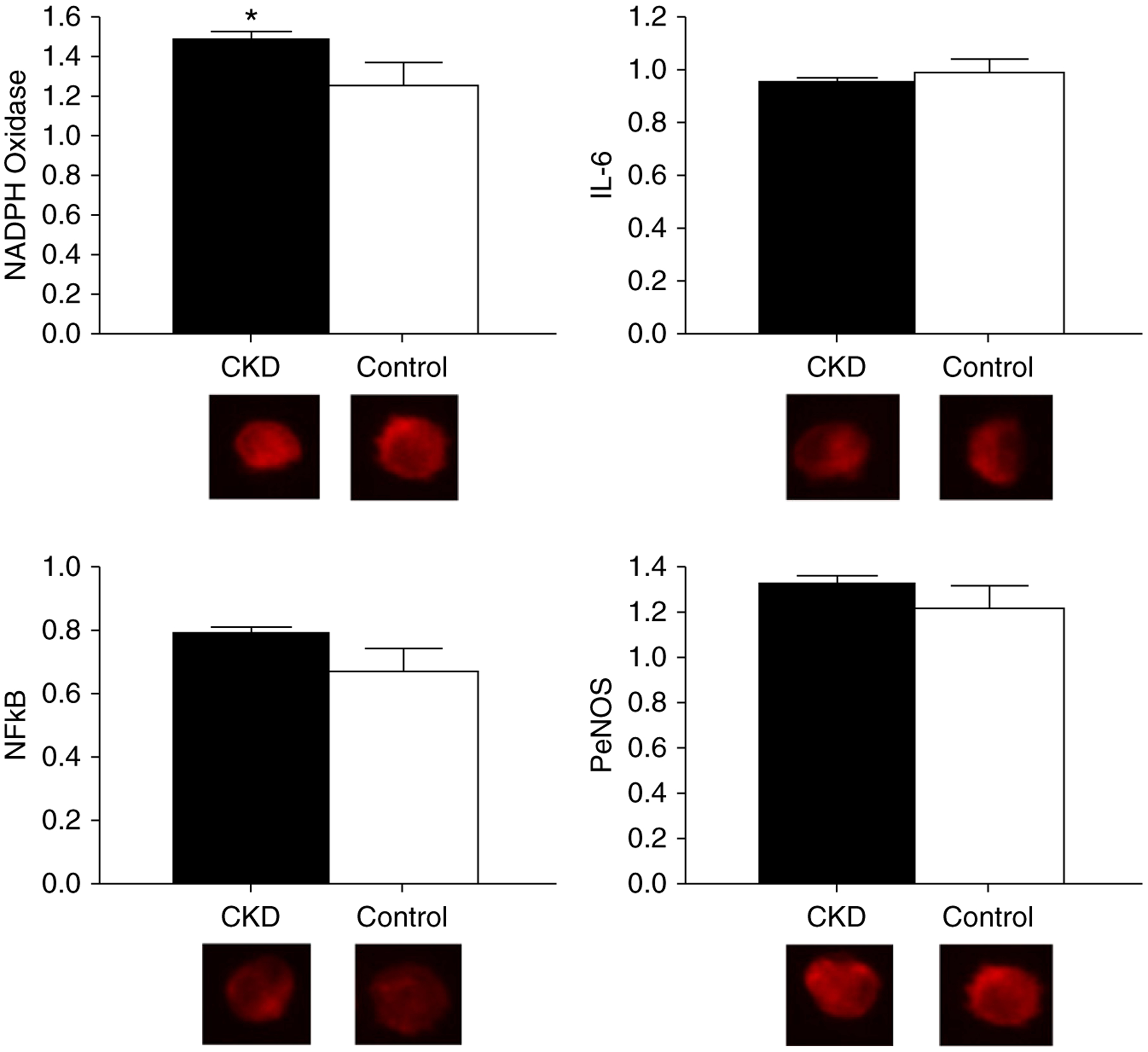
Figure 2. |. Greater vascular endothelial cell oxidative stress in participants with CKD as compared to controls.

Protein expression of NAD(P)H oxidase (CKD, 1.48±0.05; control, 1.25±0.11; P=0.05), IL-6 (CKD, 0.94±0.02; control, 0.98±0.05; P=0.43), NFκB (CKD, 0.78±0.02; control, 0.67±0.08; P=0.19), and phosphorylated endothelial cell nitric oxide synthase (PeNOS; CKD, 1.34±0.04; control, 1.23±0.10; P=0.34) in vascular endothelial cells collected from a peripheral vein of participants with CKD (black bars) compared with healthy controls (white bars). Expression is relative to human umbilical vein endothelial cell control, with representative images shown below (quantitative immunofluorescence). Values are mean±SEM. *P≤0.05.
