Abstract
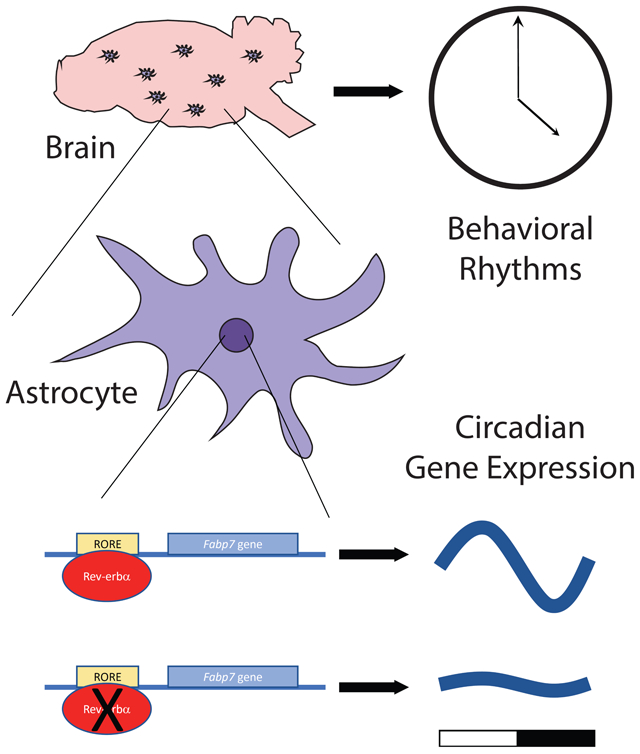
The astrocyte brain-type fatty-acid binding protein (Fabp7) circadian gene expression is synchronized in the same temporal phase throughout mammalian brain. Cellular and molecular mechanisms that contribute to this coordinated expression are not completely understood, but likely involve the nuclear receptor Rev-erbα (NR1D1), a transcriptional repressor. We performed ChIP-seq on ventral tegmental area (VTA) and identified gene targets of Rev-erbα, including Fabp7. We confirmed that Rev-erbα binds to the Fabp7 promoter in multiple brain areas, including hippocampus, hypothalamus, and VTA, and showed that Fabp7 gene expression is upregulated in Rev-erbα knock-out mice. Compared to Fabp7 mRNA levels, Fabp3 and Fabp5 mRNA were unaffected by Rev-erbα depletion in hippocampus, suggesting that these effects are specific to Fabp7. To determine whether these effects of Rev-erbα depletion occur broadly throughout the brain, we also evaluated Fabp mRNA expression levels in multiple brain areas, including cerebellum, cortex, hypothalamus, striatum, and VTA in Rev-erbα knock-out mice. While small but significant changes in Fabp5 mRNA expression exist in some of these areas, the magnitude of these effects are minimal to that of Fabp7 mRNA expression, which was over 6-fold across all brain regions. These studies suggest that Rev-erbα is a transcriptional repressor of Fabp7 gene expression throughout mammalian brain.
Keywords: lipid, metabolism, glia, BLBP, B-FABP, clock
Graphical abstract
INTRODUCTION
Fatty-acid binding proteins (Fabp) comprise a family of small (~15kDa) hydrophobic ligand binding carriers with high affinity for long-chain fatty-acids for intracellular transport, and are associated with metabolic, inflammatory, and energy homeostasis pathways (1, 2). These include three that are expressed in the adult mammalian central nervous system (CNS), and are Fabp3 (H-Fabp), Fabp5 (E-Fabp), and Fabp7 (B-Fabp). Fabp3 is primarily expressed in neurons, Fabp5 is expressed in various cell types, including both neurons and glia, and Fabp7 is most abundant in astrocytes and neural progenitors. While performing microarray analysis of transcripts in mouse brain to characterize novel diurnally regulated genes, Fabp7 was identified as a unique transcript elevated in multiple hypothalamic brain regions during the sleep phase (3). Unlike other circadian regulated gene products, Fabp7 has a synchronized pattern of global diurnal expression in adult murine brain (3-5), is regulated by the core clock gene BMAL1 (6) and has a general role in governing aspects of sleep behavior in multiple species, including flies, mice, and humans (7). Fabp7 has been shown to regulate dendritic morphology and excitatory cortical neuron synaptic function (8), as well as locomotor responses to NMDA-receptor activity (9), and other behavioral conditions including fear memory and anxiety (10). Therefore, Fabp7 may play an important role in regulating time-of-day dependent changes in astrocyte-derived and evolutionarily conserved plasticity-related processes (11-13).
Here we were interested in validating findings that Fabp7 is a target of Rev-erbα (14) and determining whether Fabp7 mRNA is regulated by Rev-erbα across multiple brain areas. We also wanted to examine whether these effects are specific to Fabp7, or whether other Fabps expressed in the CNS are similarly affected.
RESULTS
Since the time-of-day profile of Fabp7 mRNA expression is abolished in BMAL1 KO mice (6), we performed bioinformatic analysis to locate core canonical E-box elements (CACGTG) within the Fabp7 promoter. We did not detect any canonical E-box elements, so we considered whether other cis-acting elements influenced by circadian output in the Fabp7 promoter exist. Analysis of the promoter for Fabp7 gene revealed several sites known to be involved in the metabolic arm of the clock (15-17), including multiple sites for the transcriptional co-repressor nuclear receptor Rev-erbα (NR1D1), termed Rev-erbα response elements (RORE) (TABLE S1).
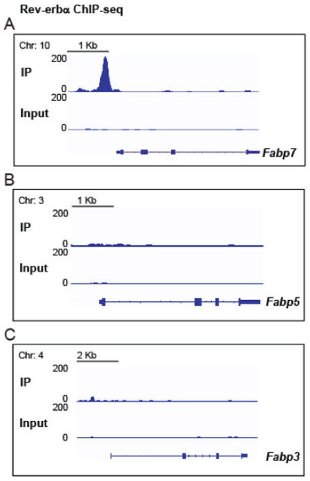
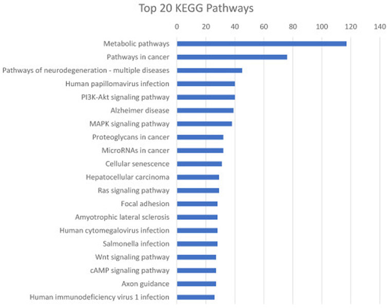
To determine whether these RORE sites were functional, we performed chromatin immunoprecipitation experiments followed by DNA-sequencing (ChIP-seq) on tissue from the ventral tegmental area (VTA), a brain region known to regulate motivational/reward behaviors (18, 19), wakefulness, and sleep (20-22). Here we identified positive Rev-erbα interactions within the first kilobase upstream of the transcription start site of the Fabp7 promoter, but not in the Fabp3 or Fabp5 promoters (Figure 1A-C). The top 20 Rev-erbα binding site loci, peak score, distance to the translational start site and gene names are listed in Table 1. Gene Ontology (GO) analysis revealed significant enrichment of several biological processes, molecular functions, and cellular components (Table 2) and Kyoto Encyclopedia of Genes and Genomes (KEGG) analysis shows the top 20 pathways in Rev-erbα ChIP-seq genes (Figure 2). The complete list of Rev-erbα ChIP-seq genes is provided in [SUPPLEMENTAL dataset 1].
Figure 1.
ChIP-seq binding profile of Rev-erbα around the Fabp7 locus (A), but not in the Fabp5 (B) or Fabp3 (C) loci in the VTA of WT mice.
Table 1.
The top 20 Rev-erbα binding site loci, peak score, distance to the translational start site (TSS), and gene names, as identified by Rev-erbα ChIP-seq.
| Chromosome | Peak Score |
Distance to TSS |
Gene Name |
|---|---|---|---|
| chr14 | 151.18628 | 506 | Nr1d2 |
| chr1 | 139.22659 | −2189 | Igsf8 |
| chr11 | 135.35667 | −27206 | Hlf |
| chr7 | 131.72737 | 2519 | Dbp |
| chr7 | 102.16678 | −206 | Dbp |
| chr2 | 102.00463 | 10184 | Cry2 |
| chr11 | 97.30782 | 1526 | Nr1d1 |
| chr6 | 95.75218 | 9516 | Bhlhe41 |
| chr3 | 94.62258 | −69 | Ciart |
| chr6 | 81.56895 | −1171 | Bhlhe40 |
| chr6 | 80.73732 | 83133 | Lsm3 |
| chr2 | 76.52149 | 401 | Aven |
| chr11 | 76.4099 | −4068 | Per1 |
| chr10 | 74.53577 | −153 | Fabp7 |
| chr15 | 71.89268 | −459 | Tef |
| chr9 | 67.80914 | −112 | Nptn |
| chr1 | 62.37303 | −197 | Coq10b |
| chr16 | 61.30798 | −793 | Ubald1 |
| chr7 | 60.32358 | 37 | Arntl |
| chr15 | 59.34713 | 45290 | Nfam1 |
Table 2.
Analysis of Gene Ontology in Rev-erbα ChIP-seq genes. Highest fold enriched Gene Ontology classes for Biological Process, Molecular Function and Cellular Component are listed with most highly enriched on top.
| PANTHER GO-Slim Biological Process | Number of Genes |
Fold Enrichment |
Raw P-value | FDR |
|---|---|---|---|---|
| circadian regulation of gene expression | 11 | 10.65 | 0.000000207 | 0.00000705 |
| neg. reg. of transforming growth factor beta receptor signaling pathway | 4 | 10.33 | 0.0021 | 0.0312 |
| regulation of circadian rhythm | 4 | 8.85 | 0.00314 | 0.0448 |
| chondroitin sulfate proteoglycan biosynthetic process | 5 | 6.46 | 0.00272 | 0.0393 |
| protein demethylation | 5 | 6.46 | 0.00272 | 0.0391 |
| protein autophosphorylation | 10 | 3.87 | 0.000719 | 0.0118 |
| regulation of actin filament organization | 15 | 2.8 | 0.000803 | 0.013 |
| response to abiotic stimulus | 16 | 2.75 | 0.000638 | 0.0106 |
| positive regulation of transcription by RNA polymerase II | 38 | 2.52 | 0.00000206 | 0.0000599 |
| regulation of cellular component size | 17 | 2.42 | 0.00196 | 0.0295 |
| positive reg. of nucleobase-containing compound metabolic process | 54 | 2.34 | 9.73E-08 | 0.00000342 |
| PANTHER GO-Slim Molecular Function | ||||
| demethylase activity | 9 | 5.17 | 0.000228 | 0.00452 |
| flavin adenine dinucleotide binding | 10 | 4.56 | 0.000242 | 0.00463 |
| transcription coregulator activity | 40 | 3.04 | 8.74E-09 | 0.000000606 |
| phosphoprotein phosphatase activity | 19 | 2.21 | 0.00295 | 0.0409 |
| small molecule binding | 44 | 1.76 | 0.000706 | 0.0112 |
| protein kinase activity | 51 | 1.62 | 0.00177 | 0.0266 |
| PANTHER GO-Slim Cellular Component | ||||
| vacuolar membrane | 13 | 2.76 | 0.00196 | 0.0344 |
| Golgi membrane | 14 | 2.55 | 0.00255 | 0.0418 |
| transcription regulator complex | 31 | 2.54 | 0.0000114 | 0.000386 |
| neuron projection | 42 | 1.72 | 0.00181 | 0.0328 |
| transferase complex | 50 | 1.69 | 0.000921 | 0.0187 |
| bounding membrane of organelle | 43 | 1.66 | 0.00248 | 0.0421 |
| nucleoplasm | 54 | 1.65 | 0.000836 | 0.0185 |
| chromatin | 67 | 1.56 | 0.000896 | 0.019 |
Figure 2.
Analysis of the top 20 KEGG pathways enriched in Rev-erb ChIP-seq genes plotted with number of hits per pathway.
To confirm that Rev-erbα binds the Fabp7 promoter in multiple brain regions, we compared Rev-erbα binding in the Fabp7 promoter against the negative control insulin, and the positive control NPAS2 in WT and Rev-erbα KO mice. We observed Rev-erbα binding to the Fabp7 and NPAS2 promoters in WT, but not Rev-erbα KO mice, in both hippocampus (Figure 3A) and hypothalamus (Figure 3B). Binding of Rev-erbα was not observed for insulin, regardless of genotype (Figure 3A, B). Since BMAL1 is known to transactivate Rev-erbα (23, 24), a transcriptional repressor, BMAL1 could influence Fabp7 gene expression (6) indirectly through Rev-erbα.
Figure 3.
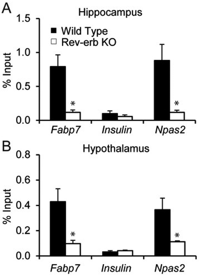
ChIP-qPCR measurements of Rev-erbα relative occupancy at Fabp7 locus, Insulin locus (negative control) and Npas2 locus (positive control) in hippocampus (A) and hypothalamus (B) of WT and Rev-erbα KO mice. Data are expressed as the percent of input and are the mean ± SEM. (Student’s t test, *p<0.05, n=3 per group).
To test the hypothesis that Rev-erbα represses Fabp7 gene expression, we examined the diurnal profile of Fabp7 mRNA in Rev-erbα KO mice. If Fabp7 expression is repressed by Rev-erbα, this predicts that Fabp7 mRNA should be elevated in the Rev-erbα KO. We confirmed that Fabp7 mRNA is elevated in hippocampus of Rev-erbα KO mice, while Fabp3 and Fabp5 mRNA levels are not affected (Figure 4A-C). To determine whether time-of-day mRNA levels are affected by Rev-erbα, we analyzed the normalized mRNA expression for Fabp3, Fabp5, and Fabp7 from six time-points over 24h of Rev-erbα KO and WT mice. While Fabp3 (Figure 4D) and Fabp5 (Figure 4E) mRNA do not oscillate in WT mice and remain unaffected in Rev-erbα KOs, the Fabp7 mRNA circadian oscillation is disrupted in the Rev-erbα KO compared to WT hippocampus (Figure 4F). Since Fabp7 expression is diurnally regulated throughout murine brain (3-5), we wanted to determine if Fabp7 mRNA levels were regulated by Rev-erbα broadly in multiple brain regions. Analysis of multiple brain regions including striatum, VTA, cerebellum, hippocampus, hypothalamus, and cortex of Rev-erbα KO compared to WT mice revealed analogous increases in Fabp7 mRNA levels (~6-15 fold), but not Fabp3 or Fabp5 mRNA levels (Figure 5). Together, these data suggest that the circadian clock control of Fabp7 mRNA expression requires Rev-erbα broadly across many brain regions.
Figure 4.
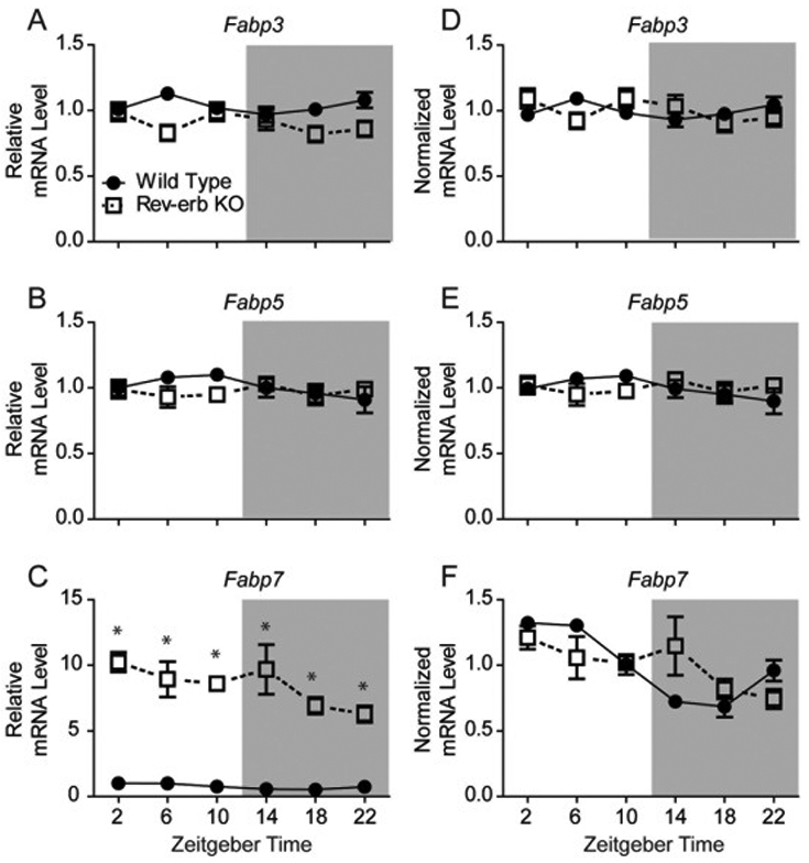
A-C: Relative hippocampal mRNA expression of various Fabps in Rev-erbα KO vs. WT mice under normal (LD) conditions. Levels of Fabp3 (A) or Fabp5 (B) are unaffected by Rev-erbα deficiency, however, Fabp7 shows a significant increase in expression based on genotype. *p<0.001, N=4-7 per group, Student’s t-test. ZT=zeitgeber time. Hippocampal mRNA expression normalized to genotype to visualize the circadian rhythmicity of Fabp3 (D), Fabp5 (E), and Fabp7 (F). Fabp7 circadian rhythmicity was significantly disrupted in Rev-erba KO mice (adj. p=0.184; JTK_Cycle) compared to WT mice (adj. p<0.001; JTK_Cycle).
Figure 5.
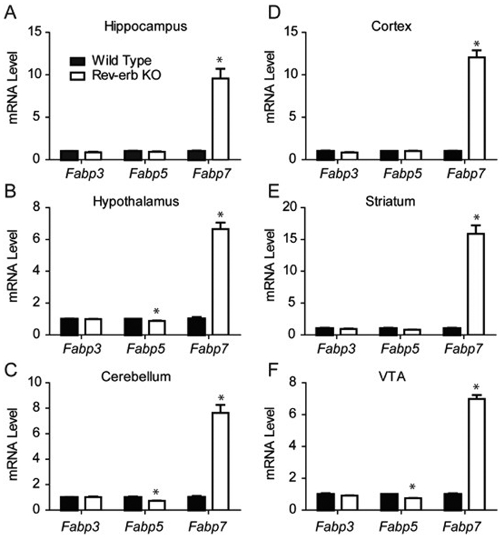
A-F: Relative mRNA expression from various brain regions of Fabps in Rev-erbα KO vs. WT mice. Levels of Fabp3 mRNA are not affected by loss of Rev-erbα, while lower levels of Fabp5 mRNA are observed in Hypothalamus (B), Cerebellum (C), and VTA (F) based on Rev-erbα deficiency compared to WT. Fabp7 mRNA, however, shows significant increases in expression in Rev-erbα KO compared to WT in all brain regions studied. *p<0.05, **p<0.01, ***p<0.001, N=3-5 per group, Student’s t-test.
DISCUSSION
The astrocyte Fabp7 gene expression is known to cycle in a synchronized fashion throughout the mammalian CNS (3-5, 14). Previous studies have shown Fabp7 circadian gene expression is under control of the core clock transcription factor BMAL1 (6), however the Fabp7 promoter lacks a canonical E-box element, suggesting that BMAL1 may indirectly exert its effects on Fabp7 circadian expression via Rev-erbα, a transcriptional repressor, and known BMAL1 target (25). Here we provide evidence that Fabp7 contains canonical ROREs and that Rev-erbα binds to the RORE regions in the Fabp7 gene locus in the VTA (Figure 1A). The current study validates a previous report that also showed Rev-erbα binding to Fabp7 in the hippocampus (14) (Figure 3A), and extends these findings to show this also occurs in the hypothalamus (Figure 3B). Taken together, these results suggest that the coordinated and synchronized expression of Fabp7 transcription is controlled by Rev-erbα direct binding in multiple brain regions throughout mammalian brain.
Rev-erbα KO mice showed a greater than 6-fold increase in Fabp7 mRNA expression across multiple brain areas, including cerebellum, cortex, hippocampus, hypothalamus, striatum, and VTA, compared to WT mice. We also observed minimal, but significant, reduction in Fabp5 mRNA in a few brain areas (cerebellum, hypothalamus, and VTA; Figure 5) and no differences in Fabp3 mRNA in any brain region, in Rev-erbα KO compared to WT mice. These reductions in Fabp5 mRNA may represent compensatory mechanisms that are in response to the large increases in Fabp7 mRNA expression in glial cells, however, to rule out a direct role of Rev-erbα in transcriptional regulation of these other Fabp types throughout brain, binding assays for Rev-erbα at their respective genetic loci across multiple brain regions would be required. Recently, local oscillators have been discovered in multiple brain regions throughout the mammalian brain (26), therefore it will be important to determine the extent to which Fabp7 oscillations require ‘global’ vs. ‘local’ coordinated control. Stability of Rev-erbα and the role of degradation processes that control the protein half-life in downstream signaling may also contribute to alterations in periodicity of gene expression (27). Future studies determining the cell-type specificity of these observations are also needed to better understand lipid-mediated signaling cascades (28, 29) downstream of circadian- and metabolically (16, 30-32) driven changes in Rev-erbα expression both within and between neurons and glia.
Understanding the molecular and cellular components that regulate Fabp7 expression will have important implications for public health. For example, pathological states associated with Fabp7 overexpression exist for a variety of diseases, including multiple types of cancer (33-38), and neurodegenerative disease, including Alzheimer’s disease (39, 40). Given the role of the circadian clock in cancer (41-43) and neurodegeneration (44-46), future studies determining the role in how circadian Fabp7 and Fabp7 lipid-signaling may feedback onto metabolic (15, 31, 47) and inflammatory pathways (48-50) may provide novel links between clock-regulated mechanisms, fatty-acid pathways, and disease.
MATERIALS AND METHODS
Animals.
The Rev-erbα knock out (KO) mice were obtained from B. Vennström and were backcrossed for >7 generations with C57/Bl6 mice. Mice (N=3-7 per group) were housed under standard 12h-light/12h-dark (LD) cycles and were sacrificed at specific times (zeitgeber time (ZT) 2, 6, 10, 14, 18, 22 with ZT0 corresponding to 7 a.m.). Animal care and use procedures followed the guidelines of the Institutional Animal Care and Use Committee of the University of Pennsylvania in accordance with the guidelines of the US National Institutes of Health.
Chromatin immunoprecipitation (ChIP).
ChIP experiments were performed as previously described (51) with minor changes. Mouse brain tissue was harvested at ZT10, minced and cross-linked in 1% formaldehyde for 20min, followed by quenching with 1/20 volume of 2.5M glycine solution for 5 minutes, and then two washes with PBS. Cell lysates with fragmented chromatin were prepared by probe sonication in ChIP dilution buffer (50mM HEPES, 155mM NaCl, 1.1% Triton X-100, 0.11% sodium deoxycholate, 0.1% SDS, 1mM phenylmethylsulfonyl fluoride [PMSF], and a complete protease inhibitor tablet [pH 7.5]). Proteins were immunoprecipitated in ChIP dilution buffer, using 1 μg of Rev-erbα antibody (Cell signaling). Cross-linking was reversed overnight at 65°C in elution buffer (50mM Tris-HCL, 10mM EDTA, 1% SDS, pH8), and DNA isolated using phenol/chloroform/isoamyl alcohol. Precipitated DNA was analyzed by quantitative PCR or high-throughput sequencing.
ChIP-qPCR.
Precipitated DNA was analyzed by quantitative PCR, using the following primers: Fabp7, forward: 5’-GGG GAT CAG GAT TGT GAT GT-3’; Fabp7, reverse: 5’-AGA TGG CTC CAA TCC TCC TT-3’; Arbp, forward: 5’- CTG GGA CGA TGA ATG AGG AT-3’; Arbp, reverse: 5’- AGC AGC TGG CAC CTA AAC AG-3’; Npas2, forward: 5’-TTG CAG AAG CTT GGG AAA AG-3’; Npas2, reverse: 5’-TTT CCT GTG GGA GGA GAC AG-3’.
ChIP-seq and cistromic analysis.
For ChIP-seq, material from three mice was pooled prior to library generation. ChIP DNA was prepared for sequencing according to the amplification protocol provided by Illumina, using adaptor oligo and primers from Illumina, enzymes from New England Biolabs and PCR Purification Kit and MinElute Kit from Qiagen. Deep sequencing was performed by the Functional Genomics Core (J. Schug and K. Kaestner) of the Penn Diabetes Endocrinology Research Center using the Illumina HiSeq2000, and sequences were obtained using the Solexa Analysis Pipeline. Sequenced reads were aligned to the mouse reference genome (mm9) and peak calling was performed with HOMER (52). ChIP-seq data are deposited in NCBI GEO GSE67973 (17), for GSM1659684 and GSM1659685 datasets.
MEME Package
Analysis of the Fabp7 promoter was done using the MEME package (http://meme.nbcr.net/meme/). 2000 base pairs upstream and 2000 base pairs downstream of the murine Fabp7 transcription start site (TSS) was used for promoter analysis. Reference to site location of cis-elements were expressed 0-4000, with 2000 being at the TSS.
GO and KEGG Analysis
Gene ontology analysis was performed on the ranked list of Rev-erbα ChIP-seq genes with peak score >2 [SUPPLEMENTAL dataset 1], using Panther GO-Slim against the mouse gene list (http://geneontology.org release 2021-01-01: 44,091; (53, 54). Top non-redundant categories are presented.
KEGG pathway analysis was performed on the same gene list using KEGG Mapper https://www.genome.jp/kegg/tool/map_pathway1.html (55) against mouse pathways.
qPCR
Total RNA was extracted from tissue using the RNeasy Mini Kit (QIAGEN) and treated with DNase (QIAGEN). The RNA was reversed transcribed using the High-Capacity cDNA Reverse Transcription kit (Applied Biosystems) and analyzed by quantitative PCR. Quantitative PCR was performed with Power SYBR Green PCR Mastermix on the PRISM 7500 (Applied Biosystems). Gene expression was normalized to mRNA levels of housekeeping gene 36B4 and the level of the gene of interest in the control samples. Circadian oscillations in gene expression were calculated using JTK_cyclev3.1 scripts (56) run on R. Amplitude confidence intervals were calculated according to Miyazaki et al., 2011 (57).
Primers:
36B4 Forward TCC-AGG-CTT-TGG-GCA-TCA-3′;
36B4 Reverse CTT-TAT-CAG-CTG-CAC-ATC-ACT-CAG-A
Fabp3 Forward CTG-ACT-CTC-ACT-CAT-GGC-AGT-GT
Fabp3 Reverse GCC-AGG-TCA-CGC-CTC-CTT
Fabp5 Forward CGA-CAG-CTG-ATG-GCA-GAA-AAA
Fabp5 Reverse GAC-CAG-GGC-ACC-GTC-TTG
Fabp7 Forward CTC-TGG-GCG-TGG-GCT-TT
Fabp7 Reverse TTC-CTG-ACT-GAT-AAT-CAC-AGT-TGG-TT
Supplementary Material
Acknowledgements:
We would like to thank Dr. A. Pack and the UPENN Center for Sleep and Circadian Neurobiology and Dr. M. Lazar and the UPENN Institute for Diabetes, Obesity, and Metabolism for advice and support.
Funding:
This work was supported by National Institute of Heath grant R35GM133440 to J.R.G.
REFERENCES
- 1.Furuhashi M, Hotamisligil GS, Fatty acid-binding proteins: role in metabolic diseases and potential as drug targets. Nat Rev Drug Discov 7, 489–503 (2008). [DOI] [PMC free article] [PubMed] [Google Scholar]
- 2.Storch J, Corsico B, The emerging functions and mechanisms of mammalian fatty acid-binding proteins. Annu Rev Nutr 28, 73–95 (2008). [DOI] [PubMed] [Google Scholar]
- 3.Gerstner JR, Vander Heyden WM, Lavaute TM, Landry CF, Profiles of novel diurnally regulated genes in mouse hypothalamus: expression analysis of the cysteine and histidine-rich domain-containing, zinc-binding protein 1, the fatty acid-binding protein 7 and the GTPase, ras-like family member 11b. Neuroscience 139, 1435–1448 (2006). [DOI] [PMC free article] [PubMed] [Google Scholar]
- 4.Gerstner JR et al. , Brain fatty acid binding protein (Fabp7) is diurnally regulated in astrocytes and hippocampal granule cell precursors in adult rodent brain. PLoS One 3, e1631 (2008). [DOI] [PMC free article] [PubMed] [Google Scholar]
- 5.Gerstner JR et al. , Time of day regulates subcellular trafficking, tripartite synaptic localization, and polyadenylation of the astrocytic Fabp7 mRNA. J Neurosci 32, 1383–1394 (2012). [DOI] [PMC free article] [PubMed] [Google Scholar]
- 6.Gerstner JR, Paschos GK, Circadian expression of Fabp7 mRNA is disrupted in Bmal1 KO mice. Mol Brain 13, 26 (2020). [DOI] [PMC free article] [PubMed] [Google Scholar]
- 7.Gerstner JR et al. , Normal sleep requires the astrocyte brain-type fatty acid binding protein FABP7. Sci Adv 3, e1602663 (2017). [DOI] [PMC free article] [PubMed] [Google Scholar]
- 8.Ebrahimi M et al. , Astrocyte-expressed FABP7 regulates dendritic morphology and excitatory synaptic function of cortical neurons. Glia 64, 48–62 (2016). [DOI] [PubMed] [Google Scholar]
- 9.Watanabe A et al. , Fabp7 maps to a quantitative trait locus for a schizophrenia endophenotype. PLoS Biol 5, e297 (2007). [DOI] [PMC free article] [PubMed] [Google Scholar]
- 10.Owada Y et al. , Altered emotional behavioral responses in mice lacking brain-type fatty acid-binding protein gene. Eur J Neurosci 24, 175–187 (2006). [DOI] [PubMed] [Google Scholar]
- 11.Lavialle M et al. , Structural plasticity of perisynaptic astrocyte processes involves ezrin and metabotropic glutamate receptors. Proc Natl Acad Sci U S A 108, 12915–12919 (2011). [DOI] [PMC free article] [PubMed] [Google Scholar]
- 12.Nagai J et al. , Behaviorally consequential astrocytic regulation of neural circuits. Neuron, (2020). [DOI] [PMC free article] [PubMed] [Google Scholar]
- 13.Gerstner JR, On the evolution of memory: a time for clocks. Front Mol Neurosci 5, 23 (2012). [DOI] [PMC free article] [PubMed] [Google Scholar]
- 14.Schnell A et al. , The nuclear receptor REV-ERBalpha regulates Fabp7 and modulates adult hippocampal neurogenesis. PLoS One 9, e99883 (2014). [DOI] [PMC free article] [PubMed] [Google Scholar]
- 15.Cho H et al. , Regulation of circadian behaviour and metabolism by REV-ERB-alpha and REV-ERB-beta. Nature 485, 123–127 (2012). [DOI] [PMC free article] [PubMed] [Google Scholar]
- 16.Bugge A et al. , Rev-erbalpha and Rev-erbbeta coordinately protect the circadian clock and normal metabolic function. Genes Dev 26, 657–667 (2012). [DOI] [PMC free article] [PubMed] [Google Scholar]
- 17.Zhang Y et al. , GENE REGULATION. Discrete functions of nuclear receptor Rev-erbα couple metabolism to the clock. Science 348, 1488–1492 (2015). [DOI] [PMC free article] [PubMed] [Google Scholar]
- 18.Morales M, Margolis EB, Ventral tegmental area: cellular heterogeneity, connectivity and behaviour. Nat Rev Neurosci 18, 73–85 (2017). [DOI] [PubMed] [Google Scholar]
- 19.Russo SJ, Nestler EJ, The brain reward circuitry in mood disorders. Nat Rev Neurosci 14, 609–625 (2013). [DOI] [PMC free article] [PubMed] [Google Scholar]
- 20.Takata Y et al. , Sleep and Wakefulness Are Controlled by Ventral Medial Midbrain/Pons GABAergic Neurons in Mice. J Neurosci 38, 10080–10092 (2018). [DOI] [PMC free article] [PubMed] [Google Scholar]
- 21.Yu X et al. , GABA and glutamate neurons in the VTA regulate sleep and wakefulness. Nat Neurosci 22, 106–119 (2019). [DOI] [PMC free article] [PubMed] [Google Scholar]
- 22.Eban-Rothschild A, Rothschild G, Giardino WJ, Jones JR, de Lecea L, VTA dopaminergic neurons regulate ethologically relevant sleep-wake behaviors. Nat Neurosci 19, 1356–1366 (2016). [DOI] [PMC free article] [PubMed] [Google Scholar]
- 23.Mohawk JA, Green CB, Takahashi JS, Central and peripheral circadian clocks in mammals. Annu Rev Neurosci 35, 445–462 (2012). [DOI] [PMC free article] [PubMed] [Google Scholar]
- 24.Albrecht U, Timing to perfection: the biology of central and peripheral circadian clocks. Neuron 74, 246–260 (2012). [DOI] [PubMed] [Google Scholar]
- 25.Guillaumond F, Dardente H, Giguère V, Cermakian N, Differential control of Bmal1 circadian transcription by REV-ERB and ROR nuclear receptors. J Biol Rhythms 20, 391–403 (2005). [DOI] [PubMed] [Google Scholar]
- 26.Paul JR et al. , Circadian regulation of membrane physiology in neural oscillators throughout the brain. Eur J Neurosci, (2019). [DOI] [PMC free article] [PubMed] [Google Scholar]
- 27.DeBruyne JP, Baggs JE, Sato TK, Hogenesch JB, Ubiquitin ligase Siah2 regulates RevErbα degradation and the mammalian circadian clock. Proc Natl Acad Sci U S A 112, 12420–12425 (2015). [DOI] [PMC free article] [PubMed] [Google Scholar]
- 28.Gooley JJ, Chua EC, Diurnal regulation of lipid metabolism and applications of circadian lipidomics. J Genet Genomics 41, 231–250 (2014). [DOI] [PubMed] [Google Scholar]
- 29.Gooley JJ, Circadian regulation of lipid metabolism. Proc Nutr Soc 75, 440–450 (2016). [DOI] [PubMed] [Google Scholar]
- 30.Kumar Jha P, Challet E, Kalsbeek A, Circadian rhythms in glucose and lipid metabolism in nocturnal and diurnal mammals. Mol Cell Endocrinol 418 Pt 1, 74–88 (2015). [DOI] [PubMed] [Google Scholar]
- 31.Bass J, Takahashi JS, Circadian integration of metabolism and energetics. Science 330, 1349–1354 (2010). [DOI] [PMC free article] [PubMed] [Google Scholar]
- 32.Eckel-Mahan K, Sassone-Corsi P, Metabolism and the circadian clock converge. Physiol Rev 93, 107–135 (2013). [DOI] [PMC free article] [PubMed] [Google Scholar]
- 33.Zhou J et al. , Overexpression of FABP7 promotes cell growth and predicts poor prognosis of clear cell renal cell carcinoma. Urol Oncol 33, 113.e119–117 (2015). [DOI] [PubMed] [Google Scholar]
- 34.Liu RZ et al. , A fatty acid-binding protein 7/RXRβ pathway enhances survival and proliferation in triple-negative breast cancer. J Pathol 228, 310–321 (2012). [DOI] [PubMed] [Google Scholar]
- 35.Mita R, Beaulieu MJ, Field C, Godbout R, Brain fatty acid-binding protein and omega-3/omega-6 fatty acids: mechanistic insight into malignant glioma cell migration. J Biol Chem 285, 37005–37015 (2010). [DOI] [PMC free article] [PubMed] [Google Scholar]
- 36.Cordero A et al. , FABP7 is a key metabolic regulator in HER2+ breast cancer brain metastasis. Oncogene 38, 6445–6460 (2019). [DOI] [PMC free article] [PubMed] [Google Scholar]
- 37.Kagawa Y et al. , Role of FABP7 in tumor cell signaling. Adv Biol Regul 71, 206–218 (2019). [DOI] [PubMed] [Google Scholar]
- 38.Ma R et al. , FABP7 promotes cell proliferation and survival in colon cancer through MEK/ERK signaling pathway. Biomed Pharmacother 108, 119–129 (2018). [DOI] [PubMed] [Google Scholar]
- 39.Teunissen CE et al. , Brain-specific fatty acid-binding protein is elevated in serum of patients with dementia-related diseases. Eur J Neurol 18, 865–871 (2011). [DOI] [PubMed] [Google Scholar]
- 40.Johnson ECB et al. , Deep proteomic network analysis of Alzheimer's disease brain reveals alterations in RNA binding proteins and RNA splicing associated with disease. Mol Neurodegener 13, 52 (2018). [DOI] [PMC free article] [PubMed] [Google Scholar]
- 41.Masri S, Sassone-Corsi P, The emerging link between cancer, metabolism, and circadian rhythms. Nat Med 24, 1795–1803 (2018). [DOI] [PMC free article] [PubMed] [Google Scholar]
- 42.Sulli G, Lam MTY, Panda S, Interplay between Circadian Clock and Cancer: New Frontiers for Cancer Treatment. Trends Cancer 5, 475–494 (2019). [DOI] [PMC free article] [PubMed] [Google Scholar]
- 43.Sulli G, Manoogian ENC, Taub PR, Panda S, Training the Circadian Clock, Clocking the Drugs, and Drugging the Clock to Prevent, Manage, and Treat Chronic Diseases. Trends Pharmacol Sci 39, 812–827 (2018). [DOI] [PMC free article] [PubMed] [Google Scholar]
- 44.Musiek ES, Holtzman DM, Mechanisms linking circadian clocks, sleep, and neurodegeneration. Science 354, 1004–1008 (2016). [DOI] [PMC free article] [PubMed] [Google Scholar]
- 45.Hood S, Amir S, Neurodegeneration and the Circadian Clock. Front Aging Neurosci 9, 170 (2017). [DOI] [PMC free article] [PubMed] [Google Scholar]
- 46.Lananna BV et al. , Chi3l1/YKL-40 is controlled by the astrocyte circadian clock and regulates neuroinflammation and Alzheimer's disease pathogenesis. Sci Transl Med 12, (2020). [DOI] [PMC free article] [PubMed] [Google Scholar]
- 47.Panda S, Circadian physiology of metabolism. Science 354, 1008–1015 (2016). [DOI] [PMC free article] [PubMed] [Google Scholar]
- 48.Carter SJ et al. , A matter of time: study of circadian clocks and their role in inflammation. J Leukoc Biol 99, 549–560 (2016). [DOI] [PubMed] [Google Scholar]
- 49.Scheiermann C, Kunisaki Y, Frenette PS, Circadian control of the immune system. Nat Rev Immunol 13, 190–198 (2013). [DOI] [PMC free article] [PubMed] [Google Scholar]
- 50.Castanon-Cervantes O et al. , Dysregulation of inflammatory responses by chronic circadian disruption. J Immunol 185, 5796–5805 (2010). [DOI] [PMC free article] [PubMed] [Google Scholar]
- 51.Feng D et al. , A circadian rhythm orchestrated by histone deacetylase 3 controls hepatic lipid metabolism. Science 331, 1315–1319 (2011). [DOI] [PMC free article] [PubMed] [Google Scholar]
- 52.Heinz S et al. , Simple combinations of lineage-determining transcription factors prime cis-regulatory elements required for macrophage and B cell identities. Mol Cell 38, 576–589 (2010). [DOI] [PMC free article] [PubMed] [Google Scholar]
- 53.Ashburner M et al. , Gene ontology: tool for the unification of biology. The Gene Ontology Consortium. Nat Genet; 25, 25–29 (2000). [DOI] [PMC free article] [PubMed] [Google Scholar]
- 54.The Gene Ontology resource: enriching a GOld mine. Nucleic Acids Res 49, D325–d334 (2021). [DOI] [PMC free article] [PubMed] [Google Scholar]
- 55.Kanehisa M, Sato Y, KEGG Mapper for inferring cellular functions from protein sequences. Protein Sci 29, 28–35 (2020). [DOI] [PMC free article] [PubMed] [Google Scholar]
- 56.Hughes ME, Hogenesch JB, Kornacker K, JTK_CYCLE: an efficient nonparametric algorithm for detecting rhythmic components in genome-scale data sets. J Biol Rhythms 25, 372–380 (2010). [DOI] [PMC free article] [PubMed] [Google Scholar]
- 57.Miyazaki M et al. , Age-associated disruption of molecular clock expression in skeletal muscle of the spontaneously hypertensive rat. PLoS One 6, e27168 (2011). [DOI] [PMC free article] [PubMed] [Google Scholar]
Associated Data
This section collects any data citations, data availability statements, or supplementary materials included in this article.


