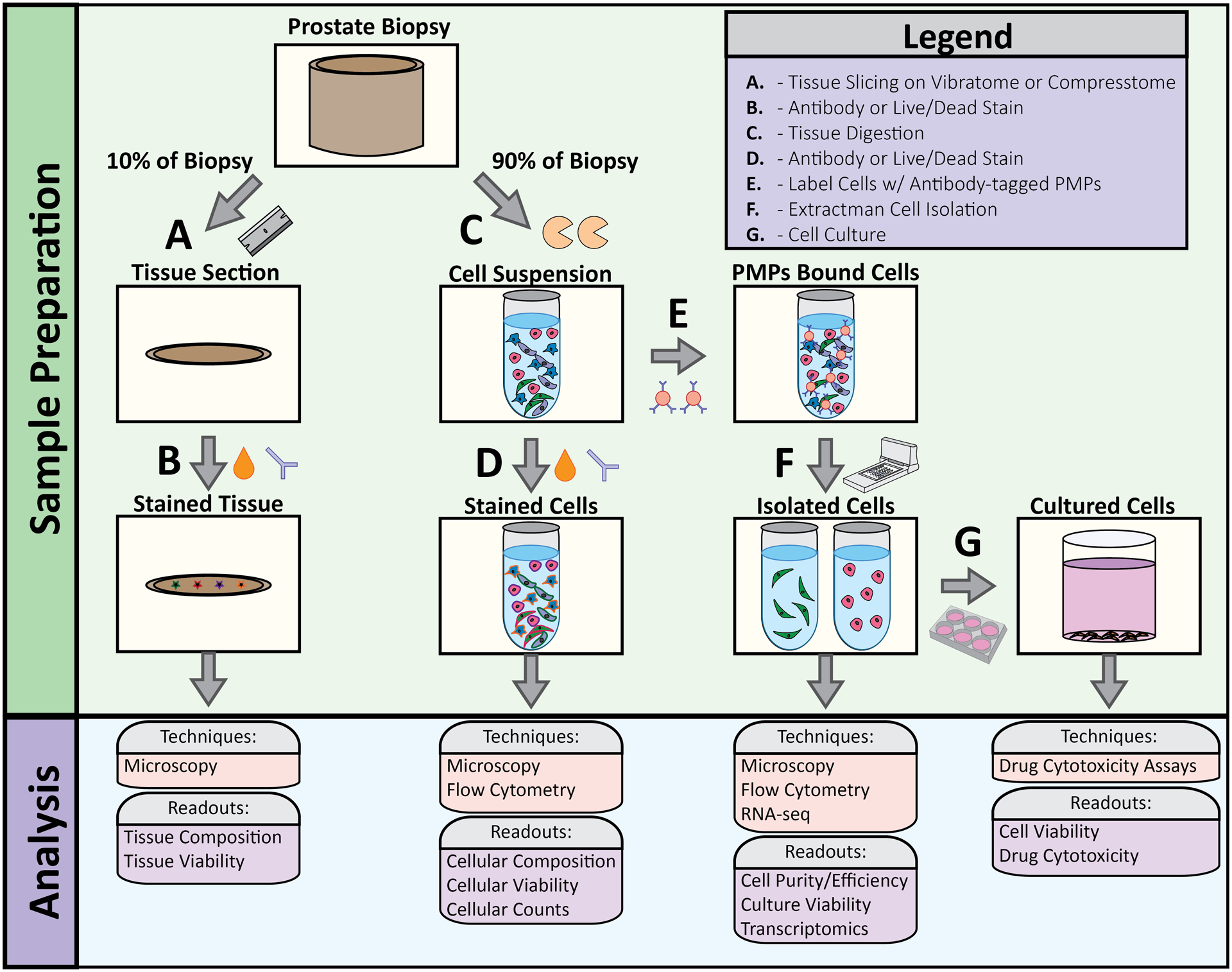
Figure 2: Overview of tissue processing workflow.

(A) 10% of the tissue biopsy was removed with a hand razor then sliced to 75um sections using a VF-300 Compresstome. (B) 75um tissue sections were stained with viability markers and cell-specific antibodies to evaluate tissue viability and cellular composition. (C) 90% of the tissue biopsy was used for enzymatic digestion to obtain single cell population. (D) Cell suspensions were stained with viability markers and cell-specific antibodies to evaluate cell viability and cell type. (E) Antibody-bound PMPs were added to cell suspension to bind specific cell types. (F) Extractman was used to isolate PMP-bound cell cells for flow analysis, RNA-seq, (G) cell culture, and cytotoxicity assays.
