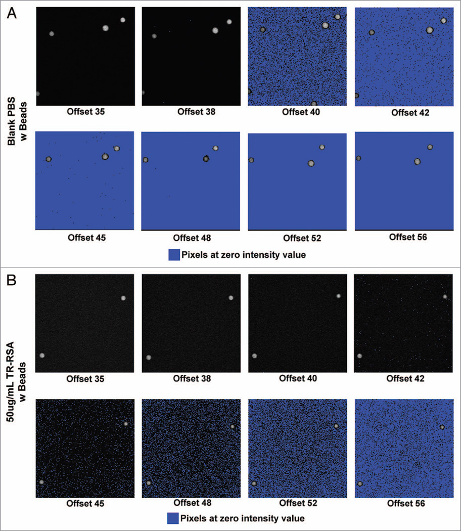
Figure 1.
Effect of increasing offset on pixel intensity values in solutions of Texas Red rat serum albumin (TR-RSA). In vitro images acquired using 2-photon microscopy display an increasingly greater number of pixel values at zero as the offset value is increased. Series (A) shows a 200×200 pixel region of fluorescent beads in a blank PBS solution with the offset values labeled from 35 through 56. Increasing numbers of pixels with values at zero (as seen by the blue warning marker) are apparent as the offset increases. Identical images in series (B) show the highest concentration of TR-RSA used (50 µg/mL). Here, a more gradual increase in the number of pixels with values at zero was seen and became prevalent in offsets of 45 and greater. For other concentrations the prevalence of pixels with values at zero were seen in numerically lower offsets as the concentration of TR-RSA decreased (image ~83 µm across).

