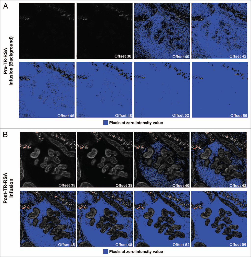
Figure 5.
Effect of increasing offset on pixel intensity values within the glomerulus in Munich Wistar rats. Two-photon intravital images display an increasingly greater number of pixels with values at zero as the offset value was increased. Series (A) shows a 250×250 pixel region of background images with a glomerulus centered and offsets labeled from 35 through 56. Concomitant with an increase in offset, more pixels are pushed to values of zero as seen with the PBS blanks in Figure 1. Images taken after infusion of Texas Red rat serum albumin (TR-RSA) show distribution throughout the peritubular vasculature and glomerular capillary loops in the center as shown in series (B). The Bowman’s Space containing the glomerular filtrate became increasingly distinguishable as the intensity values of the pixels within are pushed to zero. For this macromolecule of ~66 kDa a noticeable amount of pixels with intensity values at zero within the Bowman’s Space first occurred at offset 40. As the offset increased, additional structures such as part of the capillary loops and S1 proximal tubule segment at the upper left showed diminished fluorescent intensity (image = 104 µm across).

