To the Editor
Asthma exacerbations secondary to viral illnesses are an important cause of morbidity among children with asthma, and allergy is a risk factor for virus-induced exacerbations. Although less frequent, some exacerbations of asthma occur independently of viral illnesses. Patterns and etiology of asthma exacerbations have been investigated in cross-sectional studies1, but longitudinal data are lacking. We sought to determine if there are separate groups of children who have asthma exacerbations with viruses and children who have asthma exacerbations without viruses. In addition, we tested the hypothesis that there are distinct risk factors for virus-induced exacerbations and exacerbations not associated with viral infection.
The Childhood Origins of ASThma (COAST) study followed 259 children prospectively from birth to age 6 years, and 217 were followed until age 11 years. Of those, 102 children met criteria for asthma at 6, 8, and/or 11 years (Table I). This subset of COAST children was predominantly male (63%). Forty-nine percent had a family history of maternal asthma, and 34% had a history of paternal asthma. Forty-seven percent had aeroallergen sensitization at the age of 6, and 52% at the age of 11 years. The majority (94%) lived in a home without smoke exposure. At year 6, 15% of children were taking a daily controller either seasonally or perennially, compared to 35% of children by year 12.
Table I.
Factors associated with asthma exacerbations between ages 6 and 11 years in the COAST 206 cohort. N=102.
| All Exacerbations
|
Virus+ Exacerbations
|
Virus− Exacerbations
|
||||||
|---|---|---|---|---|---|---|---|---|
| n | mean ± SD | P-value | mean ± SD | P-value | mean ± SD | P-value | ||
| Overall Number of Exacerbations | 2.1 ± 3.3 | 1.3 ± 2.4 | 0.6 ± 1.2 | |||||
| Gender | Female | 39 | 2.3 ± 3.9 | 0.61 | 1.4 ± 2.9 | 0.64 | 0.6 ± 1.4 | 0.91 |
| Male | 63 | 2.0 ± 2.9 | 1.2 ± 2.0 | 0.6 ± 1.0 | ||||
| Year 6 Aero Sensitization | No | 35 | 1.7 ± 2.3 | 0.10 | 1.1 ± 1.8 | 0.27 | 0.4 ± 0.8 | 0.14 |
| Yes | 47 | 2.9 ± 4.1 | 1.8 ± 3.1 | 0.8 ± 1.4 | ||||
| Year 11 Aero Sensitization | No | 23 | 1.3 ± 1.2 | 0.008 | 0.8 ± 1.1 | 0.03 | 0.4 ± 0.6 | 0.11 |
| Yes | 52 | 3.2 ± 4.1 | 1.9 ± 3.0 | 0.9 ± 1.5 | ||||
| Maternal Asthma | No | 53 | 1.7 ± 2.4 | 0.17 | 1.0 ± 1.8 | 0.21 | 0.5 ± 0.9 | 0.37 |
| Yes | 49 | 2.7 ± 4.0 | 1.6 ± 2.9 | 0.7 ± 1.4 | ||||
| Paternal Asthma | No | 66 | 2.0 ± 2.9 | 0.41 | 1.2 ± 2.1 | 0.65 | 0.5 ± 1.0 | 0.17 |
| Yes | 34 | 2.5 ± 4.0 | 1.5 ± 3.0 | 0.8 ± 1.5 | ||||
| Smoke in home (year 6) | No | 94 | 2.2 ± 3.4 | 0.49 | 1.4 ± 2.5 | 0.16 | 0.6 ± 1.2 | 0.62 |
| Yes | 6 | 1.3 ± 1.5 | 0.3 ± 0.5 | 0.3 ± 0.5 | ||||
| Dog in home (year 6) | No | 64 | 2.1 ± 3.2 | 0.79 | 1.3 ± 2.2 | 0.80 | 0.6 ± 1.3 | 0.69 |
| Yes | 36 | 2.3 ± 3.5 | 1.4 ± 2.9 | 0.7 ± 1.0 | ||||
| Cat in home (year 6) | No | 75 | 2.5 ± 3.6 | 0.03 | 1.5 ± 2.7 | 0.11 | 0.7 ± 1.3 | 0.10 |
| Yes | 25 | 1.2 ± 1.9 | 0.8 ± 1.4 | 0.3 ± 0.7 | ||||
An asthma exacerbation was defined as an illness in which a child required a step-up plan of care and/or the use of oral corticosteroids. A severe asthma exacerbation was defined by the use of oral corticosteroids.
Nasal samples were collected during asthma exacerbations and analyzed for respiratory viruses. Respiratory viruses were identified using multiplex PCR.2 Allergen-specific IgE was measured by Immunocap (Phadia, Portage, MI) performed at ages 6 and 11 years, and positive results were defined as ≥ 0.35 kU/L.3
Of the 102 COAST children with asthma, 60% had at least one exacerbation between ages 6 and 11, and 38% had more than one exacerbation. When grouped by exacerbation etiology, 40 children (40%) had no exacerbations, 23 (22%) had viral only exacerbations, 13 (13%) had non-viral only exacerbations, and 19 (19%) had a mixture of both viral and non-viral exacerbations. There were 7 children (6%) who each had one exacerbation but no nasal sample. Children who had viral only or non-viral only exacerbations had fewer exacerbations on average (mean 2.4, 95% CI [1.3, 4.4] and mean 1.5, 95% CI [0.6, 3.4]) than children who experienced both viral and non-viral exacerbations (mean 6.9, 95% CI [4.7, 10.1]). Children with at least 2 exacerbations had the highest prevalence of allergic sensitization and total IgE levels at age 11 years (See Table E1 in the Online Repository).
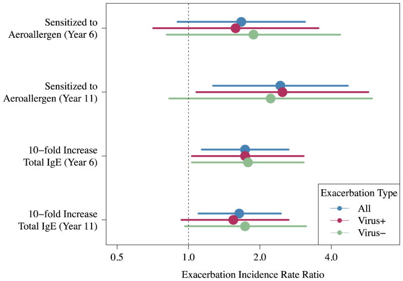
There were a total of 217 exacerbations, of which 192 had nasal samples obtained for viral analysis. Viruses were identified in 69% of exacerbations, and rhinovirus was most commonly identified (34% of exacerbations) (see Figure E1 in the Online Repository). The total number of exacerbations was positively associated with total IgE at ages 6 (p=0.009; incidence rate ratio (IRR) with respect to a one-unit increase in log10 IgE = 1.74, 95% CI [1.15, 2.64]) and 11 (p=0.02; IRR = 1.64, 95% CI [1.10, 2.46]), and with aeroallergen sensitization at age 11 (p=0.008; IRR = 2.44, 95% CI [1.27, 4.70]) (Figure 1). Having a cat in the home at year 6 was associated with a reduced number of exacerbations (IRR = 0.47, 95% CI [0.24, 0.91], p=0.03).
Figure 1.
Aeroallergen sensitization and higher total IgE associated with greater numbers of exacerbations. Aeroallergen sensitization and total IgE also associated with both the number of viral exacerbations and non-viral exacerbations.
Risk factors for viral and non-viral exacerbations were similar. For example, viral exacerbations were positively associated with total IgE levels at age 6 (p=0.04; IRR = 1.76, 95% CI [1.02, 3.02]) and aeroallergen sensitization at age 11 (p=0.03; IRR = 2.49, 95% CI [1.08, 5.73]), and non-viral exacerbations were positively associated with total IgE at age 6 (p=0.04; IRR = 1.77, 95% CI [1.02, 3.08]) and aeroallergen sensitization at age 11 (trend, p=0.11; IRR = 2.22, 95% CI [0.83, 5.94]) (Figure 1).
Finally, we compared the severity of viral vs. non-viral exacerbations. Twenty-three percent (14/60) of non-viral exacerbations, and 17% (23/132) of viral exacerbations were severe (p = 0.34). Of the 37 severe exacerbations, 14 (38%) were non-viral, 14 (38%) were associated with rhinovirus infection, and 9 (24%) were associated with other viruses. The greatest number of severe exacerbations, regardless of etiology, occurred during fall (October, November) and spring (March) (see Figures E2 and E3 in the Online Repository).
This study provides new data about patterns of asthma exacerbations over a six-year span in a cohort of suburban children. Notably, children who experienced multiple episodes had at least one viral exacerbation. Risk factors for having viral or non-viral associated asthma exacerbations were similar, and consisted of indicators of atopy (i.e. total IgE and aeroallergen sensitivity).
Previous studies have established that atopy is a risk factor for virus-induced wheezing and exacerbations of asthma. For example, wheezing illnesses in children over the age of two years are most common in children who have rhinovirus infections together with allergic sensitization, nasal eosinophilia, or elevated nasal eosinophil cationic protein.4 Additional studies have identified increased expression of the FcεR1 receptor on monocytes and dendritic cells during acute asthma exacerbations.5, One potential mechanism by which IgE could affect viral exacerbations relates to effects of the FcεR1 on interferon responses. In vitro experiments demonstrated that increased FcεR1 expression on plasmacytoid dendritic cells and IgE cross-linking strongly inhibited rhinovirus-induced interferon production in cells from children with allergic asthma.6
Strengths of the current study include the longitudinal analysis of exacerbations over a six-year period. Much of the current literature evaluates the etiology of asthma exacerbations from a cross-sectional perspective and not prospectively over multiple years. The study is limited by the relatively small numbers of children in each group, particularly the viral only and non-viral only groups, and the fact that most of the children with asthma in this cohort study have relatively mild disease.
In conclusion, our findings demonstrate that atopy is an important risk factor for both viral and non-viral exacerbations. These data are consistent with a clinical study of omalizumab, in which blocking IgE-mediated inflammation prevented both viral and non-viral exacerbations in children with moderate to severe asthma.7 One implication of these findings is that treatment of aimed at reducing IgE or perhaps other effectors of allergic inflammation could represent a common strategy to reduce morbidity due to viral and non-viral exacerbations of asthma.
Supplementary Material
Acknowledgments
Acknowledgment of Funding:
National Institutes of Health
Grant Support Numbers: P01 HL70831 and UL1TR000427
Abbreviations
- COAST
Childhood Origins of ASThma
- SABA
short-acting beta agonist
- LABA
long-acting beta agonist
- ICS
inhaled corticosteroid
- PCR
polymerase chain reaction
Footnotes
Publisher's Disclaimer: This is a PDF file of an unedited manuscript that has been accepted for publication. As a service to our customers we are providing this early version of the manuscript. The manuscript will undergo copyediting, typesetting, and review of the resulting proof before it is published in its final citable form. Please note that during the production process errors may be discovered which could affect the content, and all legal disclaimers that apply to the journal pertain.
References
- 1.Johnston SL, Pattemore PK, Sanderson G, Smith S, Lampe F, Josephs L, et al. Community study of role of viral infections in exacerbations of asthma in 9–11 year old children. BMJ. 1995;310:1225–9. doi: 10.1136/bmj.310.6989.1225. [DOI] [PMC free article] [PubMed] [Google Scholar]
- 2.Lee WM, Grindle K, Pappas T, Marshall DJ, Moser MJ, Beaty EL, et al. High-throughput, sensitive, and accurate multiplex PCR-microsphere flow cytometry system for large-scale comprehensive detection of respiratory viruses. J Clin Microbiol. 2007;45:2626–34. doi: 10.1128/JCM.02501-06. [DOI] [PMC free article] [PubMed] [Google Scholar]
- 3.Jackson DJ, Gangnon RE, Evans MD, Roberg KA, Anderson EL, Pappas TE, et al. Wheezing rhinovirus illnesses in early life predict asthma development in high-risk children. Am J Respir Crit Care Med. 2008;178:667–72. doi: 10.1164/rccm.200802-309OC. [DOI] [PMC free article] [PubMed] [Google Scholar]
- 4.Rakes GP, Arruda E, Ingram JM, Hoover GE, Zambrano JC, Hayden FG, et al. Rhinovirus and respiratory syncytial virus in wheezing children requiring emergency care. IgE and eosinophil analyses. Am J Respir Crit Care Med. 1999;159:785–90. doi: 10.1164/ajrccm.159.3.9801052. [DOI] [PubMed] [Google Scholar]
- 5.Holt PG, Strickland DH, Sly PD. Virus infection and allergy in the development of asthma: what is the connection? Curr Opin Allergy Clin Immunol. 2012;12:151–7. doi: 10.1097/ACI.0b013e3283520166. [DOI] [PubMed] [Google Scholar]
- 6.Durrani SR, Montville DJ, Pratt AS, Sahu S, DeVries MK, Rajamanickam V, et al. Innate immune responses to rhinovirus are reduced by the high-affinity IgE receptor in allergic asthmatic children. J Allergy Clin Immunol. 2012;130:489–95. doi: 10.1016/j.jaci.2012.05.023. [DOI] [PMC free article] [PubMed] [Google Scholar]
- 7.Busse WW, Morgan WJ, Gergen PJ, Mitchell HE, Gern JE, Liu AH, et al. Randomized trial of omalizumab (anti-IgE) for asthma in inner-city children. N Engl J Med. 2011;364:1005–15. doi: 10.1056/NEJMoa1009705. [DOI] [PMC free article] [PubMed] [Google Scholar]
Associated Data
This section collects any data citations, data availability statements, or supplementary materials included in this article.


