Abstract
Objective
Paraneoplastic neurologic disorders (PND) are autoimmune diseases associated with cancer and ectopic expression of a neuronal antigen in a peripheral tumor. Patients with PND harbor high titer antibodies and T cells in their serum and cerebrospinal fluid (CSF) that are specific to the tumor antigen, and treatment with the immunosuppressant FK506 (tacrolimus) decreases CSF white blood cell counts. The objective of this study was to determine the effect of FK506 on CSF chemokine levels in PND patients.
Methods
CSF samples pre- and post-FK506 treatment were tested by multiplex assay for the presence of 27 cytokines. Follow-up in vitro experiments aimed to determine whether T cells secrete CXCL10 in response to cognate antigen.
Results
Here we report that PND patients harbor high levels of the chemokine CXCL10 in their CSF. CXCL10 is a cytokine that recruits CXCR3+ cells such as activated T cells, and we found that FK506 treatment specifically decreased CSF CXCL10 from among 27 cytokines tested. In vitro, CXCL10 was only produced during antigen-specific cognate interactions between T cells and antigen presenting cells (APCs) when IFNγ receptors were present on the T cell.
Interpretation
These results support a model in which antigen-specific T cell stimulation by PND antigen-presenting APCs triggers IFNγ, followed by CXCL10 production and further lymphocyte recruitment, suggesting that treatments targeting T cells or CXCL10 in the CNS may interrupt a destructive positive feedback loop present in CNS inflammation.
INTRODUCTION
Paraneoplastic neurologic degenerations (PND) are characterized by an immune response against a neuronal antigen that is ectopically expressed by tumors outside the brain. This autoimmune response not only targets the tumor, but also the neurons that normally express the antigen. High titer antibodies to the neuronal protein are found in both the serum and cerebrospinal fluid (CSF) of patients with PND.1 However, antibodies are unlikely to be the sole cause of the neuronal destruction for those PNDs in which the target antigens are intracellular.2,3 Moreover, PND antigen-specific CD8+ T cells are present in the peripheral blood and CSF of patients with both paraneoplastic cerebellar degeneration (PCD) and the paraneoplastic Hu syndrome subtypes of PND.4–6
Our laboratory has used FK506 (tacrolimus) in the experimental treatment of patients with PND with the goal of decreasing the number of PND antigen-specific T cells in the brain and attempting to arrest disease progression. FK506 reduces the number of activated T cells in the CSF of patients with PCD.4 Not all PND patients demonstrate dramatic clinical improvements with this treatment,7 perhaps in part because most patients are diagnosed and referred after the onset of the neuronal destruction that is characteristic of PND.1 FK506, a lipid-soluble immunosuppressant used to prevent transplant rejection8, partitions well into the CNS. FK506 interferes with calcineurin phosphatase activity via FK-binding protein 12, leading to decreased activation of inflammatory genes regulated by the transcription factor NFAT,8–10 and thereby inhibits T cell function.9
Here we examined cytokine levels in the CSF of PND patients before and after treatment with FK506 after noting recurrent episodes of CSF pleiocytosis following FK506 treatments in one PCD patient, suggesting that recruitment of peripheral PCD antigen-specific T cells to the CSF resumed after treatment. Such CXCL10-dependent recruitment of T cells into the CNS has been documented previously in mice infected with West Nile Virus. In this model, neurons of infected mice generate CXCL10, which recruits CD8+ T cells into the CNS.11 Cytokines and their receptors have also been proposed to help mediate CSF entry of peripheral T cells in multiple sclerosis (MS).12 In particular, the chemokine CXCL10 is elevated in CSF during exacerbations of MS, and both CXCL10 and its receptor, CXCR3, are elevated in and co-localize with active MS lesions.13
When we examined the peripheral blood and CSF of 8 patients for the presence of 27 cytokines, CXCL10 was consistently elevated in the CSF of all patients with PND. Remarkably, CXCL10 was the only cytokine that decreased after FK506 treatment in patients, and correlated with decreases in CSF white blood cell (WBC) counts. We investigated the potential contribution of T cells in the elevation of CXCL10 levels in the CSF, since both were affected by drug treatment. We observed that T cells and APCs produce CXCL10 in an antigen-specific fashion and that production of this chemokine by T cells required expression of the IFNγ receptor on the T cell. These observations support a model in which a positive feedback develops when antigen-specific T cells encounter APCs presenting target antigen in the brain, stimulating CXCL10 production and further T cell recruitment and PND disease progression. Hence, targeting autoimmune T cells and CXCL10 together may help break this cycle, blocking further T cell homing to the CNS, suggesting a new treatment strategy for PND and allied T cell-mediated CNS disorders.
METHODS
Patient samples
Patients positive for the presence of Hu or Yo antibodies were enrolled in this Rockefeller University IRB-approved study at The Rockefeller University Hospital (RDA-572). Where noted, patients were given FK506 0.15–0.3 mg/kg/day in two divided doses to maintain plasma levels at 5–20 ng/ml and prednisone (60 mg/day, tapered to 2.5mg/day) for 10–28 days. Whole blood and CSF samples were obtained before and after treatment. PBMC and plasma were isolated from peripheral blood by Ficoll. CSF WBC counts were determined at the Memorial Sloan-Kettering Cancer Center Hematology laboratory. CSF was placed on ice immediately after lumbar puncture and centrifuged immediately at 4C to remove cells from the supernatant. Cells were re-suspended in culture medium or FACS buffer. Samples used for cytokine analysis were taken from the third or fourth collection tube. CSF supernatant and plasma were stored in aliquots at −80C until analysis.
Mouse experiments
Female C57BL/6 and IFNγR−/− mice were obtained from Jackson Labs (Bar Harbor, ME) and were housed in the Rockefeller University specific pathogen free animal facility. CXCL10−/− mice were obtained from the laboratory of Dr. A. Luster at Massachusetts General Hospital. All animal procedures were performed according to IACUC-approved animal protocols. Mice were immunized intraperitoneally with influenza A/PR/8 (Charles River Laboratory, Wilmington, MA) two weeks before spleens were harvested for isolation of CD4+ and CD8+ T cells by MACS separation (Miltenyi Biotec, Auburn, CA). Dendritic cells (DC) were generated from bone marrow cells grown in RPMI/10%FBS with GM-CSF from J558L cell culture supernatant.14 Elispot plates (Millipore, Billerica, MA) were coated with and detection performed with anti- IFNγ antibody matched pair (MabTech, Mariemont, OH). Spots were counted in a blinded fashion by Zellnet Consulting (Fort Lee, NJ).
Flow cytometry
All antibodies were purchased from BD Biosciences/Pharmingen. Cells were resuspended in FACS buffer (1%FBS/1% pooled human serum in PBS) and stained with fluorochrome-conjugated antibodies for 20 minutes at 4C, washed three times and analyzed on a Becton Dickinson FACSCaliber. Data were analyzed using FlowJo software (TreeStar, Ashland, OR).
Bead-based immunoassay of cytokines
Samples were analyzed using the Bioplex system (BioRad, Hercules, CA) 27-plex human cytokine kit according to the manufacturer’s instructions. Plasma was diluted 1:5 in the provided buffers and CSF was assayed without dilution. All patient samples were analyzed in duplicate. Tissue culture supernatants were assayed undiluted and at 1:10. Human cytokines tested were: IL-2, IL-4, IL-6, IL-8, IL-10, GM-CSF, IFNγ, TNFα, IL-1b, IL-5, IL-7, IL-12(p70), IL-13, IL-17, G-CSF, MCP-1, MIP-1β, IL-1ra, IL-9, IL-15, eotaxin, FGF-basic, IP-10 (CXCL10), MIP-1α, PDGF-bb, RANTES, VEGF. CSF CXCL10 levels were initially quantified by bead array, and confirmed by human CXCL10 ELISA.
ELISA
CSF samples and tissue culture supernatants were tested for the presence of mouse IFNγ and IL-17 and human and mouse CXCL10 using ELISA kits according to the manufacturer’s instructions (R&D Systems, Minneapolis, MN).
Statistics
Cytokine levels pre and post-FK506 treatment and the percentage of CXCR3+ cells pre- and post- FK506 were compared using the Wilcoxon matched-pairs signed rank test, with P <0.05 considered significant. Percentages of CXCR3+ cells in blood and CSF were compared using an unpaired, two-tailed t-test assuming unequal variances, with P<0.05 considered significant.
Declaration of patient and animal study approvals
The patient studies were approved by the Rockefeller University Institutional Review Board and were conducted at The Rockefeller University Hospital. Written, informed consent was obtained from all patients before enrolling in the study (RDA-572). All experiments involving animals were conducted according to a Rockefeller University IACUC-approved animal protocol and in accordance with the United States Public Health Service’s Policy on the Humane Care and Use of Laboratory Animals.
RESULTS
CXCL10 is elevated in the CSF of patients with PND
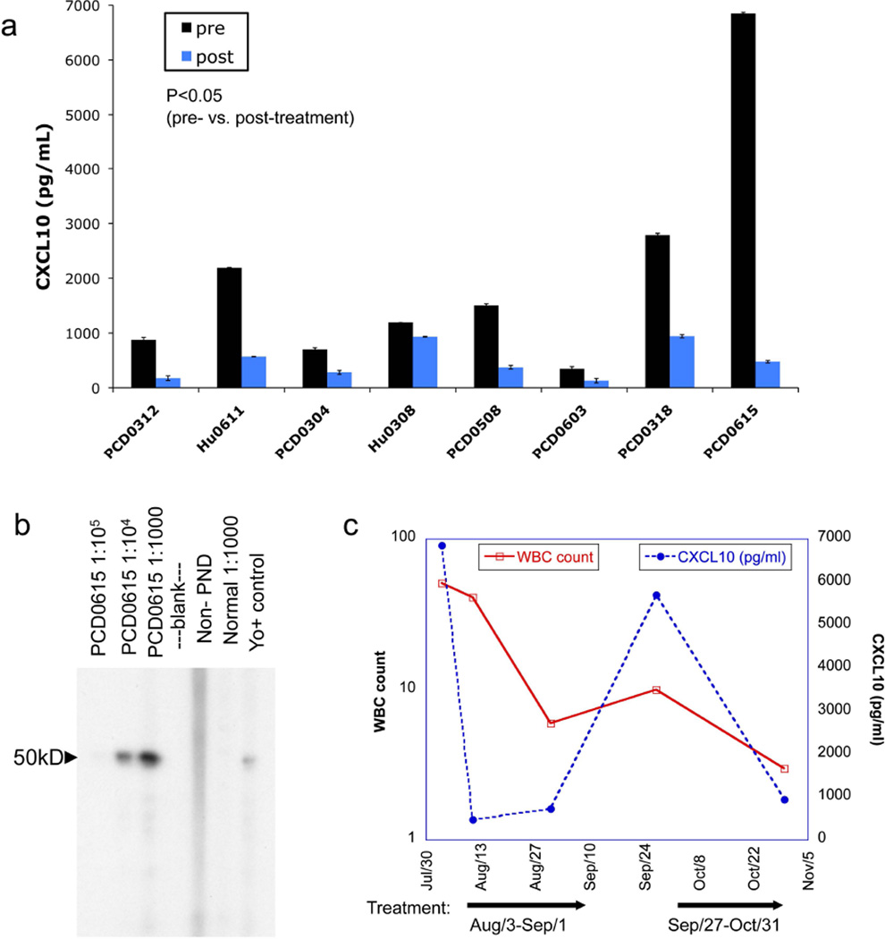
We screened CSF samples from eight patients with PND for the presence of 27 cytokines both pre- and post-FK506 treatment using a multiplex bead-based assay. Patient characteristics are outlined in Table 1. This initial bead-based screen showed high levels of CSF CXCL10 pre-treatment in these patients, so we confirmed this finding by quantifying CSF CXCL10 by ELISA. As shown in Figure 1a (dark bars), all patients with PND had elevated levels of CXCL10 (average 2054 pg/mL, range 347–6846 pg/mL). The CSF CXCL10 levels in our patients were similar to or in some cases higher than those reported previously in the literature for MS patients and were also higher than values seen in normal CSF.15 While CSF CXCL10 elevation has been reported in other disease states16,17,18 this is the first report to our knowledge that PND patients have elevated CSF CXCL10. CCL2 was also detected in PND patient CSF pre-treatment, but levels were comparable to those reported for normal individuals (data not shown). Four out of eight PND patients had less than a two-fold increase in pre-treatment CSF CXCL8 compared to controls (data not shown). Other cytokines tested were either not present or were below the limit of detection of the assay (listed in Methods).
Table 1.
PND Patient Characteristics.
| Patient ID |
Age | Antibody | Tumor type | Neurological symptoms |
Time from diagnosis |
CSF WBC (per uL) |
CSF Total cells seen |
CSF lymphocytes |
CSF RBC |
|---|---|---|---|---|---|---|---|---|---|
| 0312 | 58 | Yo | Ovarian | Cerebellar/brain stem syndrome | 8 months | 2 | 38 | 36 | 13 |
| 0611 | 73 | Hu | SCLC* | Sensory neuropathy | 6 months | 6 | 100 | 88 | None seen |
| 0304 | 57 | Yo | Ovarian | PCD | 8 months | None seen | None seen | None seen | 10 |
| 0308 | 61 | Hu | SCLC* | Peripheral neuropathy/pan cerebellar syndrome | 12 months | 2 | 82 | 72 | 1 |
| 0508 | 60 | Yo | Unknown primary | PCD | 1 month | 9 | 100 | 94 | None seen |
| 0603 | 74 | Yo | Ovarian and breast | PCD, sensory and motor neuropathy | 3 months | 3 | 24 | 14 | 3 |
| 0318 | 59 | Yo | Ovarian | PCD | 5 months | 2 | 10 | 10 | 2 |
| 0615 | 45 | Yo | Ovarian | PCD | 1 month | 51 | 100 | 87 | 10 |
SCLC= small cell lung cancer
Figure 1. CXCL10 is elevated in PND patient CSF and is decreased after FK506 treatment.
(A) Levels of CXCL10 in the CSF of 8 PND patients with the Hu syndrome or PCD pre- and post-FK506 treatment, as determined by CXCL10 ELISA. Bars represent the average of duplicate wells. p=0.011 for patients with PND pre- vs. post-treatment by Wilcoxon signed rank test with p<0.05 considered significant, indicating a decrease in CXCL10 with treatment. (B) Western blot of serum from PCD patient PCD0615 recognizing cdr2 (~50kD). (C) CSF white blood cell counts and CXCL10 levels over time in PND patient PCD0615 who was treated with repeated courses of FK506. Arrows indicate treatment periods.
We also measured cytokine and chemokine levels in these patients post-FK506 treatment. Remarkably, CXCL10 was the only CSF cytokine measured that was consistently and significantly changed after treatment with FK506 (Figure 1a dark bars vs. light bars, average fold decrease =4.56; p=0.011). CCL2 and CXCL8 were not significantly altered by the treatment (average fold decrease 1.3, p=0.401 for CCL2; average fold decrease 0.88, p=0.207 for CXCL8 pre vs. post, data not shown). Of note, IL-17, which is an important autoimmune cytokine in experimental autoimmune encephalomyelitis (EAE) mouse models of MS, was not detected in the CSF of our patients (data not shown). However, we cannot rule out the possibility that IL-17 might be bound to cell receptors or is otherwise inaccessible to our assay.
We studied the inflammatory response of one patient with Yo antibody-positive PCD (previously described in detail7; Figure 1b) over time. This patient was initially seen with relatively mild symptoms of cerebellar dysfunction that had been acutely worsening, and was treated with FK506 for 14 days, with subsequent clinical stabilization. After experiencing periods of symptom stability lasting from weeks to several months, she repeatedly developed episodes of acute symptom exacerbation. During the first two of these episodes, CSF was obtained prior to and following treatment. Analysis of her CSF revealed that during each clinical exacerbation the patient demonstrated a CSF pleiocytosis that was reduced to normal or near-normal levels following FK506 treatment, which paralleled the reduction in CXCL10 levels in her CSF (Figure 1c). This repeated elevation in CSF WBC suggested that after their elimination from the CSF, peripheral T cells were regaining access to the CNS.
CXCR3+ T cells are enriched in the CSF of PND patients compared to peripheral blood
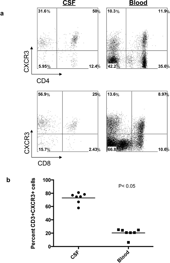
CXCL10 is an interferon γ– (IFNγ) inducible cytokine that stimulates the production and infiltration of inflammatory T cells and other cells expressing the CXCR3 receptor into inflamed sites.19 PND patients typically have abnormally high numbers of white blood cells in their CSF.1 Hence, we examined CSF of PND patients for the presence of CXCR3+ T cells. As has been reported in MS patients13, we observed enrichment of CXCR3+ T cells in PND CSF when compared to the peripheral blood CXCR3+ T cell population (Figure 2). This population was enriched nearly 3-fold for CD8+ T cells and 4-fold for CD4+ T cells for CXCR3, relative to peripheral blood T cells (Figure 2). Further, the number of CXCR3+ T cells decreased after FK506 treatment in all three patients for which these data were available, ranging from a nearly two-fold decrease in one patient to more than a seven-fold reduction in another (Table 2). These results suggest that there may be a role for a specific inflammatory pathway in patients with PND involving the cytokine CXCL10 and its receptor CXCR3 on T cells.
Figure 2. Enrichment of CXCR3+ T cells in PND patient CSF.
(A) FACS analysis of PND patient cells showing CXCR3+CD4+ T cells (top panels) and CXCR3+CD8+ T cells (bottom panels) in CSF and peripheral blood. (B) Percentage of CD3+/CXCR3+ T cells in CSF and peripheral blood from individual PND patients. Percentage of CSF CD3+/CXCR3+ cells mean=73; percentage of peripheral blood CD3+/CXCR3+ cells mean=20.4. A t-test assuming unequal variance was applied to the data with p<0.05 considered significant. P=2e10−8 for CSF vs. peripheral blood CXCR3+ cells.
Table 2.
Number of CSF CXCR3+ T cells (cells/uL)
| Patient | Pre FK506 | Post FK506 |
|---|---|---|
| Hu0308 | 1.57 | 0.83 |
| PCD0318 | 2.25 | 0.75 |
| PCD0615 | 35.19 | 4.68 |
T cells from normal donors produce CXCL10 in an antigen-specific manner
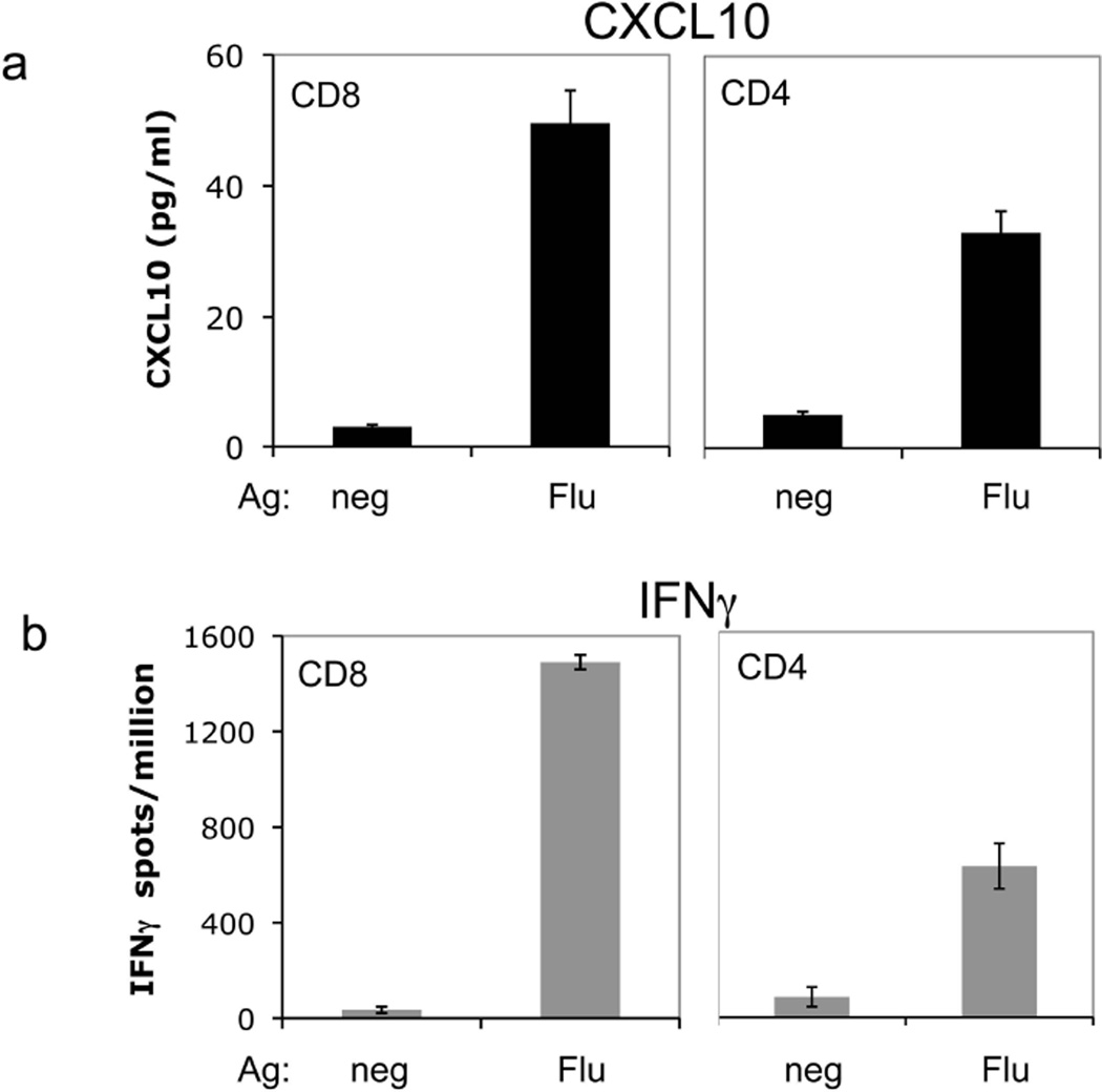
Based on the parallel decreases in patient CSF T cells and CXCL10 after FK506 treatment, we sought to determine whether T cells could produce CXCL10 and thus implicate them in production of the CXCL10 detected in the PND patient CSF. Previous work by our laboratory has shown that peripheral CD8+ T cells specific for the HuD PND antigen express CXCL10.6 Due to insufficient patient CSF T cells available for direct study, we modeled this question by assessing CXCL10 production from normal donor T cells upon encounter with specific antigen presented by dendritic cells (DC) as antigen presenting cells (APC). CD4+ or CD8+ T cells were co-cultured with DC loaded with apoptotic flu-infected 3T3 cells (for CD4+) or DCs directly infected with influenza virus (for CD8+). Analysis of supernatants from the cultures revealed that CXCL10 was only produced after a cognate CD4+ or CD8+ T cell-DC interaction (Figure 3a). CXCL10 secretion was coincident with IFNγ production, which was measured as a positive control (Figure 3b). These data indicate that CXCL10 is produced after an interaction between antigen-specific T cells and an APC but not after non-cognate interactions.
Figure 3. CXCL10 is produced in an antigen-specific fashion in cultures of human T cells and DC from normal donors.
(A) CXCL10 was measured by ELISA in supernatants from co-cultures of CD8+ or CD4+ T cells and DC presenting influenza antigen (Flu) or uninfected (neg). (B) IFNγ Elispot assay of normal donor CD8+ and CD4+ T cells cultured with DC presenting influenza antigen, performed in parallel with the experiment in (A). Error bars represent standard deviation from the mean of triplicate wells for each condition.
Production of CXCL10 in mouse T cell and DC co-cultures is antigen-specific and requires the IFNγR on the T cell
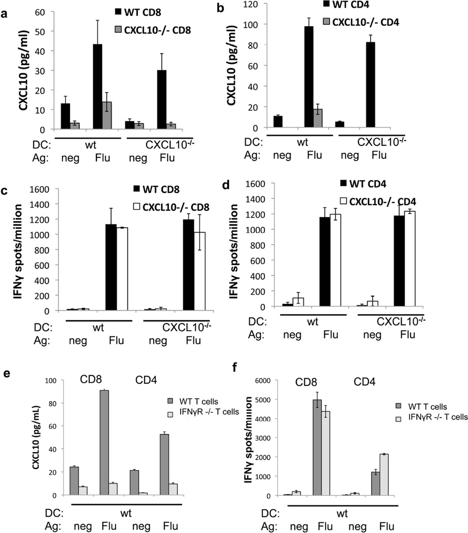
To assess whether these antigen-specific interactions led to CXCL10 production by T cells only, APC only or both cell types, we employed an assay similar to that used in the previous figure, but using cells from wild-type or CXCL10 null mice. T cells from influenza-immunized mice were co-cultured with DC pulsed with relevant influenza nucleoprotein (NP) peptide or an irrelevant control peptide. Production of CXCL10 in wild-type conditions was found to be antigen-specific (Figures 4a and 4b, black bars) and paralleled production of IFNγ (Figure 4c and d), as seen with human T cells (Figure 3). Similar results were obtained using T cells from ovalbumin-immunized mice (not shown). When influenza-specific CXCL10−/− CD4+ or CD8+ T cells were co-cultured with wild-type DCs presenting antigen, the number of IFNγ-producing cells present was similar to the number of IFNγ-producing cells in the wild-type T cell condition (Figure 4c and d, open bars). However, when CXCL10−/− T cells were co-cultured with wild-type DCs, three-fold less CXCL10 was detected (Figure 4a and b, gray bars). In comparison, no CXCL10 was detected when CXCL10−/− T cells were co-cultured with CXCL10−/− DC. Similar data were obtained using T cells from ovalbumin protein-immunized rather than virus-immunized CXCL10−/− mice (data not shown). Taken together, these results indicate that T cells produce the majority of CXCL10 in this assay and that they do so in an antigen-specific manner. DCs themselves also contribute to the production of CXCL10 after encounter with a cognate T cell in this assay, since a low level of CXCL10 is still present in the CXCL10−/− T cells with wild-type DC condition (Figure 4a and b).
Figure 4. T cell IFNγ receptor is required for antigen-specific production of CXCL10 in co-cultures of T cells and DC.
CXCL10 (A and B) and IFNγ (C and D) were measured in co-cultures of T cells from influenza-immunized CXCL10−/− or wild type C57BL/6 mice with DC from CXCL10−/− or wild type mice. For CD8+ co-cultures (A and C), DC were pulsed with influenza NP peptide (Flu) or ovalbumin peptide control (neg). For CD4+ co-cultures (B and D), DC were fed apoptotic influenza infected 3T3 cells (Flu) or uninfected 3T3 cells (neg). (E) CXCL10 measured in co-cultures set up as in A, using T cells from influenza-immunized IFNγR−/− mice. IFNγ production was determined in parallel (F). Each experiment was repeated at least three times with similar results.
DC production of CXCL10 in an antigen-specific manner suggests the presence of a signal from T cells that have encountered their cognate antigen to the DC upon which the antigen is presented. One possible mediator of such a signal from the T cells is IFNγ, since CXCL10 is known to be IFNγ-inducible20 and both cytokines are produced in an antigen-specific manner in our co-cultures (Figure 4). To address this possibility, we assessed CXCL10 production following antigen-specific T cell-DC interactions using cells from mice lacking the receptor for IFNγ (IFNγR−/−). Both IFNγ and CXCL10 were produced when wild-type T cells encountered DCs presenting influenza antigen (Figure 4e and f). However, when IFNγR−/− T cells encountered wild-type DC presenting influenza antigen, CXCL10 was not detected despite detection of expected levels of IFNγ (Figure 4e and f). These data support the idea that the IFNγR is required on the T cell for antigen-specific CXCL10 production.
DISCUSSION
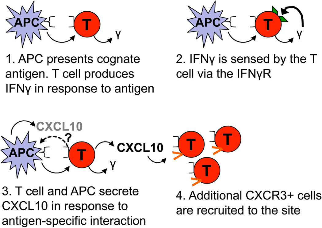
Our study began with a remarkable observation of a high CXCL10 concentration in the CSF of an acutely ill PCD patient that was decreased dramatically after treatment with the immunosuppressive drug FK506. A second course of FK506 treatment in the same patient resulted in a similar decrease in CSF CXCL10 (Figure 1c). We subsequently found that CXCL10 was decreased after FK506 treatment in seven other PND patients (Figure 1a, p=0.011). Importantly, two other chemokines also present in the CSF, CCL2 and CXCL8, were unaffected by the drug treatment in the same patients (p=0.401 and p=0.207, respectively). Moreover, the levels of twenty-four other cytokines (listed in Methods) were either undetectable or very similar to normal CSF controls, and were unaffected by FK506 treatment. The specificity of the effect of immunosuppressive therapy suggested a strong and potentially clinically relevant link to CNS inflammation in these patients that was pursued to discover a specific positive feedback loop. In this model (Figure 5), antigen-specific T cell stimulation by APCs within the CNS triggers T cell IFNγ production, followed by CXCL10 secretion by both T cells and APCs, leading to further lymphocyte recruitment to the area of inflammation.
Figure 5. Proposed model of the interactions leading to CXCL10 production by T cells and antigen presenting cells (APC).
The T cell is activated by its cognate antigen presented on an APC. The T cell secretes IFNγ in response to the antigen and senses autocrine or paracrine IFNγ via the IFNγ receptor (IFNγR). The T cell and APC secrete CXCL10 in this antigen specific interaction, which recruits additional CXCR3+ cells into the area. The signal that tells the APC to produce CXCL10 was not investigated in the current experiments, but may be related to expression of the IFNγR by APC or other signals.
Many cell types in the brain are able to produce CXCL10 such as MS patient astrocytes,13 neurons,21 and microglia.22 In addition, a correlation between CSF CXCL10 levels and CSF leukocyte count has previously been observed in MS.15 Here, we hypothesized that a key source of CXCL10 in PND patient CSF might be T cells, given that both T cells and CXCL10 were elevated in CSF from PND patients (Table 1 and Figure 1), and that we have previously seen CSF PND antigen-specific T cell responses in these patients.5, 6, 23 In support of this idea, we found that CXCL10 can indeed be produced by both human and mouse T cells, and that this production occurs specifically upon encounter with cognate antigen (Figures 3 and 4).
Interestingly, when we used CXCL10−/− T cells in this assay we found that a low level of CXCL10 production persisted in response to antigen and we attribute this production to the APC (Figure 4a and b). This result supports the possibility that the APCs are receiving antigen-specific signals from the T cells during their interaction, inducing them to elaborate the same chemokine, a potential component of the positive feedback loop (Figure 5) that would be location-specific. The additional release of CXCL10 would further increase recruitment of T cells to the local area. A recently published study demonstrated that CXCL10 production by DC is important in DC-T cell interactions during priming of T cells in the lymph node24. It is possible that CXCL10 production by APCs is similarly important for reactivation and further recruitment of memory T cells in the brain in PND, and that CXCL10 production by responding T cells augments the response.
It has been shown previously that microglia are able to produce CXCL10 and express its receptor CXCR3.25 Our observation of CXCL10 production by antigen-specific T cells uncovers a new, potentially clinically relevant dimension to T cell inflammation in the brain. Indeed, it has been shown previously that CXCL10 deficient mice infected with mouse hepatitis virus show decreased migration of effector T cells to the brain.19 In light of our observation that CXCL10 production is antigen specific, and given cells capable of presenting antigen within the brain13,26 we suggest that PND T cells encountering antigen in the CNS may be the source of the high CXCL10 levels in CSF from patients with PND.
A leading candidate for mediating the signal for CXCL10 production between the APC and the T cell was the receptor for IFNγ (IFNγR) since IFNγ is known to induce CXCL10 secretion.20 Indeed, when T cells lacking the IFNγR were co-cultured with DC, antigen-specific production of CXCL10 was completely obliterated, while IFNγ production remained unchanged (Figure4e and 4f). This suggests that there is a T cell intrinsic feedback loop for CXCL10 production, and that the low level of CXCL10 produced by the DC may require the IFNγR-mediated T cell feedback loop as well (Figure 5). CXCL10 and the CXCR3 receptor have been proposed to work in an inflammation-promoting loop in allergy27 and to modulate IFNγ production in EAE.28 CXCR3 has been suggested to not only play a role in T cell priming in the lymph node24 but also in the recruitment of T cells into peripheral target tissues.29 Taken together, these data suggest that the high concentration of CXCL10 present in PND patient CSF may be due to production of this chemokine by infiltrating autoimmune T cells interacting with APC in the CNS.30 However, EAE can be induced in CXCL10−/− mice, indicating that this chemokine is not essential for the development of this MS-like disease in mice.30
It is important to note that previous in vivo studies of the inflammatory response to CNS virus infection and chronic EAE have demonstrated that astrocytes are a major source of CXCL10.31,32,13 Our observations of CXCL10 production by DCs and T cells in an antigen-specific in vitro system do not rule out the possible additional production of CXCL10 by astrocytes or other cells in patients with PND. Indeed, it may even be that one cell type is responsible for CXCL10 production early during disease initiation, and other cell types such as T cells and APCs participate in CXCL10 production to propagate the disease state. This could lead to recruitment of additional T cells into the CNS in general or into the parenchyma.
In a clinical study, 10 out of 16 of our PND patients treated with FK506 showed improvement of their symptoms7 which, when combined with observed decreases in CSF WBC count and CXCL10 levels reported here, suggests that this treatment strategy warrants further study, particularly if used early in the disease. In addition, the specific mechanism by which FK506 (tacrolimus) decreases CXCL10 in PND patient CSF is of interest. Although the inhibition of calcineurin activity in T cells is a known effect of tacrolimus, additional intracellular targets such as p38 and JNK have been suggested.33 Interestingly, our laboratory has recently shown that FK506 preferentially accumulates in DCs, thereby targeting T cells engaged in antigen-specific T cell/APC interactions.34 It is possible that FK506 may similarly act upon APCs in the brain. Inhibition of the EAE animal model of MS has been observed with FK506 treatment.35,36 It would be interesting to determine whether treatment of EAE or MS patients with tacrolimus decreases CXCL10 levels in a similar fashion to that shown in our PND patients. A limitation of our study is that patients were treated with both FK506 and a tapered prednisone dose. Thus, it is possible that some of the effect of treatment on CSF CXCL10 may be due to prednisone. However, based on studies by others,37,38 it is reasonable to expect FK506 to be responsible for most or all of the effect.
In conclusion, we have shown a significant effect of FK506 immunosuppressive therapy on the important inflammatory chemokine CXCL10 in patients with PND. As elevated levels of CXCL10 are also found in other autoimmune neurological disorders, the presence of this chemokine in inflammatory CSF is significant and may be directly involved in the autoimmune response. It is not known whether the arrival of T cells or peripheral antigen presenting cells in the CNS or another process altogether is the initiating event for diseases such as PND. Interestingly, Bailey and colleagues have shown that CNS myeloid DC present endogenous peptide to T cells in the brain to drive EAE relapses in mouse models.39 The mechanism of immunosuppression and CXCL10 reduction in PND patients treated with FK506 may include decreased recruitment of T cells and/or APC from the periphery to the CNS, or interruption of the interaction of these two cell types or their intracellular signaling. Inhibition of T cell access to the brain might consequently slow the progression of disease, and may argue for combination therapy with agents that prevent T cell access to the CNS.
Acknowledgements
We thank the patients with paraneoplastic neurologic disorders who participated in this study and their families, The Rockefeller University Hospital staff for facilitating patient studies, and Dr. Jerome Posner for referring many of the patients. We also thank Dr. Andrew Luster for kindly providing CXCL10−/− mice, Dr. Chunhao Tu for statistics advice and members of our laboratory for many helpful discussions of this work.
Funding
This study was supported in part by grants UL1RR024143 8 and KL2TR000151 from the National Institutes of Health National Center for Research Resources and National Center for Advancing Sciences to The Rockefeller University Hospital. Robert Darnell is an Investigator of the Howard Hughes Medical Institute.
Footnotes
Conflict of Interest Statement
The authors declare no relevant conflicts of interest.
Author contributions
WR designed and performed the experiments, analyzed the data and wrote the manuscript. NB helped with designing and performing mouse experiments, data analysis and helped write the manuscript. MF recruited, enrolled and followed patients in the study, obtained patient samples, and helped with patient data analysis and writing the manuscript. AD recruited, enrolled, and followed patients in the study, obtained patient samples and edited the manuscript. RR provided advice about chemokine analysis and experiments and edited the manuscript. RD supervised the project, helped with the design of experiments and data analysis, followed patients in the study and wrote the manuscript.
REFERENCES
- 1.Darnell RB, Posner JB. Paraneoplastic syndromes affecting the nervous system. Semin Oncol. 2006;33:270–298. doi: 10.1053/j.seminoncol.2006.03.008. [DOI] [PubMed] [Google Scholar]
- 2.Darnell RB. Onconeural antigens and the paraneoplastic neurologic disorders: At the intersection of cancer, immunity, and the brain. Proceedings of the National Academy of Sciences. 1996;93:4529–4536. doi: 10.1073/pnas.93.10.4529. [DOI] [PMC free article] [PubMed] [Google Scholar]
- 3.Roberts WK, Darnell RB. Neuroimmunology of the paraneoplastic neurological degenerations. Curr Opin Immunol. 2004;16:616–622. doi: 10.1016/j.coi.2004.07.009. [DOI] [PubMed] [Google Scholar]
- 4.Albert ML, Austin LM, Darnell RB. Detection and treatment of activated T cells in the cerebrospinal fluid of patients with paraneoplastic cerebellar degeneration. Ann Neurol. 2000;47:9–17. [PubMed] [Google Scholar]
- 5.Albert ML, Darnell JC, Bender A, et al. Tumor-specific killer cells in paraneoplastic cerebellar degeneration. Nat Med. 1998;4:1321–1324. doi: 10.1038/3315. [DOI] [PubMed] [Google Scholar]
- 6.Roberts WK, Deluca IJ, Thomas A, et al. Patients with lung cancer and paraneoplastic Hu syndrome harbor HuD-specific type 2 CD8+ T cells. J Clin Invest. 2009;119:2042–2051. doi: 10.1172/JCI36131. [DOI] [PMC free article] [PubMed] [Google Scholar]
- 7.D O, M F, S T, et al. Cellular immune suppression in paraneoplastic neurologic syndromes targeting intracellular antigens. Arch Neurol. 2012;69:1132–1140. doi: 10.1001/archneurol.2012.595. [DOI] [PMC free article] [PubMed] [Google Scholar]
- 8.Schreiber S, Crabtree G. The mechanism of action of cyclosporin A and FK506. Immunology Today. 1992;13:136–142. doi: 10.1016/0167-5699(92)90111-J. [DOI] [PubMed] [Google Scholar]
- 9.Sawada S, Suzuki G, Kawase Y, Takaku F. Novel immunosuppressive agent, FK506. In vitro effects on the cloned T cell activation. J Immunol. 1987;139:1797–1803. [PubMed] [Google Scholar]
- 10.Snyder SH, Lai MM, Burnett PE. Immunophilins in the nervous system. Neuron. 1998;21:283–294. doi: 10.1016/s0896-6273(00)80538-3. [DOI] [PubMed] [Google Scholar]
- 11.Klein RS, Lin E, Zhang B, et al. Neuronal CXCL10 directs CD8+ T-cell recruitment and control of West Nile virus encephalitis. J Virol. 2005;79:11457–11466. doi: 10.1128/JVI.79.17.11457-11466.2005. [DOI] [PMC free article] [PubMed] [Google Scholar]
- 12.Engelhardt B, Ransohoff RM. The ins and outs of T-lymphocyte trafficking to the CNS: anatomical sites and molecular mechanisms. Trends Immunol. 2005;26:485–495. doi: 10.1016/j.it.2005.07.004. [DOI] [PubMed] [Google Scholar]
- 13.Sørensen T, Trebst C, Kivisakk P, et al. Multiple sclerosis: a study of CXCL10 and CXCR3 co-localization in the inflamed central nervous system. Journal of Neuroimmunology. 2002;127:59–68. doi: 10.1016/s0165-5728(02)00097-8. [DOI] [PubMed] [Google Scholar]
- 14.Blachere NE, Darnell RB, Albert ML. Apoptotic cells deliver processed antigen to dendritic cells for cross-presentation. PLoS Biol. 2005;3:e185. [Google Scholar]
- 15.Sorensen TL, Tani M, Jensen J, et al. Expression of specific chemokines and chemokine receptors in the central nervous system of multiple sclerosis patients. J Clin Invest. 1999;103:807–815. doi: 10.1172/JCI5150. [DOI] [PMC free article] [PubMed] [Google Scholar]
- 16.Chang CC, Omarjee S, Lim A, et al. Chemokine levels and chemokine receptor expression in the blood and the cerebrospinal fluid of HIV-infected patients with cryptococcal meningitis and cryptococcosis-associated immune reconstitution inflammatory syndrome. J Infect Dis. 2013;208:1604–1612. doi: 10.1093/infdis/jit388. [DOI] [PMC free article] [PubMed] [Google Scholar]
- 17.Kamat A, Lyons JL, Misra V, et al. Monocyte activation markers in cerebrospinal fluid associated with impaired neurocognitive testing in advanced HIV infection. J Acquir Immune Defic Syndr. 2012;60:234–243. doi: 10.1097/QAI.0b013e318256f3bc. [DOI] [PMC free article] [PubMed] [Google Scholar]
- 18.Henningsson AJ, Tjernberg I, Malmvall BE, et al. Indications of Th1 and Th17 responses in cerebrospinal fluid from patients with Lyme neuroborreliosis: a large retrospective study. J Neuroinflammation. 2011;8:36. doi: 10.1186/1742-2094-8-36. [DOI] [PMC free article] [PubMed] [Google Scholar]
- 19.Dufour JH, Dziejman M, Liu MT, et al. IFN-gamma-inducible protein 10 (IP-10; CXCL10)-deficient mice reveal a role for IP-10 in effector T cell generation and trafficking. J Immunol. 2002;168:3195–3204. doi: 10.4049/jimmunol.168.7.3195. [DOI] [PubMed] [Google Scholar]
- 20.Luster AD, Ravetch JV. Biochemical characterization of a gamma interferon-inducible cytokine (IP-10) J Exp Med. 1987;166:1084–1097. doi: 10.1084/jem.166.4.1084. [DOI] [PMC free article] [PubMed] [Google Scholar]
- 21.Wang J, Campbell IL. Innate STAT1-dependent genomic response of neurons to the antiviral cytokine alpha interferon. J Virol. 2005;79:8295–8302. doi: 10.1128/JVI.79.13.8295-8302.2005. [DOI] [PMC free article] [PubMed] [Google Scholar]
- 22.Balashov KE, Rottman JB, Weiner HL, Hancock WW. CCR5(+) and CXCR3(+) T cells are increased in multiple sclerosis and their ligands MIP-1alpha and IP-10 are expressed in demyelinating brain lesions. Proc Natl Acad Sci U S A. 1999;96:6873–6878. doi: 10.1073/pnas.96.12.6873. [DOI] [PMC free article] [PubMed] [Google Scholar]
- 23.Santomasso BD, Roberts WK, Thomas A, et al. A T cell receptor associated with naturally occurring human tumor immunity. Proceedings of the National Academy of Sciences. 2007;104:19073–19078. doi: 10.1073/pnas.0704336104. [DOI] [PMC free article] [PubMed] [Google Scholar]
- 24.Groom JR, Richmond J, Murooka TT, et al. CXCR3 chemokine receptor-ligand interactions in the lymph node optimize CD4+ T helper 1 cell differentiation. Immunity. 2012;37:1091–1103. doi: 10.1016/j.immuni.2012.08.016. [DOI] [PMC free article] [PubMed] [Google Scholar]
- 25.Flynn G, Maru S, Loughlin J, et al. Regulation of chemokine receptor expression in human microglia and astrocytes. J Neuroimmunol. 2003;136:84–93. doi: 10.1016/s0165-5728(03)00009-2. [DOI] [PubMed] [Google Scholar]
- 26.Millward JM, Caruso M, Campbell IL, et al. IFN-gamma-induced chemokines synergize with pertussis toxin to promote T cell entry to the central nervous system. J Immunol. 2007;178:8175–8182. doi: 10.4049/jimmunol.178.12.8175. [DOI] [PubMed] [Google Scholar]
- 27.Gangur V, Simons FE, Hayglass KT. Human IP-10 selectively promotes dominance of polyclonally activated and environmental antigen-driven IFN-gamma over IL-4 responses. FASEB J. 1998;12:705–713. doi: 10.1096/fasebj.12.9.705. [DOI] [PubMed] [Google Scholar]
- 28.Liu L, Huang D, Matsui M, et al. Severe disease, unaltered leukocyte migration, and reduced IFN-gamma production in CXCR3−/− mice with experimental autoimmune encephalomyelitis. J Immunol. 2006;176:4399–4409. doi: 10.4049/jimmunol.176.7.4399. [DOI] [PubMed] [Google Scholar]
- 29.Groom JR, Luster AD. CXCR3 in T cell function. Exp Cell Res. 2011;317:620–631. doi: 10.1016/j.yexcr.2010.12.017. [DOI] [PMC free article] [PubMed] [Google Scholar]
- 30.Klein RS, Izikson L, Means T, et al. IFN-inducible protein 10/CXC chemokine ligand 10-independent induction of experimental autoimmune encephalomyelitis. J Immunol. 2004;172:550–559. doi: 10.4049/jimmunol.172.1.550. [DOI] [PubMed] [Google Scholar]
- 31.Park C, Lee S, Cho IH, et al. TLR3-mediated signal induces proinflammatory cytokine and chemokine gene expression in astrocytes: differential signaling mechanisms of TLR3-induced IP-10 and IL-8 gene expression. Glia. 2006;53:248–256. doi: 10.1002/glia.20278. [DOI] [PubMed] [Google Scholar]
- 32.Glabinski AR, Tani M, Strieter RM, et al. Synchronous synthesis of alpha- and beta-chemokines by cells of diverse lineage in the central nervous system of mice with relapses of chronic experimental autoimmune encephalomyelitis. Am J Pathol. 1997;150:617–630. [PMC free article] [PubMed] [Google Scholar]
- 33.Matsuda S, Shibasaki F, Takehana K, et al. Two distinct action mechanisms of immunophilin-ligand complexes for the blockade of T-cell activation. EMBO Rep. 2000;1:428–434. doi: 10.1093/embo-reports/kvd090. [DOI] [PMC free article] [PubMed] [Google Scholar]
- 34.Orange DE, Blachere NE, Fak J, et al. Dendritic cells loaded with FK506 kill T cells in an antigen-specific manner and prevent autoimmunity in vivo. Elife. 2013;2:e00105. doi: 10.7554/eLife.00105. [DOI] [PMC free article] [PubMed] [Google Scholar]
- 35.Gold BG, Voda J, Yu X, et al. FK506 and a nonimmunosuppressant derivative reduce axonal and myelin damage in experimental autoimmune encephalomyelitis: neuroimmunophilin ligand-mediated neuroprotection in a model of multiple sclerosis. J Neurosci Res. 2004;77:367–377. doi: 10.1002/jnr.20165. [DOI] [PubMed] [Google Scholar]
- 36.Inamura N, Hashimoto M, Nakahara K, et al. Immunosuppressive effect of FK506 on experimental allergic encephalomyelitis in rats. International Journal of Immunopharmacology. 1988;10:991–995. doi: 10.1016/0192-0561(88)90046-x. [DOI] [PubMed] [Google Scholar]
- 37.Chang KT, Lin HY, Kuo CH, Hung CH. Tacrolimus suppresses atopic dermatitis-associated cytokines and chemokines in monocytes. J Microbiol Immunol Infect. 2014 doi: 10.1016/j.jmii.2014.07.006. [DOI] [PubMed] [Google Scholar]
- 38.Cristillo AD, Macri MJ, Bierer BE. Differential chemokine expression profiles in human peripheral blood T lymphocytes: dependence on T-cell coreceptor and calcineurin signaling. Blood. 2003;101:216–225. doi: 10.1182/blood-2002-03-0697. [DOI] [PubMed] [Google Scholar]
- 39.Bailey SL, Schreiner B, McMahon EJ, Miller SD. CNS myeloid DCs presenting endogenous myelin peptides 'preferentially' polarize CD4+ T(H)-17 cells in relapsing EAE. Nat Immunol. 2007;8:172–180. doi: 10.1038/ni1430. [DOI] [PubMed] [Google Scholar]


