Summary
This study examined recent trends in type-specific HPV infection rates in women referred for HPV typing as part of cervical cancer screening in the United States. HPV analyses were performed from March 2004 to March 2011. Women were aged 21–65 years at testing. The 18 most prevalent HPV types were analysed. Type-specific HPV infection rates were estimated in 3-month blocks. Lowess smoothing was used to examine time trends in infection rates for each HPV type, both combined, and separated by age group (younger women 21–30 years, older women 31–64 years). A total of 220914 women were included in the final analysis. The number of HPV tests performed on the younger age group increased, with the number of HPV infections and multiple type HPV infections decreasing. When separated by HPV type-specific analysis, the majority of HPV infection rates decreased; however, HPV types 61 and 83 increased. When analysing the older age group, there was a marked increase of the number of HPV tests. Overall, the rates of any HPV infection, as well as multiple type infections, were lower compared to the younger age group. The change in type-specific HPV rates in the older age group was minimal, with many rates remaining the same. In this population of women, overall rates of HPV infection decreased, while the number of HPV tests increased. Younger women had a more marked decrease in HPV infection rates, while for older women type-specific HPV infection rates appear consistent.
Keywords: Epidemiology, HPV infection rates, type-specific HPV
Introduction
Human papillomavirus (HPV) infection continues to be the most common sexually transmitted disease and a crucial link to cervical cancer [1]. According to the National Cancer Institute, an estimated 4200 women in the United States died of cervical cancer in 2011, and cervical cancer continues to be the second most common cancer in women worldwide [2]. As such, HPV infection continues to cause significant burden on both men and women. There are over 100 different HPV genotypes, separated into low and high cancer risk types. The highest prevalence of HPV infection has been documented in persons aged between 20 and 24 years [3].
Even though the decrease of cervical cancer in most high-income countries has been attributed to Pap test screening, there are drawbacks to this screening method. While highly specific, Pap tests have low sensitivity, primarily due to the subjective nature of reading samples [4]. HPV testing has emerged as a possible candidate to replace cytology for primary screening; however, the debate is still ongoing. The American College of Obstetrics and Gynecology in a recent practice bulletin states that at this time there is not enough evidence for HPV testing as primary screening; however, recent studies suggest that co-testing may have minimal added benefit to HPV typing alone [5]. As such, there has been movement to incorporate HPV genotyping in cervical cancer screening strategies.
This paper provide an exploratory analysis of recent trends in HPV type-specific infections during the same time period in which HPV vaccines became available. This examination is timely because it is unclear how vaccination may change the trends in HPV infections in recent years.
Materials and Methods
Study population
Prior to the initiation of this investigation, appropriate approval was granted by the University of Minnesota's Institutional Review Board. We analysed data from women who had HPV typing performed by Access Genetics (Eden Prairie, USA) between March 2004 and March 2011. Access Genetics offers medical diagnostic services, including HPV testing. In addition to reporting the presence or absence of high-risk HPV types they perform PCR-based HPV typing. Although there are over 442 laboratories that perform HPV analysis for Access Genetics, only laboratories that submitted samples throughout the 7-year period were included. These laboratories were located in New Hampshire, Iowa, and California. Patients' age at testing, laboratory location, and test media type were the only demographic information available.
Specimen analysis
Specimen analysis was performed at Access Genetics as previously described [6]. Samples were processed within 48 h of being received in liquid cytology medium. DNA was extracted by salt precipitation, and amplification of the genomic DNA was achieved by using modification of the method of Resnick et al. [7]. Next, samples positive for HPV were subjected to genotyping by restriction endonuclease fragment analysis. Interpretation of the HPV genotypes was based on the pattern of resulting bands for each enzyme, which was compared to the genomic maps of each viral type. The 18 most prevalent HPV types in this age group were considered for analysis: 6, 11, 16, 18, 31, 52, 53, 54, 58, 59, 61, 62, 66, 68, 82, 83, 84, and 89. We considered high-risk types as HPV types 16, 18, 31, 52, 53, 58, 59, 66, 68 and 82 (MM4); and low-risk types as HPV types 6, 11, 54, 61, 62, 83 (MM7), 84 (MM8), 89 (CP6108). The subtypes listed in parentheses were combined with their primary type for analysis.
Statistical methods
As this was an exploratory analysis, no formal sample size or power calculations were performed prior to the analysis. Women with an adequate sample for HPV type evaluation between March 2004 and March 2011 were considered (n=413447). Evaluation was then limited to the first available sample from each individual (subsequent results were removed) and to those aged 21–65 years at the time of testing (n= 374267). When further limiting the samples to those laboratories that submitted samples throughout the time-frame, the final analysis included 220914 women.
Demographic information available for these women was summarized. Type-specific HPV infection rates were estimated in 3-month periods over time. To explore the data, we used Lowess smoothing (locally weighted scatterplot smoothing) to examine infection rates over time for each of the HPV types without making assumptions about the form of the relationship. Infection rates are presented stratified by age group (21–30, 31–64 years). These age groups were chosen based on the American Society for Colposcopy and Cervical Pathology (ASCCP) guidelines [8]. All statistical analyses were performed using SAS version 9.2 (SAS Institute, USA) and R version 2.7 (R Development Core Team, http://www.r-project.org/).
Results
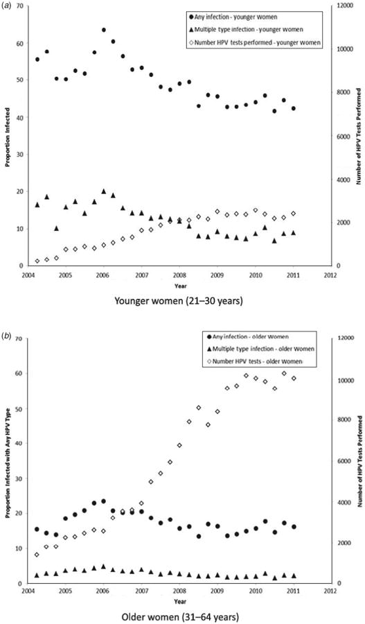
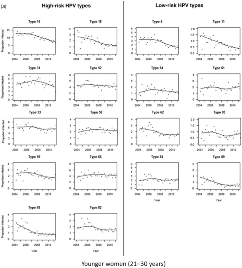
This exploratory analysis included 220914 women; 47461 aged 21–30 years and 173453 aged 31–64 years. Over the study period of 2004–2011, the number of HPV tests performed increased, while the number of HPV infections decreased in both age groups (Fig. 1a, b). The number of HPV tests performed in the older age group (31–64 years) markedly increased beginning in 2007, with more than double the number of HPV tests performed by the end of the time-frame (Fig. 1b). However, the change in number of HPV tests performed on the younger age group (21–30 years), increased more modestly and appeared to level around 2009. As expected, the proportion of women who were HPV positive during the time-frame was larger in the younger age group than in the older group (47.3% vs. 16.5%, P<0.0001). The number of multiple type HPV infections appears to have decreased among young women but remained relatively constant among older women over the time-frame of the study. HPV type-specific infection rates by age group are presented in Figure 2(a, b). In the younger age group, the majority of high-risk HPV types decreased over time, with the most marked decreases noted in HPV types 16, 18, and 68. The decrease of HPV types 18 and 68 stabilized towards the end of the study period. Interestingly, there were several HPV types that increased prior to 2007 and then began to decrease in infection rates; most notably HPV type 31 A few HPV types remained relatively constant, including types 52, 58, and 66. When evaluating the low-risk HPV types, a similar sharp decrease was noted in HPV types 6 and 11. HPV types 62 and 89 declined prior to 2008 and then stabilized.
Fig. 1.

Rates of HPV Infection (any type) and the number of tests performed at these three laboratories from 2004–2011. (a) Younger women (21–30 years); (b) older women (31–64 years).
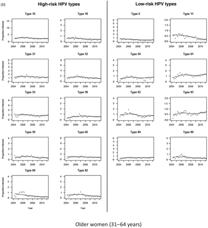
Fig. 2.


Infection rates over time by human papillomavirus (HPV) type. Infection rates by HPV type are defined as the number of new positive infections per total number of women tested within a 3-month period. Changes over time are represented by Lowess smoothing curves (solid black line). (a) Younger women (21–30 years); (b) older women (31–64 years).
The majority of type-specific HPV infection rates declined in older women as well, although starting infection rates were lower than in younger women. Among the older women, the infection rates of all high-risk HPV types remained constant over the study period. Among low-risk HPV types, infection rates showed more change over time, particularly HPV type 11, which steadily declined over the study period.
Discussion
In this study population, HPV infection rates overall decreased over time for both age groups. This suggests that there may be a change in HPV infection rates in the United States. A marked increase in HPV testing of older women occurred after 2007; this may be due to increased availability of HPV genotyping or increasing education on the impact of HPV testing of certain cytology results. This could increase the number of low-risk women tested, resulting in lower infection rates. This same increase in genotyping was not seen with the younger age group.
It is unclear how the incidence rates of type-specific HPV infection will change as vaccination becomes more widespread. The Victorian Cervical Cytology Registry study showed a decrease in the incidence of high-grade cervical abnormalities over time in a population that received the Gardasil vaccination [6]. While we do not know the vaccination status of the women in our study, it is interesting that the HPV type-specific analysis of infection rates appeared different between the age groups. While the older women type-specific analysis was relatively constant over time for each HPV type, the younger women group had more marked changes. This may be due to the natural history of HPV infection in younger women or possibly by the overall impact of vaccination.
As mentioned earlier, the vaccination status of the women in this study is unknown. Therefore, we cannot make statements on the possible direct or indirect effect of vaccination on the decrease in HPV infection rates over time. This information would have been beneficial for the analysis because it would have allowed for inference about the impact of vaccination in the study population and especially in women aged <31 years. However, we know that it is likely that those women in the 31–64 years age group did not receive the vaccine because the vaccine had not yet received FDA approval for those patient populations. Furthermore, it is unknown whether the incidence of the HPV types was also due to persistent infection. Since only the first noted infection during the time of analysis was included, women who were diagnosed before March 2004 could not be identified.
In the current study, rates of HPV infection decreased in both young and older women, even as the number of HPV tests increased. However, over the next decade HPV infection rates may vary due to vaccination and the magnitude of this change is unknown, due to social, cultural, and geographical differences. Moreover, additional studies are needed with available data about vaccination status, cytological examination and HPV types in the female population.
Acknowledgments
The authors acknowledge Elizabeth Ralston Howe and Dr Ronald McGlennen, of Access Genetics, for their contribution to the background work and data used in this study. This research was funded by NIH T-32 training grant no. 5T32-CA132715. Research reported in this publication was supported by NIH grant P30 CA77598 utilizing the Biostatistics and Bioinformatics Core shared resource of the Masonic Cancer Center, University of Minnesota and by the National Center for Advancing Translational Sciences of the National Institutes of Health Award Number UL1TR000114. The content is solely the responsibility of the authors and does not necessarily represent the official views of the National Institutes of Health.
Footnotes
Declaration of Interest: Levi S. Downs Jr., MS, MD. Glaxo Smith Kline: research support for HPV vaccine clinical trials, advisory board (honoraria); Merck Corporation: advisory board (honoraria).
References
- 1.Wells M, et al. Tumors of the breast and female genital organs. World Health Organization classification of tumors. Pathology and Genetics. 2003:259–261. [Google Scholar]
- 2.National Cancer Institute. Cervical cancer statistics. [Accessed December 2 2013]; http://www.cancer.gov/cancertopics/types/cervical.
- 3.Dunne EF, et al. Prevalence of HPV infection among females in the United States. Journal of the American Medical Association. 2007;297:813–819. doi: 10.1001/jama.297.8.813. [DOI] [PubMed] [Google Scholar]
- 4.Tota JE, et al. Epidemiology and burden of HPV infection and related diseases: Implications for prevention strategies. Preventive Medicine. 2011;53:S12–S21. doi: 10.1016/j.ypmed.2011.08.017. [DOI] [PubMed] [Google Scholar]
- 5.Chelmow D, et al. ACOG practice bulletin: Screening for cervical cancer. ACOG clinical management guidelines for the obstetrician gynecologist. 2012 Nov;Number 131 [Google Scholar]
- 6.Dickson EL, et al. Multiple-type HPV infections: a cross-sectional analysis of the prevalence of specific types in 309,000 women referred for HPV testing at the time of cervical cytology. International Journal of Gynecologic Cancer. 2013;23:1295–1302. doi: 10.1097/IGC.0b013e31829e9fb4. [DOI] [PMC free article] [PubMed] [Google Scholar]
- 7.Resnick RM, et al. Detection and typing of human papillomavirus in archival cervical cancer specimens by DNA amplification with consensus primers. Journal of the National Cancer Institute. 1990;82:1477–1484. doi: 10.1093/jnci/82.18.1477. [DOI] [PubMed] [Google Scholar]
- 8.American Society for Colposcopy and Cervical Pathology. Management guidelines. [Accessed December 2 2013]; http://www.asccp.org/Guidelines-2/Management-Guidelines-2.


