Abstract
In T cells, the Tec kinases ITK and RLK are activated by TCR stimulation, and are required for optimal downstream signaling. Studies of CD4+ T cells from Itk−/− and Itk−/− Rlk−/− mice have indicated differential roles of ITK and RLK in TH1, TH2, and TH17 differentiation and cytokine production. However, these findings are confounded by the complex T cell developmental defects in these mice. Here, we examine the consequences of ITK and RLK inhibition using a highly selective and potent small molecule covalent inhibitor PRN694. In vitro TH polarization experiments indicate that PRN694 is a potent inhibitor of TH1 and TH17 differentiation and cytokine production. Using a T cell adoptive transfer model of colitis, we find that in vivo administration of PRN694 markedly reduces disease progression, T cell infiltration into the intestinal lamina propria, and IFN-γ production by colitogenic CD4+ T cells. Consistent with these findings, TH1 and TH17 cells differentiated in the presence of PRN694 show reduced P-selectin binding and impaired migration to CXCL11 and CCL20, respectively. Together, these data indicate that ITK plus RLK inhibition may have therapeutic potential in TH1-mediated inflammatory diseases.
Introduction
Tec family tyrosine kinases play a key role in antigen receptor-mediated signaling pathways in lymphocytes. Among these kinase family members, T cells express IL-2-inducible kinase (ITK), resting lymphocyte kinase (RLK), and tyrosine kinase expressed in hepatocellular carcinoma (TEC) (1). Although each of these kinases is expressed in mature naïve T cells, ITK is the most predominant. Based on mRNA analysis, RLK is expressed at 3- to 10-fold lower levels than ITK, and Tec is 30- to 100-fold reduced compared to ITK (2, 3). Following T cell receptor (TCR) stimulation, ITK is activated, and directly phosphorylates phospholipase Cγ1 (PLCγ1). Activated PLCγ1 hydrolyzes phosphatidylinositol 4,5-biphosphate (PIP2) to produce inositol triphosphate (IP3) and diacylglycerol (DAG), secondary messengers that lead to Ca2+ influx and mitogen-activated protein kinase (MAPK) and Protein kinase C (PKC) activation (4). As a consequence, Itk−/− T cells have significant defects in T cell activation and differentiation (5–8). For RLK, some evidence supports a role in TCR signaling, as Itk−/− Rlk−/− double-deficient T cells are more impaired than those lacking only ITK (5, 9). Nonetheless, based on current data, the precise functions of RLK and TEC in T cell activation are unclear.
To elucidate the role of Tec kinases in TCR signaling, several studies have addressed the impact of a deficiency in ITK, or ITK plus RLK, in CD4+ helper T (TH) cell differentiation and function. Initial studies showed that Itk−/− mice exhibited impaired TH2 differentiation and TH2-biased responses to parasitic infection, with little effect on protective TH1 responses to intracellular protozoans (2, 10). These data were further supported by controlled in vitro studies that demonstrated that naïve Itk−/− CD4+ T cells were defective in TH2 but not TH1 differentiation, in part due to the fact that differentiated TH2 cells fail to express any RLK protein, as do TH1 cells (2). Additionally, ITK and RLK functions in helper T cells are at least partially redundant, as RLK overexpression in Itk−/− mice was able to restore TH2 responses in animal models of allergic asthma and schistosome egg-induced lung granuloma formation (11). Nonetheless, it has been difficult to distinguish which phenotypes observed in these mice are due to the functions of ITK and/or RLK in mature naïve CD4+ T cells, and which are the consequence of altered T cell development generating an abnormal cytokine environment in the Itk−/− or Itk−/−Rlk−/− mice.
More recently, studies by P. Schwartzberg and colleagues have indicated an additional role for ITK in TH17 differentiation. Specifically, Itk−/− T cells showed reduced IL-17A production and increased forkhead box P3 (Foxp3) expression following in vitro polarization (12, 13). In addition, Itk−/− T cells provided enhanced regulatory T cell (Treg)-mediated protection in an adoptive transfer model of colitis, due to their increased potential to upregulate Foxp3 (13), although another study found that Itk−/− Tregs were unable to protect against T cell mediated colitis (14). In spite of some of disparities between studies, in general, these findings have provided impetus for the development of small molecule ITK kinase inhibitors, with the intent of using them as treatments for atopic diseases, as well as for their potential as an immunosuppressant to block graft rejection or autoimmunity.
The complex phenotype of Itk−/− mice, including defects in T cell development, activation, differentiation, and effector function, has made it difficult to precisely assess the function of ITK in each lineage of T cells and at different stages of an immune response. It has also been challenging to distinguish functions of ITK in T cell activation and differentiation from effects due to altered T cell development in Itk−/− mice. A more direct strategy to address ITK and/or RLK function in T cells is to utilize a selective small molecule inhibitor of these Tec kinases. PRN694 is a small molecule that forms an irreversible covalent bond with C442 in ITK or C350 in RLK, and has recently been shown to selectively inhibit ITK and RLK in T cells (15). To date, the inhibitory effects of PRN694 on CD4+ T helper cell differentiation and effector function have not been tested.
In this study, we examined the effects of PRN694 on CD4+ T cell differentiation and function in vitro and in vivo. Surprisingly, we found that PRN694 showed potent inhibitory effects on TH1 differentiation and IFN-γ production as well as on TH17 differentiation and IL-17A production, with decreased potency on TH2 differentiation. To test the relevance of this inhibitory activity in vivo, we utilized the T cell adoptive transfer model of colitis, an inflammatory condition mediated by IFN-γ-producing TH1 cells (16, 17). Consistent with our in vitro data, PRN694 administration ameliorated colitis disease progression and markedly inhibited colonic inflammation. Together, these data indicate that simultaneous inhibition of ITK and RLK by a small molecule inhibitor may be an effective treatment for TH1-biased inflammatory diseases.
Materials and Methods
Mice
C57BL/6 wild-type (WT), C57BL/6 Rag2−/−, C57BL/6 OT-II Rag1−/− TCR transgenic (Tg), and B10.A 5C.C7 Rag2−/− TCR Tg mice were purchased from Taconic Biosciences and housed in SPF conditions at University of Massachusetts Medical School (UMMS) in accordance with Institutional Animal Care and Use Committee (IACUC) guidelines. CD-1 mice were purchased from Charles River; all housing and procedures with these mice were in accordance with the guidelines approved by Principia Biopharma’s IACUC.
T helper cell polarization and CFSE dilution assay
Naïve CD4+ T cells (3.0 × 10⁵) from B10.A 5C.C7 Rag2−/− TCR Tg or C57BL/6 OT-II Rag1−/− TCR Tg mice were isolated using CD4 (L3T4) MicroBeads (Miltenyi Biotec). Isolated cells were plated in 12- or 24-well plates and activated by plate-bound anti-CD3 (1.0 μg/mL) and anti-CD28 (4.0 μg/mL) (BD Biosciences) for 72hr in the presence of following conditions: TH0 (anti-CD3/CD28 with no cytokines); TH1 (IL-12, 10 ng/mL plus anti-IL-4, 10 μg/mL); TH2 (IL-4, 10 ng/mL plus anti-IFN-γ, 10 μg/mL); TH17 (IL-6, 20 ng/mL, TGF-β, 5.0 ng/mL, IL-1β, 20 ng/mL, plus anti-IFN-γ, anti-IL-4, and anti-IL-2, each at 10 μg/mL) (all anti-cytokine antibodies were purchased from R&D Systems and BD Biosciences). For CFSE dilution assay, CD4+ T cells isolated by magnetic separation were resuspended in 1.0 mL of 1X PBS with 0.1% BSA at 5.0 × 10⁶ cells/mL. Then, cells were stained with CFSE at a final concentration of 2.0μM at 37°C for 10min. After incubation, stained cells were quenched by adding 5 volume of ice-cold RPMI 1640 complete medium (10% FBS) for 5min on ice. Cells were washed three times with complete medium and then plated for TH polarization for 72hr.
Cytokine and transcription factor analysis
T cells were stimulated with PMA (50 ng/mL) and Ionomycin (1.0 μg/mL) for 5hr with protein transport inhibitors, GolgiStop™ and GolgiPlug™ (BD Biosciences), each at 1.0 μg/mL. All cells were first stained with antibodies to CD4 and CD44 (BD Biosciences), and LIVE/DEAD® Fixable Aqua Dead Cell Stain Kit (Life Technologies). Stained cells were then fixed and permeabilized by using BD Cytofix/Cytoperm™ Kit (BD Biosciences). For cytokine staining, cells were stained with antibodies to IFN-γ, IL-4, IL-17A, or IL-2 (BD Biosciences). For transcription factor staining, cells were fixed and permeabilized using Foxp3/Transcription Factor Staining Buffer Set (eBioscience), and stained with antibodies to T-bet, GATA-3, RORγt, or Foxp3 (BD Biosciences). Cells were analyzed on an LSRII flow cytometer (BD Biosciences) and data were analyzed with FlowJo (Tree Star).
Measurements of cytokine production by human PBMC and in mouse plasma
Human peripheral blood mononuclear cells (PBMC) isolated from whole blood were incubated for 1hr with and without PRN694 and stimulated with plate-bound anti-CD3 (2.5 μg/mL) and soluble anti-CD28 (1.0 μg/mL) (BD Biosciences) for 18hr. Supernatants were collected, frozen, and analyzed using the Human InflammationMap® v.1.0 biomarker panel (Myriad RBM). For the IL-2 inhibition dose-response, PBMC were incubated for 1hr with a concentration range of PRN694 and stimulated either with anti-CD3/CD28 or 10 μM of thapsigargin (Sigma-Aldrich) for 18hr. Supernatants were analyzed for IL-2 using the AlphaLISA IL-2 kit (Perkin Elmer). For cytokine inhibition in vivo, triplicate CD-1 mice were administered a 20 mg/kg intraperitoneal dose of PRN694 followed either 1 or 6hr later by administration of 10 μg/mouse of anti-CD3 (R&D Systems). Plasma was collected 2hr after anti-CD3 injection and analyzed for cytokines using the RodentMap® v.3.0 biomarker panel (Myriad RBM).
Mouse adoptive transfer colitis model and PRN694 administration
CD4+ CD45RBhi T cells were sorted from spleens of B6 WT mice and 4.0 × 10⁵ cells were injected i.p. into Rag2−/− hosts. Recipient mice were weighed daily and sacrificed 7 weeks post-transfer, at which time colons were removed for length measurement, histologic analysis, and lymphocyte isolation, and lymphoid organs harvested for flow cytometry analysis. Vehicle (5% ethanol/95% Captex 355 EP/NF, ABITEC) or PRN694 was orally administered according to the following regimen: once a day (40 mg/kg) for weeks 0–2 and 4–7, and twice a day (20 mg/kg) for weeks 2–4.
Histologic examination of colon
Colon specimens obtained from vehicle-treated or PRN694-treated hosts were fixed in 10% buffered formalin and stained with hematoxylin and eosin. To assess the severity of colitis, histologic scores of the proximal, middle, and distal colon were examined.
In vivo PRN694 target engagement assay and assessments of toxicity
Naïve B6 mice were dosed with vehicle or PRN694 (40 mg/kg). After 2hr or 6hr of oral administration, the dosed mice were sacrificed, and CD4+ T cells were isolated from the spleen by using EasySep™ Mouse CD4+ T cell Isolation Kit (StemCell Technologies). Then, isolated CD4+ T cells were stimulated with anti-CD3 (10 μg/mL) and anti-CD28 (5.0 μg/mL) for 5min at 37°C. After stimulation, CD4+ T cells (4.0 × 10⁶) were lysed by CelLytic M buffer (Sigma-Aldrich) containing proteinase and phosphatase inhibitor. The expression of phosphorylated-PLCγ1 and total PLCγ1 was examined by western blot using anti-p-PLCγ1 (Tyr783) mAb and anti-PLCγ-1 mAb (D9H10) (Cell Signaling) at 1:1000 dilution. The secondary antibody was AlexaFluor647-conjugated anti-rabbit IgG (H+L) (Invitrogen) at 1:1000 dilution, and the signal was evaluated using Typhoon Biomolecular Imager (GE Healthcare Life Sciences) with signal quantification using ImageQuant TL 7.0 software (GE Healthcare Life Sciences). Following long-term in vivo administration, we noted that the mice treated with PRN694 gained weight comparable to control untreated Rag2−/− controls and showed no behavioral indications of drug-mediated toxicity. Furthermore, to address whether PRN694 might be generally cytotoxic, we examined the inhibition of proliferation of the HCT116 colorectal carcinoma cell line in the presence or absence of PRN694, and found an IC50 of 10μM in this assay. As this is a concentration higher than the maximum concentration of PRN694 in plasma following a 40 mg/kg oral dose, we concluded that PRN694 had no apparent toxicity during the 7 week dosing time frame.
Isolation of intraepithelial lymphocytes (IELs) and lamina propria (LP) lymphocytes from the colon
Colons were opened longitudinally and then cut into 1–2 cm small pieces. After incubation with EDTA and DTT in HBSS at 37°C for 20min with vigorous vortexing, the cell suspension was passed through a 70 μm strainer, and the flow-through containing IELs was collected by centrifugation. For LP lymphocytes, the remaining tissues were digested with Collagenase D (Roche), Dispase II (Roche), and DNase I (Roche) at 37°C for 20min and then vortexed vigorously. Final cell suspensions were isolated using 40%/80% Percoll (GE Healthcare Life Sciences) density gradients, and the viability of extracted cells was tested using Trypan Blue.
P-selectin glycoprotein ligand-1 binding and cell migration assays
Naïve CD4+ T cells were isolated with CD4 (L3T4) MicroBeads (Miltenyi Biotec) and then cultured in the presence or absence of PRN694 (25 or 50nM) for 72hr in each TH polarization condition as described above. For PSGL-1 binding assay, cultured and polarized CD4+ T cells were first treated with 2.4G2 Fc block (anti-CD16/CD32) for 10min at 4°C and then washed with FACS buffer (1X PBS containing 5.0% FBS). Recombinant P-selectin-human IgG fusion protein (eBioscience) was added to the cells (1:200 dilution) at 4°C for 30min. Cells were washed, then stained with APC-conjugated anti-human IgG (Jackson ImmunoResearch Laboratories) and surface marker antibodies. The cell migration assay was performed by using HTS Transwell-96 well permeable supports with 3.0 μm pore (Corning). Cultured CD4+ T cells were washed twice and resuspended at 2.5 × 10⁶ cells/mL with serum-free RPMI 1640 containing 0.1% BSA. Lower chambers were loaded with 200 μL of diluted chemokines (CXCL11 100 ng/mL; CCL20 100 ng/mL; CCL25 300 ng/mL) (PeproTech and R&D Systems). 25 μL of cell suspension (5.0 × 10⁵ cells) was loaded in the upper chamber polycarbonate filter. Cell migration was performed at 37°C, 5% CO2 for 3hr, and non-migrated cells on the upper chamber were rinsed off with 1X PBS containing 0.1% BSA. After centrifugation (1500 rpm for 10min), the upper chamber was removed, and the migrated cells were resuspended in 1X PBS with 0.1% BSA. The numbers of migrated cells were counted by the flow cytometer. To calculate the chemotaxis index, the numbers of cells migrated in response to each chemokine were divided by the numbers of spontaneously migrated cells.
Statistical analysis
Data are represented as means ± SEM. Statistically significant differences were determined by Student’s t tests.
Results
Inhibition of ITK and RLK kinases potently inhibits CD4+ helper T cell differentiation
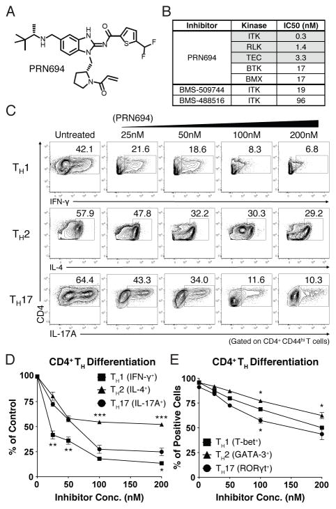
To determine the consequences of Tec kinase inhibition on TH differentiation, we utilized a recently reported small molecule covalent inhibitor of ITK and RLK, PRN694 (Fig. 1A) (15). Based on in vitro kinase assays, PRN694 is a potent inhibitor of all three Tec kinases expressed in T cells, demonstrates less potency toward the other Tec kinases BTK and BMX (Fig. 1B), and shows excellent kinome-wide selectivity (15). As reported, PRN694 binds covalently to a conserved cysteine residue in the ATP binding sites of ITK and RLK (ITK C442, RLK C350), and is highly selective for these two kinases in T cells (15). Unlike its binding to ITK and RLK, PRN694 does not appear to bind covalently to BTK and hence displays a limited duration of BTK inhibition biochemically and in cells (15). Furthermore, PRN694 is 63- to 320-fold more potent in inhibiting ITK than previously-described ITK inhibitors, e.g. BMS-509744 and BMS-488516 (Fig. 1B) (18–22).
Figure 1. PRN694 inhibits CD4+ helper T cell differentiation.
(A) Chemical structure of PRN694. (B) The selectivity and potency of PRN694 was examined by in vitro kinase assay. The table shows the IC50 values for the kinases indicated; for complete data set, see (15). (C–D) Purified naïve mouse splenic CD4+ T cells were stimulated in TH polarizing conditions in the presence of PRN694 at the indicated concentrations for 72hr. CD4+ T cells were then re-stimulated with PMA and Ionomycin for 5hr and analyzed by intracellular cytokine staining. The untreated controls were cultured in the presence of dimethyl sulfoxide (DMSO). The percentages of cytokine-producing CD4+ T cells (gated on CD4+ CD44hi) are shown (C). Compilation of data from three independent experiments indicating the inhibitory effect of PRN694 on the cytokine production are shown. For each cytokine, the data were normalized to the percentage of cytokine-producing cells in the absence of inhibitor (D). (E) Data from three experiments were compiled, and for each TH transcription factor, the data were normalized to the percentage of positive cells in the absence of inhibitor.
To assess the potential effect of PRN694 on CD4+ TH cell differentiation, we utilized naïve CD4+ T cells from B10.A 5C.C7 Rag2−/− TCR Tg mice, and stimulated these cells under TH1-, TH2-, or TH17-polarizing conditions. Cells were cultured for 3 days in the presence of increasing doses of PRN694, and then analyzed for cytokine production after restimulation with PMA and Ionomycin for 5hr. Among these conditions, PRN694 inhibited TH1 differentiation more potently than was observed for TH2 and TH17 differentiation. Whereas a low concentration of PRN694 (25nM) inhibited IFN-γ production by TH1 cells by > 50%, 50–100nM PRN694 was required to achieve a similar level of inhibition of IL-17A production by TH17 cells (Fig. 1C–D). Furthermore, none of the doses tested achieved 50% inhibition of IL-4 production by TH2 cells. The effects of PRN694 on cytokine production did not correlate with effects on cell proliferation (Supplemental Fig. 1A–B), indicating that the inhibition of IFN-γ production by TH1 cells was not due to reduced proliferation of these cells in the presence of PRN694.
We also examined the expression of TH cell lineage-determining transcription factors. Overall, the expression levels of all transcription factors tested (T-bet, GATA-3, and RORγt) were decreased upon the inhibitor treatment; however, the modest differences observed between the three TH cell lineages did not correlate precisely with diminished cytokine production (Fig. 1E). Collectively, these data indicate that PRN694 specifically targets ITK and RLK kinase activity and also impairs CD4+ T helper cell differentiation and cytokine production, with a range of potency as follows: TH1 > TH17 > TH2.
PRN694 inhibits TCR-induced cytokine production from human PBMC and in mouse plasma
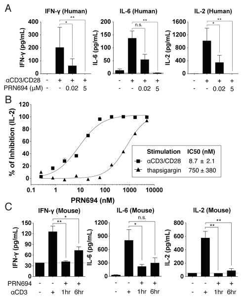
To further study the effect of dual ITK/RLK inhibition on T cell function, human PBMC were stimulated with anti-CD3/CD28 in both the absence and presence of PRN694, and the cytokines produced after 18hr were measured using a biomarker panel. We utilized both a moderate (20nM) and high (5.0μM) concentration of PRN694 and focused on cytokines that were robustly induced in this short-term assay, including IFN-γ, IL-6, and IL-2. The increase in each of these cytokines induced by anti-CD3/CD28 stimulation was completely blocked by 5.0μM of PRN694 and was strongly inhibited by 20nM of PRN694 (Fig. 2A; Supplemental Table 1). To quantify the potency of inhibition of IL-2 production, a 12-concentration dose-range of PRN694 was tested. These data indicated an IC50 value of 8.7 ± 2.1nM (Fig. 2B). In order to confirm that the inhibition of IL-2 production was due to selective ITK and RLK inhibition, we bypassed the requirement for these kinases by stimulating PBMC with the calcium pump inhibitor thapsigargin, and observed that the ability of PRN694 to inhibit IL-2 production was strongly impaired (Fig. 2B). To determine whether PRN694 inhibited these cytokine responses in vivo, mice were injected with a 20 mg/kg intraperitoneal dose of PRN694, followed by injection of anti-CD3 either 1hr or 6hr later to stimulate cytokine production. Plasma was collected 2hr post-anti-CD3 injection and cytokines were analyzed using a cytokine biomarker panel (Fig. 2C). The robust increase in IFN-γ, IL-6, and IL-2 induced by anti-CD3 stimulation was inhibited by PRN694 at both time points (Fig. 2C; Supplemental Table 2).
Figure 2. PRN694 inhibits TCR-induced cytokine production from human PBMC and in mouse plasma.
(A) Human PBMC were stimulated in vitro with anti-CD3/CD28 for 18hr in the absence or presence of PRN694 at 20nM or 5.0μM, and levels of IFN-γ, IL-6, and IL-2 in the supernatants were measured as part of the Human InflammationMap® v.1.0 biomarker panel (Myriad RBM). Complete list of all biomarkers of the panel is found in Supplemental Table 1. Data are shown as mean ± SEM (n=3 per group). (B) A dose-response of PRN694 was used to assess the potency of inhibition of IL-2 production by PBMC. Cells were stimulated with anti-CD3/CD28 (Square) or thapsigargin (Triangle). IL-2 was quantified using an AlphaLISA IL-2 immunoassay. (C–D) Mice were injected i.p. with PRN694 (20 mg/kg) followed by anti-CD3 (10 μg/mouse) 1 or 6hr later. 2hr post-anti-CD3 injection, plasma were collected and cytokines were measured as part of the RodentMap® v.3.0 biomarker panel (Myriad RBM). Complete list of for all biomarkers of the panel is found in Supplemental Table 2. Dotted lines indicate the detection limit for each cytokine in the assay. Data are shown as mean ± SEM (n=3 per group).
PRN694 treatment in vivo ameliorates symptoms in the CD4+ CD45RBhi T cell transfer model of colitis
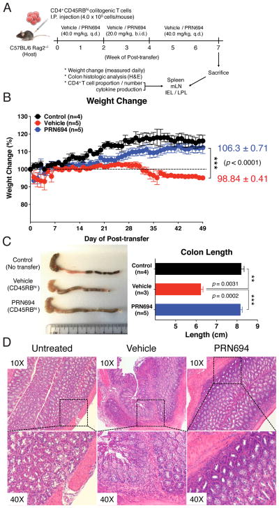
Due to the potent inhibitory effect of PRN694 on IFN-γ production by TH1 cells in in vitro culture experiments, we considered whether PRN694 might function in vivo to suppress a TH1/ IFN-γ-mediated disease. To test this hypothesis, we adoptively transferred WT colitogenic CD4+ CD45RBhi T cells from C57BL/6 mice into RAG2-deficient hosts and monitored the mice for weight loss as a surrogate for disease progression. Previous studies have demonstrated that the colitis induced in this model is due to TH1-mediated inflammation, with little involvement of TH2 or TH17 effector responses (16, 23, 24). In addition to the transferred cells, mice received vehicle alone or PRN694 by oral gavage. Based on studies of ITK target occupancy in thymocytes following in vivo administration of PRN694 in mice (15), we dosed mice daily, and for a two-week period (weeks 2–4), twice daily (Fig. 3A). As expected, recipient mice receiving vehicle alone failed to gain weight and progressively lost weight, beginning 4 weeks after T cell transfer. In contrast, PRN694-treated mice exhibited no weight loss and remained similar to control RAG2-deficient mice that did not receive a colitogenic T cell transfer (Fig. 3B). Consistent with these data, analysis of colon length at 7 weeks post-transfer indicated that the reduced colon length seen in the vehicle-treated mice was prevented in the PRN694-treated mice (Fig. 3C). In addition, histological analysis of the colonic epithelium revealed lymphocytic infiltration in the colon of the vehicle-treated mice (Fig. 3D), in contrast to the reduced inflammation seen in the colon of PRN694-treated mice. To further confirm target engagement of PRN694 with ITK in vivo, splenic CD4+ T cells were isolated from vehicle-dosed or PRN694-dosed mice (2hr or 6hr post-treatment) and then stimulated with anti-CD3/CD28 mAb to examine PLCγ1 phosphorylation. As shown, the phosphorylation of PLCγ1 was completely blocked at both time points following PRN694 administration (Supplemental Fig. 1C). Together, these data demonstrate that PRN694 reduces T cell-mediated colonic inflammation by inhibiting ITK/RLK.
Figure 3. PRN694 administration ameliorates colitis disease progression.
(A–D) Colitogenic CD4+ CD45RBhi splenic T cells (4.0 × 10⁵ cells/mouse) from B6 WT mice were injected i.p. into B6 Rag2−/− hosts. Recipients were treated with vehicle (Red, n=5) or PRN694 (Blue, n=5) with the indicated regimen (A), and disease progression of dosed recipients and untreated (No CD4+ CD45RBhi T cell transfer) Rag2−/− controls (Black, n=4) was monitored by weight change (B). Data are shown as mean ± SEM. At 7 weeks post-transfer, untreated Rag2−/− control, vehicle-dosed, and PRN694-dosed mice were sacrificed for the measurement of colon length (C) and histologic analysis using H&E staining (10X and 40X) (D). Data are compiled from two independent experiments.
PRN694 impairs IFN-γ-mediated TH1 responses and prevents T cell migration to the inflamed colon
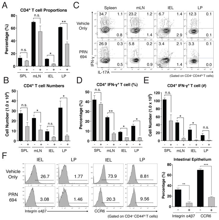
To determine the basis of PRN694-mediated inhibition of colitis, we analyzed the T cell populations in the spleen, mesenteric lymph node (mLN), intestinal epithelium (intraepithelial lymphocytes, IEL), and lamina propria (LP) of recipient Rag2−/− mice at 7 weeks post-transfer. Assessment of CD4+ T cell proportions did not reveal any significant differences in the spleens or mLNs between the vehicle- or PRN694-treated mice, although a modest but significant difference in absolute numbers of CD4+ T cells was observed in mLN (Fig. 4A and 4B). However, CD4+ T cell proportions in intestinal epithelium and LP of PRN694-treated mice were significantly less than those seen in the vehicle-treated mice, along with a statistically significant difference in absolute numbers in the LP (Fig. 4A and 4B). To test whether PRN694 exerted any inhibitory effect on T cell function, we examined cytokine production from transferred colitogenic T cells in several different organs after a brief in vitro stimulation. As expected, CD4+ T cells showed robust IFN-γ production in all analyzed sites of vehicle-treated mice, whereas no IL-17A production was observed (Fig. 4C). Interestingly, PRN694 administration markedly reduced the proportions of CD4+ T cells producing IFN-γ in mesenteric LN, IEL and LP, although this effect was less prominent in the spleen (Fig. 4C and 4D). There was also a large decrease in the numbers of IFN-γ-producing CD4+ T cells in all organs following PRN694 treatment, although the suppression did not reach significance in the LP (Fig. 4E). Consistent with our in vitro studies, the reduced production of IFN-γ by T cells in the PRN694-treated mice could not be accounted for simply by defects in expression of the TH1 transcription factor, T-bet (Supplemental Fig. 2A–B).
Figure 4. PRN694 treatment reduces colitogenic T cell proportions and IFN-γ production in the intestinal epithelium.
(A–B) The proportions and absolute numbers of CD4+ T cells in spleen, mesenteric lymph nodes, intestinal epithelium, and colon lamina propria are shown. (C–E) Isolated lymphocytes from various sites were stimulated with PMA and Ionomycin, and analyzed for IL-17A and IFN-γ production. Dot-plots show gated CD4+ CD44hi T cells (C). The proportions (D) and numbers (E) of IFN-γ-producing CD4+ T cells are shown. Data are compiled from two independent experiments. (F) Histograms show the expression of gut-homing receptors, integrin α4β7 (LPAM-1) and CCR6, on colonic IEL and LP CD4+ CD44hi T cells. Numbers indicate the percentages of cells in each region. Graph shows a compilation of data from 3–5 mice in each group.
Due to the reduced proportion of CD4+ T cells in the inflamed intestines of PRN694-treated mice, we also investigated expression of gut-homing receptors, integrin α4β7 (LPAM-1) and CCR6, on colitogenic T cells (25–27). Consistent with the proportions of gut-infiltrating CD4+ T cells, expression of both integrin α4β7 and CCR6 were reduced on intestinal epithelial CD4+ T cells when PRN694 was administered (Fig. 4F). In contrast, we did not detect any significant level of expression of these receptors on LP-isolated CD4+ T cells from either group of mice (Fig. 4F). Together, these data strongly suggest that PRN694 attenuates in vivo TH1-biased intestinal inflammation through the inhibition of IFN-γ production and the expression of intestine-homing surface receptors.
PRN694 impairs iTreg differentiation and IL-2 production by transferred colitigenic CD4+ T cells
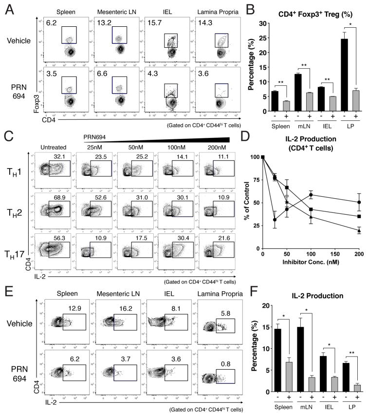
A recent study has shown that Itk−/− CD4+ T cells have an increased propensity to upregulate Foxp3 and differentiate into iTreg cells when stimulated under TH17-polarizing conditions (13). We therefore considered whether the inhibitory effect of PRN694 on colitis might be due to enhanced differentiation of Foxp3+ iTreg cells in the inhibitor-treated mice. Instead, analysis of T cells at 7-weeks post-transfer indicated that PRN694 significantly reduced the proportions of CD4+ T cells expressing Foxp3 compared to controls, a result consistent with the findings of Huang et al. (14). This was evident in all organs examined (Fig. 5A–B). Examination of surface markers commonly found on the majority of Treg cells, including CTLA-4, PD-1, and GITR, also showed reduced expression on Foxp3+ CD4+ T cells in PRN694-treated mice (Supplemental Fig. 2C–E).
Figure 5. PRN694 treatment decreases Treg frequencies and IL-2 production from CD4+ T cells in vitro and in vivo.
(A–B) CD4+ T cells from spleen, mLN, IEL, and lamina propria from vehicle-treated or PRN694-treated mice at 7 weeks of post-transfer were examined for Foxp3 expression. (A) Dot plots show CD4 vs. Foxp3 staining, with numbers indicating the percentages of CD4+ Foxp3+ Treg cells, and (B) the graph shows a compilation of data from 3–5 mice in each group. (C–D) Naïve CD4+ T cells were stimulated in TH polarizing conditions with PRN694 at the indicated concentrations for 72hr. Cells were then re-stimulated with PMA and Ionomycin for 5hr and then analyzed for IL-2 production. (C) The percentages of IL-2-producing CD4+ T cells (gated on CD4+ CD44hi) are shown. (D) Compilation of data from 3 independent experiments is shown, with the data for each TH subset normalized to the percentage of positive cells in the absence of inhibitor. (E–F) Isolated lymphocytes from vehicle-treated or PRN694-treated mice at 7 weeks post-transfer were stimulated with PMA and Ionomycin, and analyzed for IL-2 production. Plots show gated CD4+ CD44hi T cells, and the graph shows a compilation of data from 3–5 mice in each group.
Since upregulation of Foxp3 and iTreg differentiation are dependent on IL-2 (28–31), we examined the effects of PRN694 on IL-2 production by in vitro polarized CD4+ T cells. As shown, PRN694 inhibited IL-2 production by all three lineages of T helper cells (Fig. 5C–D). Consistent with these data, T cells from the adoptive transfer colitis studies also showed reduced proportions of cells capable of producing IL-2 following treatment with PRN694 (Fig. 5E–F). These data indicate that reduced disease progression in mice treated with PRN694 is not a consequence of enhanced differentiation of Foxp3+ iTreg cells, but rather a result of inhibition of differentiation and activation of the IFN-γ-producing TH1 effector cell population.
PRN694 inhibits P-selectin binding activity and chemokine-induced migration of polarized TH1 and TH17 cells
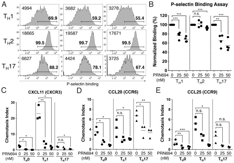
Our results in the adoptive transfer colitis studies showed reduced numbers of CD4+ T cells present in the colonic epithelium of PRN694-treated mice compared to controls. As previous studies using naïve T cells from Itk−/− and Itk−/−Rlk−/− mice had shown a role for these kinases in CCL19- and CXCL12-induced migration (7, 32), we considered whether CD4+ effector T cells might also require ITK and RLK for efficient migration into tissues. Using CD4+ T cells polarized in vitro to TH1, TH2, or TH17 lineages in the presence or absence of PRN694, we first examined binding to P-selectin, a ligand expressed on activated endothelium (33). Both TH1 and TH17 cells showed reduced P-selectin binding following differentiation in the presence of both doses of PRN694 tested; in contrast, TH2 cells showed only a modest reduction, and this reduction was only visible in the higher dose of inhibitor (Fig. 6A–B).
Figure 6. PRN694 leads to impaired P-selectin binding and inhibits chemokine-induced CD4+ T cell migration.
(A–E) Isolated naïve CD4+ T cells were cultured in the presence or absence of PRN694 (25 or 50nM) for 72hr in each TH polarization condition. (A) Histograms show polarized CD4+ T cells stained with recombinant P-selectin-human IgG fusion protein as an assay for PSGL-1 binding. Bold numbers above region indicate the percentages of cells staining positively for P-selectin-Ig based on comparison to an isotype control stain; numbers at the left of each histogram indicate the mean fluorescence intensity (MFI) of P-selectin-Ig staining in each panel. (B) Graph shows a compilation of data from two independent experiments The data were normalized to the percentage of P-selectin binding cells among each TH polarizing condition in the absence of inhibitor. (C–E) Cultured CD4+ T cells (5.0 × 10⁵ cells) were plated in the upper chamber of a HTS Transwell-96 well permeable supports (3.0 μm pore); bottom chambers were loaded with diluted CXCL11 (100 ng/mL) (C), CCL20 (100 ng/mL) (D), or CCL25 (300 ng/mL) (E). After 3hr at 37°C, the numbers of migrated cells were counted by the flow cytometer. The chemotaxis index was calculated by dividing the number of cells that migrated in response to each chemokine by the number of spontaneously migrated cells.
As a second approach, we tested migration of CD4+ effector T cells to chemokines implicated in trafficking to the gastrointestinal epithelium. We focused on responses to CXCL11, a ligand for CXCR3 commonly found on effector TH1 cells (34), CCL20, a ligand for CCR6 found on TH17 cells (35, 36), and CCL25, a ligand for CCR9 (37, 38), which is expressed on gut-resident T cells. Using Transwell migration assay, we found that PRN694 inhibited TH1 migration to CXCL11, with little migration observed by other subsets to this chemokine. CCL20 induced a modest degree of migration of TH17 cells, and this migration was reduced approximately two-fold by PRN694. TH0 and TH1 cells showed fewer cells migrating in response to CCL20, and these responses were further reduced by PRN694. Overall, few cells of any lineage migrated in response to CCL25, with significant inhibition seen only in TH1 and TH0 cells. Polarized TH2 cells showed no migration to any of the chemokines tested (not shown). Overall, these data confirm that PRN694 impacts differentiating TH1 and TH17 cells to reduce their ability to bind to activated endothelium and to migrate to inflammatory chemokines.
Discussion
In this report, we show that inhibition of ITK and RLK by PRN694 leads to a significant impairment in CD4+ TH cell differentiation and to protection from TH1-mediated colitis. Although PRN694 treatment inhibited the differentiation of all CD4+ TH subsets in vitro, the most potent inhibitory effect we observed was on TH1 differentiation. These findings are consistent with a recent study using an allele-sensitive mutant of ITK coupled with a selective inhibitor, which also revealed an important role for ITK kinase activity in TH1 differentiation and cytokine production (39). Our data with PRN694 are also consistent with previous studies showing that Itk−/− Rlk/− mice mounted a normal protective type II cytokine response against Schistosoma mansoni, but were highly susceptible to the intracellular protozoan, Toxoplasma gondii (5, 6). Furthermore, these results strongly suggest that a dual inhibitor of ITK and RLK would likely have a different effect on TH2-skewed inflammation such as asthma or atopic diseases compared to an inhibitor that was selective for ITK alone.
Also consistent with previous studies using Itk−/− T cells (12), we observed that PRN694 was a potent inhibitor of TH17 differentiation and IL-17A production in vitro. Nonetheless, it is unlikely that inhibition of TH17 responses can account for the effects of PRN694 in ameliorating disease in our colitis experiments. While some studies have shown a role for TH17 cells in colitis (40–42), these effects were seen either when colitogenic BALB/c T cells were transferred to syngeneic hosts (41) or when the cecal bacterial antigen-specific C3H/HeJBir CD4+ T cell lines were transferred to C3H.SCID hosts and then restimulated for 10 days in the presence of bacterial antigen-pulsed DCs and with or without IL-23 prior to analysis (42). In contrast, a study using a protocol most similar to our own found that colitis was actually enhanced when TH17 responses were eliminated (40), and the transfer of naïve CD4+ T cells from Il17a−/− mice still induced a severe colitis (41). Furthermore, several other reports suggest that TH17 responses arise early on during colitis disease progression and that in vitro polarized TH17 cells are eventually converted into TH1 cells after adoptive transfer to lymphopenic hosts (43, 44). Using this CD4+ CD45RBhi adoptive transfer model of T cell-mediated colitis, we were unable to detect IL-17A production from colitogenic T cells in mice that were treated with vehicle alone, suggesting that TH17 responses were not contributing to disease progression in our studies. Thus, we were unable to assess the efficacy of PRN694 to inhibit IL-17A production in vivo in this disease model.
In the current study, we cannot rule out an effect of PRN694 on the Tec kinase family member, Tec. Previous studies have indicated that Tec mRNA expression is negligible in primary T cells examined ex vivo, including memory phenotype cells (45) (www.immgen.org) and that only modest increases in Tec protein expression (< 2-fold) are observed even after TH1 polarization in vitro (46). Furthermore, studies of T cell responses in Tec−/− mice have failed to reveal any significant defects due to the absence of Tec. Therefore, it seems unlikely that the inhibitory effects of PRN694 observed in our studies are due to its effect on Tec activity in T cells.
Interestingly, we observed reduced differentiation of Foxp3+ iTreg cells in PRN694-treated mice compared to controls. This is in contrast to a previous study showing increased differentiation Itk−/− naïve CD4+ cells into of Foxp3+ iTreg cells in a similar adoptive transfer model of colitis (13). The difference in these two sets of findings could be due to a number of factors. First, the numbers of cells transferred and the time points analyzed were each different in the two studies. More importantly, there may be an unappreciated difference in the starting populations of CD4+ CD45RBhi CD25− T cells isolated from WT mice, as in our studies, compared to those isolated from Itk−/− mice. In addition, PRN694 is a potent inhibitor of both ITK and RLK, and this dual inhibition may lead to a different outcome than the single genetic deficiency in Itk. It is also important to consider that a small molecule kinase inhibitor might have a different effect than a genetic deficiency that causes a complete absence of ITK protein, as the former situation would not abolish any kinase-independent functions of ITK. Finally, it is important to consider the possibility that mice housed in two different environments have significant differences in their microbiota, and that these differences may impact iTreg differentiation in vivo.
Our studies indicate that PRN694 treatment reduces the numbers of colitogenic CD4+ T cells in the intestinal epithelium at 7 weeks post-transfer. It is possible that one component of this decrease is reduced proliferation of the T cells in the presence of PRN694, although our in vitro studies examining proliferation suggest that this explanation is unlikely. Instead, we propose that impaired T cell migration to the gastrointestinal (GI) tract is contributing to this deficit. We find that PRN694 treatment during T helper cell polarization leads to reduced P-selectin binding on TH1 and TH17 cells, an alteration that could impact T cell rolling on the activated endothelium prior to extravasation into the GI tract. This possibility is consistent with our previous studies showing impaired tissue homing and transendothelial migration of Itk−/− T cells in an autoimmune disease model (47). Furthermore, we observed reduced expression of integrin α4β7 and CCR6, and reduced in vitro chemotaxis to inflammatory chemokines, by PRN694-treated CD4+ T cells. Together, these data provide strong support for the conclusion that ITK/RLK inhibition leads to impaired T cell migration into the GI tissue.
Overall, we show that PRN694 inhibits CD4+ effector T cell differentiation and cytokine production, particularly for TH1 and TH17 lineage cells. Together with the inhibitory effect of PRN694 on T cell migration and on colitis disease progression, the data presented here provide strong support for the development of a combined ITK/RLK inhibitor as a potential therapeutic strategy for TH1-mediated, and possibly TH17-mediated, inflammatory diseases.
Supplementary Material
Acknowledgments
We thank all members of the Berg lab, especially Regina Whitehead and Sharlene Hubbard for their technical assistance. We also thank UMMS Department of Animal Medicine for the maintenance of mouse colonies and Flow Cytometry Core Facility for the assistance in cell sorting.
This work was supported by NIH Grants AI084987 (to L.J.B.) and AI083505 (to L.J.B.).
Footnotes
Disclosures
Authors H-S.C., H.M.S., and L.J.B. have no financial conflicts of interest. Authors H.H, Y.X., T.O., J.O.F., R.H., and J.M.B. declare competing financial interests. They are members of a biotechnology company developing molecules for inflammatory and autoimmune diseases.
References
- 1.Berg LJ, Finkelstein LD, Lucas JA, Schwartzberg PL. Tec family kinases in T lymphocyte development and function. Annu Rev Immunol. 2005;23:549–600. doi: 10.1146/annurev.immunol.22.012703.104743. [DOI] [PubMed] [Google Scholar]
- 2.Miller AT, Wilcox HM, Lai Z, Berg LJ. Signaling through Itk promotes T helper 2 differentiation via negative regulation of T-bet. Immunity. 2004;21:67–80. doi: 10.1016/j.immuni.2004.06.009. [DOI] [PubMed] [Google Scholar]
- 3.Colgan J, Asmal M, Neagu M, Yu B, Schneidkraut J, Lee Y, Sokolskaja E, Andreotti A, Luban J. Cyclophilin A regulates TCR signal strength in CD4+ T cells via a proline-directed conformational switch in Itk. Immunity. 2004;21:189–201. doi: 10.1016/j.immuni.2004.07.005. [DOI] [PubMed] [Google Scholar]
- 4.Andreotti AH, Schwartzberg PL, Joseph RE, Berg LJ. T-cell signaling regulated by the Tec family kinase, Itk. Cold Spring Harb Perspect Biol. 2010;2:a002287–a002287. doi: 10.1101/cshperspect.a002287. [DOI] [PMC free article] [PubMed] [Google Scholar]
- 5.Schaeffer EM, Debnath J, Yap G, McVicar D, Liao XC, Littman DR, Sher A, Varmus HE, Lenardo MJ, Schwartzberg PL. Requirement for Tec kinases Rlk and Itk in T cell receptor signaling and immunity. Science. 1999;284:638–641. doi: 10.1126/science.284.5414.638. [DOI] [PubMed] [Google Scholar]
- 6.Schaeffer EM, Yap GS, Lewis CM, Czar MJ, McVicar DW, Cheever AW, Sher A, Schwartzberg PL. Mutation of Tec family kinases alters T helper cell differentiation. Nat Immunol. 2001;2:1183–1188. doi: 10.1038/ni734. [DOI] [PubMed] [Google Scholar]
- 7.Takesono A, Horai R, Mandai M, Dombroski D, Schwartzberg PL. Requirement for Tec kinases in chemokine-induced migration and activation of Cdc42 and Rac. Curr Biol. 2004;14:917–922. doi: 10.1016/j.cub.2004.04.011. [DOI] [PubMed] [Google Scholar]
- 8.Ellmeier W, Jung S, Sunshine MJ, Hatam F, Xu Y, Baltimore D, Mano H, Littman DR. Severe B cell deficiency in mice lacking the tec kinase family members Tec and Btk. J Exp Med. 2000;192:1611–1624. doi: 10.1084/jem.192.11.1611. [DOI] [PMC free article] [PubMed] [Google Scholar]
- 9.Schaeffer EM, Broussard C, Debnath J, Anderson S, McVicar DW, Schwartzberg PL. Tec family kinases modulate thresholds for thymocyte development and selection. J Exp Med. 2000;192:987–1000. doi: 10.1084/jem.192.7.987. [DOI] [PMC free article] [PubMed] [Google Scholar]
- 10.Fowell DJ, Shinkai K, Liao XC, Beebe AM, Coffman RL, Littman DR, Locksley RM. Impaired NFATc translocation and failure of Th2 development in Itk-deficient CD4+ T cells. Immunity. 1999;11:399–409. doi: 10.1016/s1074-7613(00)80115-6. [DOI] [PubMed] [Google Scholar]
- 11.Sahu N, Venegas AM, Jankovic D, Mitzner W, Gomez-Rodriguez J, Cannons JL, Sommers C, Love P, Sher A, Schwartzberg PL, August A. Selective expression rather than specific function of Txk and Itk regulate Th1 and Th2 responses. J Immunol. 2008;181:6125–6131. doi: 10.4049/jimmunol.181.9.6125. [DOI] [PMC free article] [PubMed] [Google Scholar]
- 12.Gomez-Rodriguez J, Sahu N, Handon R, Davidson TS, Anderson SM, Kirby MR, August A, Schwartzberg PL. Differential expression of interleukin-17A and -17F is coupled to T cell receptor signaling via inducible T cell kinase. Immunity. 2009;31:587–597. doi: 10.1016/j.immuni.2009.07.009. [DOI] [PMC free article] [PubMed] [Google Scholar]
- 13.Gomez-Rodriguez J, Wohlfert EA, Handon R, Meylan F, Wu JZ, Anderson SM, Kirby MR, Belkaid Y, Schwartzberg PL. Itk-mediated integration of T cell receptor and cytokine signaling regulates the balance between Th17 and regulatory T cells. J Exp Med. 2014;211:529–543. doi: 10.1084/jem.20131459. [DOI] [PMC free article] [PubMed] [Google Scholar]
- 14.Huang W, Jeong A-R, Kannan AK, Huang L, August A. IL-2-inducible T cell kinase tunes T regulatory cell development and is required for suppressive function. J Immunol. 2014;193:2267–2272. doi: 10.4049/jimmunol.1400968. [DOI] [PMC free article] [PubMed] [Google Scholar]
- 15.Zhong Y, Dong S, Strattan E, Ren L, Butchar JP, Thornton K, Mishra A, Porcu P, Bradshaw JM, Bisconte A, Owens TD, Verner E, Brameld KA, Funk JO, Hill RJ, Johnson AJ, Dubovsky JA. Targeting Interleukin-2-inducible T-cell Kinase (ITK) and Resting Lymphocyte Kinase (RLK) Using a Novel Covalent Inhibitor PRN694. J Biol Chem. 2015 doi: 10.1074/jbc.M114.614891. jbc.M114.614891. [DOI] [PMC free article] [PubMed] [Google Scholar]
- 16.Powrie F, Leach MW, Mauze S, Menon S, Caddle LB, Coffman RL. Inhibition of Th1 responses prevents inflammatory bowel disease in scid mice reconstituted with CD45RBhi CD4+ T cells. Immunity. 1994;1:553–562. doi: 10.1016/1074-7613(94)90045-0. [DOI] [PubMed] [Google Scholar]
- 17.Read S, Powrie F. Induction of inflammatory bowel disease in immunodeficient mice by depletion of regulatory T cells. Curr Protoc Immunol. 2001;Chapter 15(Unit 15.13–15.13.10) doi: 10.1002/0471142735.im1513s30. [DOI] [PubMed] [Google Scholar]
- 18.Lin T-A, McIntyre KW, Das J, Liu C, O’Day KD, Penhallow B, Hung C-Y, Whitney GS, Shuster DJ, Yang X, Townsend R, Postelnek J, Spergel SH, Lin J, Moquin RV, Furch JA, Kamath AV, Zhang H, Marathe PH, Perez-Villar JJ, Doweyko A, Killar L, Dodd JH, Barrish JC, Wityak J, Kanner SB. Selective Itk inhibitors block T-cell activation and murine lung inflammation. Biochemistry. 2004;43:11056–11062. doi: 10.1021/bi049428r. [DOI] [PubMed] [Google Scholar]
- 19.Das J, Furch JA, Liu C, Moquin RV, Lin J, Spergel SH, McIntyre KW, Shuster DJ, O’Day KD, Penhallow B, Hung C-Y, Doweyko AM, Kamath A, Zhang H, Marathe P, Kanner SB, Lin T-A, Dodd JH, Barrish JC, Wityak J. Discovery and SAR of 2-amino-5-(thioaryl)thiazoles as potent and selective Itk inhibitors. Bioorg Med Chem Lett. 2006;16:3706–3712. doi: 10.1016/j.bmcl.2006.04.060. [DOI] [PubMed] [Google Scholar]
- 20.Snow RJ, Abeywardane A, Campbell S, Lord J, Kashem MA, Khine HH, King J, Kowalski JA, Pullen SS, Roma T, Roth GP, Sarko CR, Wilson NS, Winters MP, Wolak JP, Cywin CL. Hit-to-lead studies on benzimidazole inhibitors of ITK: discovery of a novel class of kinase inhibitors. Bioorg Med Chem Lett. 2007;17:3660–3665. doi: 10.1016/j.bmcl.2007.04.045. [DOI] [PubMed] [Google Scholar]
- 21.Riether D, Zindell R, Kowalski JA, Cook BN, Bentzien J, Lombaert SD, Thomson D, Kugler SZ, Skow D, Martin LS, Raymond EL, Khine HH, O’Shea K, Woska JR, Jeanfavre D, Sellati R, Ralph KLM, Ahlberg J, Labissiere G, Kashem MA, Pullen SS, Takahashi H. 5-Aminomethylbenzimidazoles as potent ITK antagonists. Bioorg Med Chem Lett. 2009;19:1588–1591. doi: 10.1016/j.bmcl.2009.02.012. [DOI] [PubMed] [Google Scholar]
- 22.Sahu N, August A. ITK inhibitors in inflammation and immune-mediated disorders. Curr Top Med Chem. 2009;9:690–703. doi: 10.2174/156802609789044443. [DOI] [PMC free article] [PubMed] [Google Scholar]
- 23.Neurath MF, Weigmann B, Finotto S, Glickman J, Nieuwenhuis E, Iijima H, Mizoguchi A, Mizoguchi E, Mudter J, Galle PR, Bhan A, Autschbach F, Sullivan BM, Szabo SJ, Glimcher LH, Blumberg RS. The transcription factor T-bet regulates mucosal T cell activation in experimental colitis and Crohn’s disease. J Exp Med. 2002;195:1129–1143. doi: 10.1084/jem.20011956. [DOI] [PMC free article] [PubMed] [Google Scholar]
- 24.Strober W, Fuss IJ. Proinflammatory cytokines in the pathogenesis of inflammatory bowel diseases. Gastroenterology. 2011;140:1756–1767. doi: 10.1053/j.gastro.2011.02.016. [DOI] [PMC free article] [PubMed] [Google Scholar]
- 25.Petrovic A, Alpdogan O, Willis LM, Eng JM, Greenberg AS, Kappel BJ, Liu C, Murphy GJ, Heller G, van den Brink MRM. LPAM (alpha 4 beta 7 integrin) is an important homing integrin on alloreactive T cells in the development of intestinal graft-versus-host disease. Blood. 2004;103:1542–1547. doi: 10.1182/blood-2003-03-0957. [DOI] [PubMed] [Google Scholar]
- 26.Varona R, Cadenas V, Gómez L, Martínez-A C, Márquez G. CCR6 regulates CD4+ T-cell-mediated acute graft-versus-host disease responses. Blood. 2005;106:18–26. doi: 10.1182/blood-2004-08-2996. [DOI] [PubMed] [Google Scholar]
- 27.Monteiro P, Gosselin A, Wacleche VS, El-Far M, Said EA, Kared H, Grandvaux N, Boulassel M-R, Routy J-P, Ancuta P. Memory CCR6+CD4+ T cells are preferential targets for productive HIV type 1 infection regardless of their expression of integrin β7. J Immunol. 2011;186:4618–4630. doi: 10.4049/jimmunol.1004151. [DOI] [PubMed] [Google Scholar]
- 28.Boyman O, Sprent J. The role of interleukin-2 during homeostasis and activation of the immune system. Nat Rev Immunol. 2012;12:180–190. doi: 10.1038/nri3156. [DOI] [PubMed] [Google Scholar]
- 29.Malek TR, Castro I. Interleukin-2 receptor signaling: at the interface between tolerance and immunity. Immunity. 2010;33:153–165. doi: 10.1016/j.immuni.2010.08.004. [DOI] [PMC free article] [PubMed] [Google Scholar]
- 30.Yu A, Zhu L, Altman NH, Malek TR. A low interleukin-2 receptor signaling threshold supports the development and homeostasis of T regulatory cells. Immunity. 2009;30:204–217. doi: 10.1016/j.immuni.2008.11.014. [DOI] [PMC free article] [PubMed] [Google Scholar]
- 31.Fontenot JD, Rasmussen JP, Gavin MA, Rudensky AY. A function for interleukin 2 in Foxp3-expressing regulatory T cells. Nat Immunol. 2005;6:1142–1151. doi: 10.1038/ni1263. [DOI] [PubMed] [Google Scholar]
- 32.Fischer AM, Mercer JC, Iyer A, Ragin MJ, August A. Regulation of CXC chemokine receptor 4-mediated migration by the Tec family tyrosine kinase ITK. J Biol Chem. 2004;279:29816–29820. doi: 10.1074/jbc.M312848200. [DOI] [PubMed] [Google Scholar]
- 33.Ley K, Kansas GS. Selectins in T-cell recruitment to non-lymphoid tissues and sites of inflammation. Nat Rev Immunol. 2004;4:325–335. doi: 10.1038/nri1351. [DOI] [PubMed] [Google Scholar]
- 34.Qin S, Rottman JB, Myers P, Kassam N, Weinblatt M, Loetscher M, Koch AE, Moser B, Mackay CR. The chemokine receptors CXCR3 and CCR5 mark subsets of T cells associated with certain inflammatory reactions. J Clin Invest. 1998;101:746–754. doi: 10.1172/JCI1422. [DOI] [PMC free article] [PubMed] [Google Scholar]
- 35.Esplugues E, Huber S, Gagliani N, Hauser AE, Town T, Wan YY, O’Connor W, Rongvaux A, Van Rooijen N, Haberman AM, Iwakura Y, Kuchroo VK, Kolls JK, Bluestone JA, Herold KC, Flavell RA. Control of TH17 cells occurs in the small intestine. Nature. 2011;475:514–518. doi: 10.1038/nature10228. [DOI] [PMC free article] [PubMed] [Google Scholar]
- 36.Wang C, Kang SG, Lee J, Sun Z, Kim CH. The roles of CCR6 in migration of Th17 cells and regulation of effector T-cell balance in the gut. Mucosal Immunol. 2009;2:173–183. doi: 10.1038/mi.2008.84. [DOI] [PMC free article] [PubMed] [Google Scholar]
- 37.Stenstad H, Ericsson A, Johansson-Lindbom B, Svensson M, Marsal J, Mack M, Picarella D, Soler D, Márquez G, Briskin M, Agace WW. Gut-associated lymphoid tissue-primed CD4+ T cells display CCR9-dependent and -independent homing to the small intestine. Blood. 2006;107:3447–3454. doi: 10.1182/blood-2005-07-2860. [DOI] [PubMed] [Google Scholar]
- 38.Cassani B, Villablanca EJ, Quintana FJ, Love PE, Lacy-Hulbert A, Blaner WS, Sparwasser T, Snapper SB, Weiner HL, Mora JR. Gut-tropic T cells that express integrin α4β7 and CCR9 are required for induction of oral immune tolerance in mice. Gastroenterology. 2011;141:2109–2118. doi: 10.1053/j.gastro.2011.09.015. [DOI] [PMC free article] [PubMed] [Google Scholar]
- 39.Kannan A, Lee Y, Qi Q, Huang W, Jeong A-R, Ohnigian S, August A. Allele-sensitive mutant, Itkas, reveals that Itk kinase activity is required for Th1, Th2, Th17, and iNKT-cell cytokine production. Eur J Immunol. 2015;45:2276–2285. doi: 10.1002/eji.201445087. [DOI] [PMC free article] [PubMed] [Google Scholar]
- 40.O’Connor W, Kamanaka M, Booth CJ, Town T, Nakae S, Iwakura Y, Kolls JK, Flavell RA. A protective function for interleukin 17A in T cell-mediated intestinal inflammation. Nat Immunol. 2009;10:603–609. doi: 10.1038/ni.1736. [DOI] [PMC free article] [PubMed] [Google Scholar]
- 41.Noguchi D, Wakita D, Tajima M, Ashino S, Iwakura Y, Zhang Y, Chamoto K, Kitamura H, Nishimura T. Blocking of IL-6 signaling pathway prevents CD4+ T cell-mediated colitis in a T(h)17-independent manner. Int Immunol. 2007;19:1431–1440. doi: 10.1093/intimm/dxm114. [DOI] [PubMed] [Google Scholar]
- 42.Elson CO, Cong Y, Weaver CT, Schoeb TR, McClanahan TK, Fick RB, Kastelein RA. Monoclonal anti-interleukin 23 reverses active colitis in a T cell-mediated model in mice. Gastroenterology. 2007;132:2359–2370. doi: 10.1053/j.gastro.2007.03.104. [DOI] [PubMed] [Google Scholar]
- 43.Feng T, Qin H, Wang L, Benveniste EN, Elson CO, Cong Y. Th17 cells induce colitis and promote Th1 cell responses through IL-17 induction of innate IL-12 and IL-23 production. J Immunol. 2011;186:6313–6318. doi: 10.4049/jimmunol.1001454. [DOI] [PMC free article] [PubMed] [Google Scholar]
- 44.Harbour SN, Maynard CL, Zindl CL, Schoeb TR, Weaver CT. Th17 cells give rise to Th1 cells that are required for the pathogenesis of colitis. Proc Natl Acad Sci USA. 2015;112:7061–7066. doi: 10.1073/pnas.1415675112. [DOI] [PMC free article] [PubMed] [Google Scholar]
- 45.Felices M, Yin CC, Kosaka Y, Kang J, Berg LJ. Tec kinase Itk in gammadeltaT cells is pivotal for controlling IgE production in vivo. Proc Natl Acad Sci USA. 2009;106:8308–8313. doi: 10.1073/pnas.0808459106. [DOI] [PMC free article] [PubMed] [Google Scholar]
- 46.Tomlinson MG, Kane LP, Su J, Kadlecek TA, Mollenauer MN, Weiss A. Expression and function of Tec, Itk, and Btk in lymphocytes: evidence for a unique role for Tec. Mol Cell Biol. 2004;24:2455–2466. doi: 10.1128/MCB.24.6.2455-2466.2004. [DOI] [PMC free article] [PubMed] [Google Scholar]
- 47.Jain N, Miu B, Jiang J-K, McKinstry KK, Prince A, Swain SL, Greiner DL, Thomas CJ, Sanderson MJ, Berg LJ, Kang J. CD28 and ITK signals regulate autoreactive T cell trafficking. Nat Med. 2013;19:1632–1637. doi: 10.1038/nm.3393. [DOI] [PMC free article] [PubMed] [Google Scholar]
Associated Data
This section collects any data citations, data availability statements, or supplementary materials included in this article.


