Abstract
MicroRNAs (miRNAs) are important post-transcriptional regulators of gene expression and are implicated in the etiology of several neuropsychiatric disorders, including substance use disorders (SUDs). Using in silico genome-wide sequence analyses, we identified miR-495 as a miRNA whose predicted targets are significantly enriched in the Knowledgebase of Addiction-Related Genes (ARG) database (KARG; http://karg.cbi.pku.edu.cn). This small non-coding RNA is also highly expressed within the nucleus accumbens (NAc), a pivotal brain region underlying reward and motivation. Using luciferase reporter assays, we found that miR-495 directly targeted the 3’UTRs of Bdnf, Camk2a, and Arc. Furthermore, we measured miR-495 expression in response to acute cocaine in mice and found that it is downregulated rapidly and selectively in the NAc, along with concomitant increases in ARG expression. Lentiviral-mediated miR-495 overexpression in the NAc shell (NAcsh) not only reversed these cocaine-induced effects, but also downregulated multiple ARG mRNAs in specific SUD-related biological pathways, including those that regulate synaptic plasticity. miR-495 expression was also downregulated in the NAcsh of rats following cocaine self-administration. Most importantly, we found that NAcsh miR-495 overexpression suppressed the motivation to self-administer and seek cocaine across progressive ratio, extinction, and reinstatement testing, but had no effect on food reinforcement, suggesting that miR-495 selectively affects addiction-related behaviors. Overall, our in silico search for post-transcriptional regulators identified miR-495 as a novel regulator of multiple ARGs that play a role in modulating motivation for cocaine.
Keywords: microRNA, addiction, BDNF, Camk2a, self-administration
INTRODUCTION
Substance use disorder (SUD) is a chronic, debilitating condition characterized by compulsive drug use despite negative consequences and a high recurrence of relapse even after prolonged periods of abstinence (1, 2). SUD is believed to be a dysfunction of neuroplasticity (3), whereby altered gene expression impacts neuronal function and subsequent behavior (4). Drugs of abuse cause widespread epigenetic changes to chromatin accessibility, thereby altering the transcriptional activity of several genes (5–7). However, less is known about the post-transcriptional processes that control mRNA dynamics and, ultimately, translation into functional proteins. Among the non-coding RNAs, microRNAs (miRNAs) play a critical role in the post-transcriptional control of a large number of transcripts. These small RNAs typically guide the RNA-induced silencing complex (RISC) through the binding of complementary sequences in the 3’ untranslated region (3’UTR) of the target mRNAs, leading to mRNA degradation or translational repression (8). A single miRNA is predicted to target hundreds of different mRNAs, and a single mRNA can be regulated by multiple miRNAs. Therefore, dysregulation of these “master” regulators impacts several cellular processes simultaneously and has been linked to many diseases and neurological disorders (9–13).
Recent studies indicate that several drugs of abuse regulate miRNA expression in the nucleus accumbens (NAc) and other regions of the brain reward pathway (14–18). In turn, in vivo manipulations of specific miRNAs or subsets of miRNAs alter the development of addiction-like behaviors in rodents (18–22). Thus, we sought to identify a candidate miRNA that targets multiple addiction-related genes (ARGs) and may provide a novel mechanism for - reverting drug-induced aberrant gene expression. In this study, we used bioinformatics analyses of the 3’ untranslated regions (3’UTRs) of transcripts in the Knowledgebase of Addiction-Related Genes database (KARG; 23) to identify miR-495, a miRNA that targets many ARGs in regulatory networks previously implicated in SUDs. We found that miR-495 is enriched within the NAc and is downregulated by acute cocaine administration and during cocaine self-administration. Viral-mediated miR-495 overexpression not only robustly downregulated ARG expression but more importantly, diminished motivation for cocaine.
MATERIALS AND METHODS
Animals
Male 2-month-old C57BL/6J mice (Jackson Labs, Bar Harbor, ME, USA) and adult 2-month-old Sprague Dawley rats (Charles River, San Diego, CA, USA) were maintained on a 12-h and 14/10-h reverse light/dark cycle, respectively. Animal studies were performed in accordance with NIH Animal Welfare guidelines under protocols approved by the Institutional Animal Care and Use committees at the University of New Mexico and Arizona State University.
Bioinformatics analyses
The lists for mouse, human, and rat ARGs were retrieved from the KARG database (http://karg.cbi.pku.edu.cn, 23). Lists of KARG genes with evidence number scores ≥2 (23) were used to acquire the 3'UTRs sequences from ENSEMBL BioMart. The frequencies of predicted targets of miR-495 and two previously identified addiction-related miRNAs, let-7 and miR-212 (20, 21), in these KARG lists vs. the respective genomes were calculated using TargetScan 6.2 (http://www.targetscan.org) conserved sites. miR-495 binding sites in the 3’UTR of the KARG/TargetScan dataset were further validated using miRanda (24).
NAc shell (NAcsh) viral injections and cocaine self-administration, extinction, and reinstatement
Rats were trained to self-administer cocaine, infused with lentiviruses (LV) containing either green fluorescent protein (GFP; LV-GFP) or GFP+miR-495 (LV-miR-495) into the NAcsh, and then tested on a fixed ratio (FR) 5 and progressive ratio (PR) schedule of cocaine reinforcement, as well as during extinction and cue and cocaine-primed reinstatement, as described in Supplementary Information.
Data Analysis
Power analyses were performed to determine adequate sample sizes (PASS, NCSS software, Kaysville, UT, USA). Behavioral and biochemical measures were analyzed using Student t tests or ANOVAs followed by tests for simple effects, where appropriate, using SPSS 24.0 (IBM, Armonk, NY, USA). Adjustments to degrees of freedom were made when unequal variances between groups existed (e.g., Welch’s correction, Huynh-Feldt correction).
Full materials and methods for miRNA fluorescent in situ hybridization, dual luciferase assays, acute cocaine treatment, reverse transcription and qPCR, western blotting, intracranial virus injections, cocaine self-administration, food reinforcement, and histology are provided in the Supplementary Information.
RESULTS
All statistical values are reported in Supplementary Table 1.
In silico analyses identify miR-495 as a putative post-transcriptional regulator of addiction-related genes in the nucleus accumbens
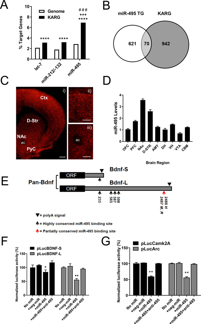
Initial bioinformatics analyses were aimed at identifying miRNAs that target addiction-associated mRNAs expressed in the NAc. The 3’UTR sequences of mouse, human, and rat gene sets of the KARG database (http://karg.cbi.pku.edu.cn) with evidence scores ≥ 2 (23) were used to determine the prevalence of miRNA binding sites predicted by TargetScan (http://www.targetscan.org). Among the ARG-targeting miRNAs, we found that miR-495, a microRNA expressed in the adult rat striatum (http://miRBase.org, 25, 26), is predicted to target several ARG mRNAs, such as Bdnf, Camk2a, Arc, and others (Supplementary Table S2). The percentage of mouse KARG genes containing conserved 3’UTR miR-495 binding sites (7%, 70 genes) is significantly higher than in the entire genome (2.5%), and similar results were obtained using human or rat KARG gene sets (83 genes, 7.7% and 55 genes, 5.6 %, respectively). To confirm this method, we assessed the proportion of KARG gene targets of two miRNAs previously associated with cocaine addiction, miR-212/-132 (21) and let-7 (14, 20). As expected, both miRNAs targeted a higher % of KARG genes than in the genome (miR-212/-132: 2.8%; let-7: 3%), confirming the utility of this approach to identify miRNAs associated with addiction. Given that the frequency of miR-495 targets in KARG was significantly higher (~2-fold) than those for miR212/-132 and let-7 (Figure 1A, B), it is likely that miR-495 targets may impact a wider variety of functions involved in addiction than those that were established for miR-212/-132 and let-7. Furthermore, the average evidence scores for miR-495 KARG targets were significantly higher than those for the whole KARG set, suggesting the association of predicted miR-495 KARG targets with addiction is heavily supported by previous research (Supplementary Figure S1). Using miRanda, we further validated the presence of high affinity 3’UTR miR-495 binding sites (ΔG ≤ -15 kcal/mol) in the mouse dataset, including Bdnf and Camk2a (Supplementary Table S2; 24). Fluorescent in situ hybridization (FISH) confirmed brain-wide miR-495 expression, including the NAc and medial prefrontal cortex (mPFC), as previously reported in human mPFC tissue (Figure 1C; scrambled locked nucleic acid (LNA) control in Supplementary Figure S2; 27). Using qRT-PCR, we confirmed miR-495 expression in these regions, with the highest expression within the NAc (Figure 1D). Thus, miR-495 is a candidate regulator of a set of ARGs conserved in mammals.
Figure 1. miR-495 targets several addiction-related genes (ARGs) and is expressed in addiction-related brain regions.
(A) Although the frequencies of miR-495, miR-212/132 and let-7 putative targets are all enriched in the KARG database compared to the entire genome, the frequency of miR-495 targets in KARG is significantly higher (~2-fold) than those of miR-212/132 and let-7. ****p < 0.0001 vs. genome, +++p < 0.001 miR-495 vs. miR-212/132 and # # # p< 0.001 miR-495 vs. let-7, two-tailed Chi square test. (B) Number of genes with putative miR-495 target sites (miR-495 TG) in the mouse KARG set. (C) Representative images of a coronal mouse brain section where miR-495 was visualized using fluorescent in situ hybridization at 4× (i), with insets at 10× focusing on the PFC (ii) and the NAc (iii). Scale bars 500 µm in panel i and 200 µm in ii and iii. (D) qRT-PCR analysis of miR-495 levels in different brain regions (n = 3). (E) Schematic representation of the short and long 3’UTR transcripts of BDNF including the positions of conserved and partially conserved miR-495 binding sites (M = Mus musculus, R = Rattus norvegicus, H = Homo sapiens). For in vitro target validation, HeLa cells were transfected with a firefly luciferase reporter containing the 3’UTR of Bdnf and Camk2a. A Renilla vector was co-transfected with the firefly reporter. Pre-miR-495, anti-miR-495 and pre-miR™ miRNA precursor negative control #2 were transfected as described in Supplementary Information. The alternative 3’UTRs of BDNF (F), as well as the 3’UTRs of Camk2a and Arc, were assayed (G). n = 4 *p < 0.05, **p < 0.01. Error bars indicate SEM. Ctx = neocortex, PyC = pyriform cortex, ac = anterior commissure. OFC = orbitofrontal cortex, PFC = prefrontal cortex, NAc = nucleus accumbens, D-STR = dorsal striatum, AMY = amygdala, DH = dorsal hippocampus, VH = ventral hippocampus, VTA = ventral tegmental area, CBM = cerebellum.
miR-495 directly targets the 3’UTRs of Bdnf, Camk2a, and Arc
To validate direct miR-495 binding to predicted target ARG mRNAs, we utilized luciferase reporter constructs containing target mRNA 3’UTRs. Due to differential poly(A) site usage, the predicted miR-495 target, Bdnf, is present in vivo as two different transcripts with a short (Bdnf-S) or long 3’UTR (Bdnf-L) produced from the same promoter. The long form contains more miR-495 binding sites (Figure 1E; http://www.targetscan.org/mmu_50/), suggesting that miR-495 preferentially regulates Bdnf-L. The binding sites within the 3’UTR at nucleotide (nt) positions 233, 565, 587, and 598 are highly conserved between mouse, rat and human, while the last binding site at nt 2487 in mouse and rat or nt 2488 in human is partially conserved. Indeed, dual-luciferase assays showed that miR-495 significantly reduced the activity of the reporter containing the 3’UTR for Bdnf-L by ~50% and for Bdnf-S by ~20% (Figure 1F). Given that these isoforms have been hypothesized to have different functions and localization within the neuron, these results suggest that miR-495 may preferentially regulate Bdnf-L and its associated functions (28). Additionally, miR-495 significantly reduced the activity of reporters containing the 3’UTRs of Camk2a and Arc by ~40% and 45%, respectively (Figure 1G). All effects were blocked by anti-miR-495, and miR-495 had no effect on empty vectors (Supplementary Figure S3). These in vitro studies demonstrate that the predicted miR-495 binding sites in these ARGs are indeed functional.
miR-495 and target mRNA expression in response to acute cocaine administration
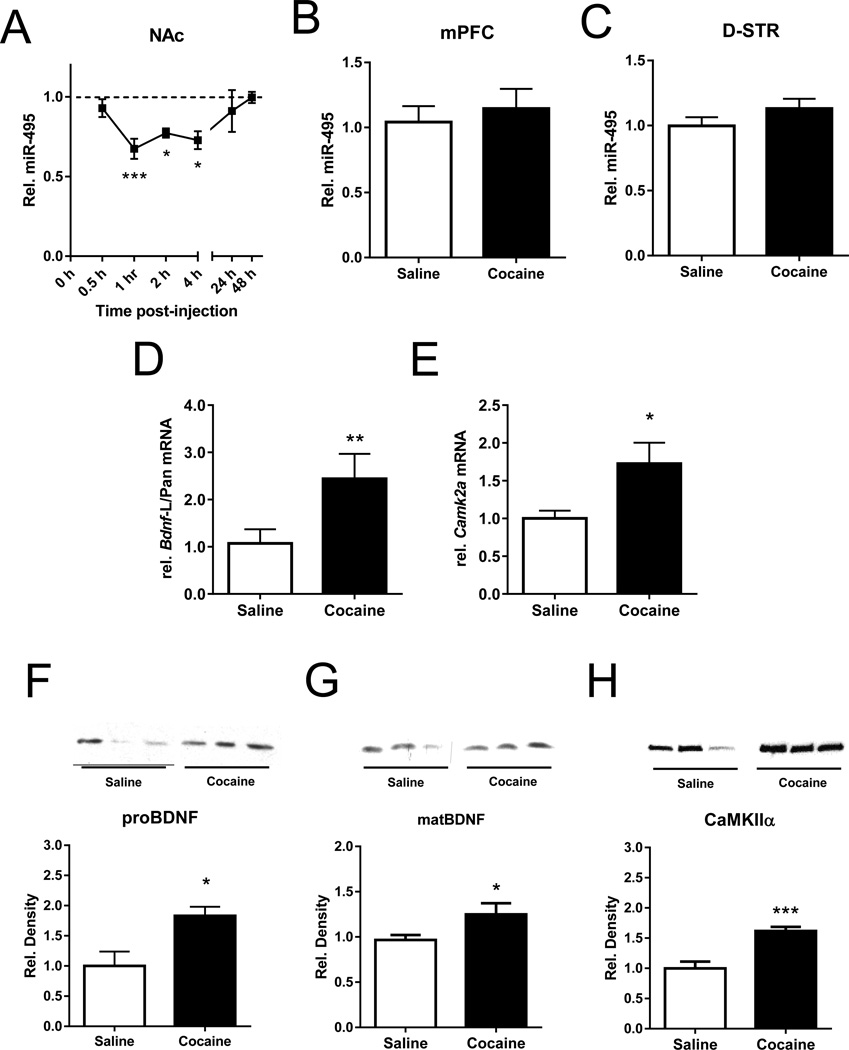
The anatomical localization and targets of miR-495 suggest that it may play a role in the post-transcriptional mechanisms underlying addiction-related plasticity. To examine this further, we determined the effect of an acute cocaine injection (15 mg/kg, i.p.) in mice on NAc miR-495 expression at different time points. NAc miR-495 was significantly downregulated between 1–4 h post-injection (Figure 2A). This effect was brain region-specific, as miR-495 expression was not significantly altered by cocaine 2 h post-acute cocaine in the mPFC or dorsal striatum (Figure 2B, C).
Figure 2. Acute cocaine effects on NAc miR-495 and target mRNA expression.
Male C57Bl/6 mice received an acute injection of saline or cocaine (15 mg/kg, i.p.) and NAc tissue was processed for qRT-PCR and Western blot. (A) NAc miR-495 levels were found to be downregulated rapidly after acute cocaine (0.5 h, n = 5; 1 h, n = 10; 2 h, n = 4; 4 h, n = 6; 24 h, n = 6; 48 h, n = 5). miR-495 expression was not altered by acute cocaine 2h after within the medial prefrontal cortex (B; mPFC; coc n = 5, sal n = 6) or dorsal striatum (C; DS; coc n = 6, sal n = 8). Acute cocaine increases expression of NAc Bdnf-L relative to pan-Bdnf nearly two-fold, as measured by qRT-PCR (D; coc n = 4, sal n = 5). (E) Acute cocaine also increased Camk2a mRNA at 2 h (n = 5). NAc pro-BDNF (F), mature BDNF (G) and CaMKIIα (H) increases in protein levels were also found by Western blot 2 h post-cocaine or saline injection, corrected for total protein by Coomassie Brilliant Blue staining (proBDNF, coc n = 9, sal n = 7; matBDNF, coc n = 9, sal n = 8; CaMKIIα n = 6/group, representative blot with each lane representing individual animals; bars represent quantification of average density of each sample from duplicate blots). Error bars indicate SEM. *p < 0.05, **p < 0.01 vs. saline.
Next, we assessed the expression of two luciferase validated miR-495 targets, Bdnf and Camk2a, at the middle of this timeframe: 2 h post-injection. We found that both NAc Bdnf-Pan mRNA, which is the sum of Bdnf-S and Bdnf-L isoforms, and Bdnf-L mRNA were significantly increased 2 h post-injection (Supplementary Figure S4). Although this demonstrates that Bdnf mRNA is upregulated by acute cocaine, it does not point to the mechanism involved in this cocaine-induced upregulation. Since both Bdnf transcripts originate from the same promoter, differences between the two isoforms would suggest regulation at the post-transcriptional level. To evaluate the possibility, we calculated the ratio between Bdnf-L and Bdnf-Pan and found that it was significantly increased by ~two-fold (Figure 2D), indicating that acute cocaine preferentially upregulates the long 3’ UTR variant that contains a greater number of miR-495 binding sites than the short form (Figure 1E, F). Additionally, we found that both proBDNF and mature BDNF protein were significantly increased within the NAc 2 h after cocaine treatment (Figure 2F, G). Another luciferase-validated miR-495 target, Camk2a, was found to be regulated 2 h post-injection within the NAc as both mRNA (Figure 2E) and protein (Figure 2H) were increased. Thus, NAc miR-495 expression is rapidly decreased by exposure to cocaine concomitantly with increased expression of its ARG targets, Bdnf and Camk2a. This inverse relationship in cocaine-induced gene expression suggests a functional link between miR-495 and its target ARGs in vivo.
Overexpression of miR-495 within the NAc shell reverses cocaine-induced ARG expression
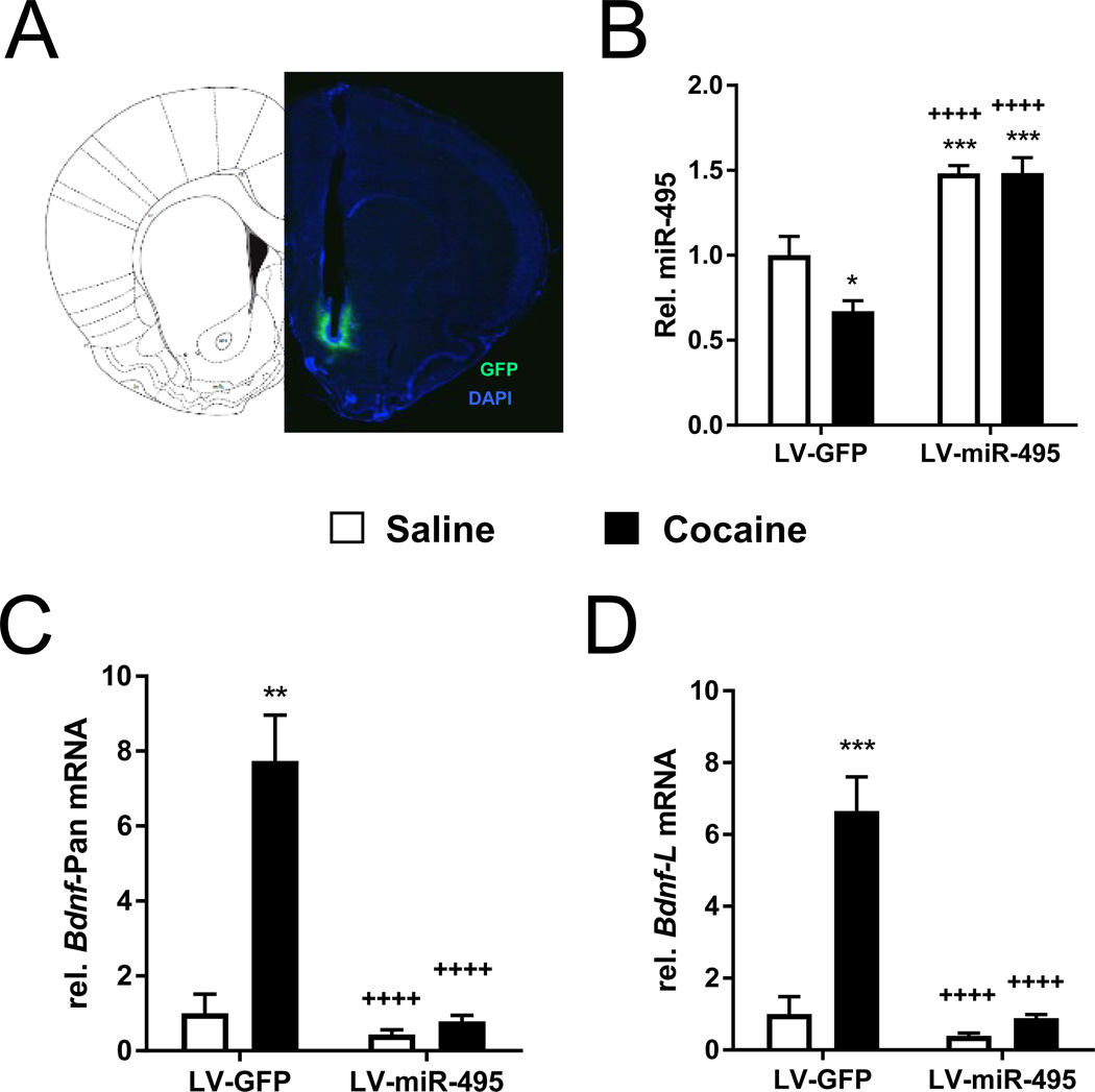
To further examine the regulatory relationship between cocaine-induced NAc miR-495 downregulation and upregulation of target ARG mRNAs, we next tested whether these changes could be reversed by restoring miR-495 levels in the NAc with viral-mediated overexpression. Lentivirus (LV) encoding pri-miR-495+GFP (LV-miR-495) or GFP (LV-GFP) was infused into the NAc shell (NAcsh; Figure 3A) of male Sprague-Dawley rats who were treated 2 weeks later with saline or cocaine (15 mg/kg, i.p.). Cocaine-treated LV-GFP rats were found to express significantly lower NAc miR-495 levels compared to saline-treated LV-GFP controls (Figure 3B), replicating the cocaine-induced downregulation of NAc miR-495 in mice without lentiviral infusion (Figure 2A). LV-miR-495 rats exhibited significantly greater NAc miR-495 expression compared to LV-GFP-infused animals in either treatment group, effectively reversing the cocaine-induced decrease in miR-495 (Figure 3B). Similarly, cocaine-treated LV-GFP rats were found to express significantly higher levels of both Bdnf variants compared to saline treated LV-GFP controls and LV-miR-495 animals in either drug treatment group (Figure 3C, D). Thus, the ability of miR-495 overexpression to reduce the cocaine-induced upregulation of both Bdnf transcripts suggests a functional link between miR-495 and its targets in vivo.
Figure 3. NAc miR-495 overexpression counteracts cocaine-induced changes in gene expression.
Two weeks following infusion of either LV-GFP or LV-miR-495 into the NAcsh (A), rats received an injection of saline or cocaine (15 mg/kg, i.p.). LV-miR-495 blocked the cocaine-induced decrease in NAc miR-495 expression 2-h post-injection (B) and prevented the cocaine-induced increase in NAc pan-Bdnf (C) and Bdnf-L (D) expression. LV-GFP-saline: n = 3; LV-miR-495-saline: n = 5; LV-GFP-cocaine: n = 8; LV-miR-495-cocaine: n = 6. Error bars indicate SEM. *p < 0.05, ** < 0.01, *** < 0.001 vs. saline treated LV-GFP rats and ++++p < 0.0001 vs. cocaine treated LV-GFP.
Pathway analysis of mRNAs downregulated by miR-495 overexpression reveals multiple regulatory networks involved in SUDs
Since multiple ARGs are predicted targets of miR-495 (Supplementary Table S2), we used microarray analysis to determine the global effects of NAcsh miR-495 overexpression on mRNA expression in vivo. We found that 1027 mRNAs were significantly decreased after LV-miR-495 treatment. Of the previously identified 691 miR-495 targets, 76 were significantly downregulated by miR-495 overexpression, 15 of which, including Bdnf, Camk2a, Arc, Gria3 and Stmn2, were also present in KARG (Supplementary Table S3). A complete list of miR-495 regulated transcripts on the arrays has been deposited in the Gene Expression Omnibus (GEO) database (GSE85500).
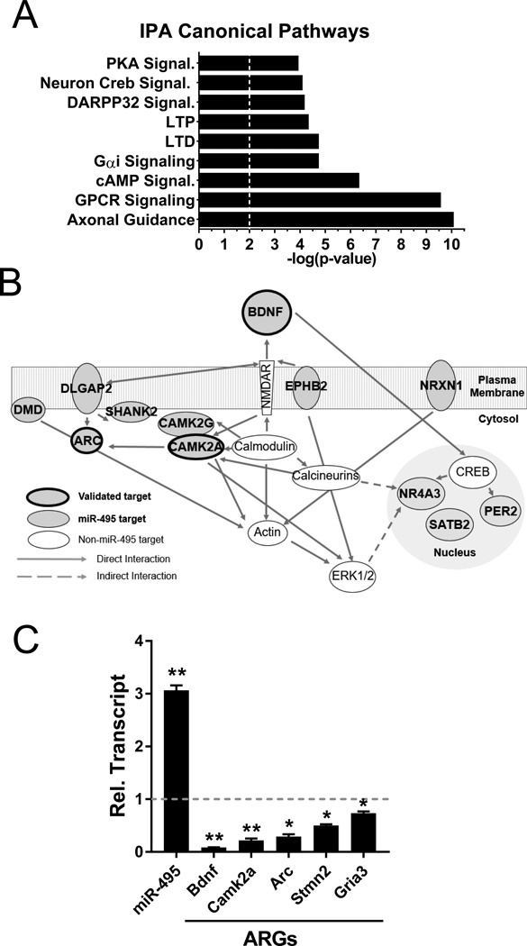
Using Ingenuity Pathway Analysis (IPA), we searched for canonical pathways enriched with NAc mRNAs downregulated by miR-495 overexpression. We found multiple pathways that were previously characterized in addiction-related behavior, including GPCR signaling, cAMP-mediated signaling, synaptic long-term depression (LTD) and potentiation (LTP), CREB, and PKA signaling (Figure 4A). The top biological network of interacting molecules regulated by miR-495 contained 21 of the original 76 molecules (selections in Figure 4B, for complete list see Supplementary Table S4) with functions related to drug-evoked synaptic plasticity (e.g., Gria3, Shank2, Arc, Ephb2, Camk2a), transcription factors (e.g., Satb2, Per2), and chromatin remodeling (e.g., Satb2; 6, 30). Using qRT-PCR, we confirmed miR-495 overexpression decreased selected target mRNAs (Figure 4C, Supplementary Table S3). Altogether, these results indicate that miR-495 regulates multiple target ARG mRNAs both in vitro (Figure 1F, G) and in vivo.
Figure 4. miR-495 regulates genes involved in several addiction-related networks.
Rats were infused with either LV-GFP or LV-miR-495 into the NAcsh and 1 week later NAc RNA was collected for both microarray and qRT-PCR analysis (n = 5/group). (A) Top canonical pathways enriched in genes downregulated by miR-495 overexpression (vertical dotted line represents threshold, p < 0.01) were determined using Ingenuity Pathway Analysis (IPA). (B) A graphical representation of the genes associated with the top signaling network denotes the presence of predicted and validated miR-495 targets. (C) LV-miR-495 infusion increased miR-495 expression and decreased expression of several ARGs compared to LV-GFP controls (dotted line), as measured by qRT-PCR. Error bars indicate SEM. *p < 0.05, **p < 0.01 compared to controls.
NAcsh miR-495 expression decreases following cocaine self-administration
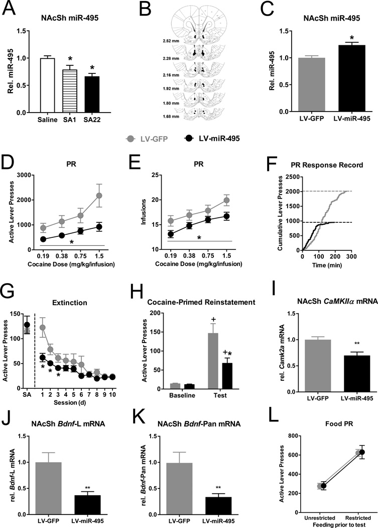
Using qRT-PCR, NAc miR-495 expression was measured in rats that self-administered cocaine for either 1 or 22 days (SA1 vs. SA22). The cocaine groups did not differ in total cocaine infusions during the test session (Supplementary Figure S5). Saline-yoked controls at each time point did not significantly differ in NAcsh miR-495 and were combined. NAcsh miR-495 was significantly decreased in the SA1 and SA22 group compared to the saline group (Figure 5A). Furthermore, a significant linear trend was found across time, where NAcsh miR-495 levels decreased as the number of cocaine self-administration sessions increased. Although baseline miR-495 expression in both NAc subregions are similar (Supplementary Figure S6), no effect on miR-495 expression was found in the adjacent NAc core after short or long-term self-administration (Supplementary Figure S7), demonstrating NAc subregion-specific effects.
Figure 5. miR-495 overexpression in the NAcsh suppresses motivation to self-administer and seek cocaine in rats.
(A) Endogenous NAcsh miR-495 levels are suppressed following 1 and 22 days of cocaine self-administration, as measured by qRT-PCR (Saline: n = 7; SA1: n = 6; SA22: n = 7). *p < 0.05 vs. Saline-yoked. (B) Histological verification of NAcsh cannula placement. (C) NAcsh miR-495 levels are increased in the LV-miR-495 group compared to the LV-GFP group following behavioral testing (n = 4/group). NAcsh miR-495 overexpression reduced responding (D) and intake (E) on a PR schedule of cocaine reinforcement. (F) Cumulative response records of representative rats tested at 1.5 mg/kg/infusion that were closest to their respective group means. Dotted line represents the total number of lever presses emitted once break point was achieved. (G) NAcsh miR-495 overexpression reduced cocaine-seeking behavior during the first three 1-h extinction sessions. (H) NAcsh miR-495 overexpression reduced cocaine-primed reinstatement during the 1-h test session (10 mg/kg, i.p.). LV-GFP: n = 12, LV-miR-495: n = 10. (I–K) Downregulation of Camk2a (LV-GFP: n = 7, LV miR-495: n = 8, p < 0.01) and Bdnf-L and -Pan (n = 6/group, p < 0.01) in NAcsh of miR-495 OE animals after behavioral testing. (L) NAcsh miR-495 overexpression had no effect on responding under a PR schedule of food reinforcement, regardless of whether the rats were food restricted (18g/day) or not (n = 8/group). Error bars indicate SEM. Panels C-H: *p < 0.05 vs. LV-GFP. +p < 0.05 vs. extinction baseline.
miR-495 overexpression in the NAcsh reduces motivation to self-administer and seek cocaine
To test the role of NAc miR-495 in models of addiction-related behavior, we next examined the effect of NAcsh miR-495 overexpression on self-administration and seeking behavior. Two weeks following NAcsh infusions of LV-miR-495 or LV-GFP (Figure 5B), rats were given access to varying doses of cocaine delivered on a FR5 and PR schedule of reinforcement. NAcsh miR-495 overexpression was persistent to the end of our behavioral experiments (Figure 5C). As such, NAcsh miR-495 overexpression had no effect on responding or intake on the low effort FR5 schedule of cocaine reinforcement (Supplementary Figure S8). However, NAcsh miR-495 overexpression significantly decreased responding and intake on the high effort PR schedule compared to controls across all cocaine doses tested (Figure 5D, E), without effects on inactive lever pressing (Supplementary Figure S9A). The cumulative response records in Figure 5F show a lower break point in a representative LV-miR-495 rat compared to a LV-GFP rat, consistent with a decrease in motivation.
Next, we tested the effect of NAcsh miR-495 overexpression on other measures of motivation, including extinction and reinstatement. Before extinction testing, rats received >3 sessions on an FR5 schedule of cocaine reinforcement on the training dose. We found no group difference in active lever pressing during this baseline before extinction (Figure 5G). During extinction, NAcsh miR-495 overexpression decreased active lever pressing during the first 3 sessions compared to LV-GFP controls (Figure 5G), without effects on inactive lever pressing (Supplementary Figure S9B). We then tested rats for both cue and cocaine-primed reinstatement of cocaine seeking. While the virus groups did not differ in the degree of cue reinstatement (Supplementary Figure S10), the LV-miR-495 group exhibited significantly reduced cocaine-primed reinstatement compared to the LV-GFP controls (Figure 5H), without differences in inactive lever pressing in either case (Supplementary Figure S9C, D). To confirm the effect of miR-495 overexpression on target regulation during drug-seeking behavior, we measured the levels of Camk2a, Bdnf-L, and Bdnf-Pan mRNAs in the NAcsh of rats 1 week after extinction and reinstatement experiments. As shown in Figures 5 I–K the levels of these three target mRNAs were significantly decreased by miR-495 OE. Collectively, these results suggest that NAcsh miR-495 overexpression decreases motivation to both self-administer and seek cocaine. The additional decrease in target gene expression suggests that the behavioral effect of miR-495 overexpression is mediated through these ARGs.
To test the specificity for cocaine and evaluate any potential locomotor-suppressing effects of miR-495 overexpression, a separate group of rats were trained to lever press for food pellets while receiving either LV-GFP or LV-miR-495. LV-miR-495 had no effect on responding or intake on an FR5 schedule of food reinforcement (Supplementary Figure S11). To parallel differences in motivational value of low and high cocaine doses on a PR schedule, we subjected rats to varying levels of food restriction. Rats that had been food-restricted (18g/day) exhibited higher PR measures than those that had been unrestricted, but both virus groups exhibited similar levels of motivation for food under both feeding conditions (Figure 5L, Supplementary Figure S12). This suggests a selective effect of miR-495 overexpression on motivation for cocaine.
DISCUSSION
Here we established NAc miR-495 as a novel post-transcriptional regulator of both ARG expression and motivation for cocaine. Initial bioinformatics analyses identified miR-495 as a miRNA with predicted targets enriched in the KARG database and with preferential expression in the brain reward and motivation pathway. Among these, we validated that miR-495 targeted Bdnf and Camk2a both in vitro and in vivo. Next, we found that cocaine decreased miR-495 expression along with concomitant increases in ARG targets in the NAc. When the cocaine-mediated miR-495 downregulation was blocked by lentiviral-mediated overexpression, cocaine-induced upregulation of ARG target mRNAs in the NAc was also prevented. From the miR-495 overexpression microarray analysis, we found that several of the downregulated target genes formed networks involved in receptor signaling, gene regulation, and synaptic plasticity. Importantly, we found that NAc miR-495 overexpression reduced motivation to self-administer and seek cocaine, without effects on food reinforcement, suggesting that NAc miR-495 selectively regulates genes involved in motivation for cocaine. Given that motivation for drug is a key factor involved in human drug relapse, miR-495 may have translational value as a novel therapeutic target.
In contrast to other addiction-related miRNAs whose expression levels were shown to increase in response to drugs of abuse (14, 17, 18, 21), miR-495 expression in the NAc decreased shortly after exposure to cocaine. This rapid downregulation could be due to several factors. While miRNAs are stable in non-neuronal cell types, some miRNAs decay at faster rates in neurons via activity-dependent processes (31), which could be triggered by cocaine administration. The decrease in miR-495 expression may also be due to transcriptional repression. miR-495 is located within miRNA cluster B of the Dlk1-Dio3 maternally imprinted region that is under the control of Methyl-CpG-binding protein 2 (MeCP2) and other transcription factors (32). Using Mecp2-null mice, Wu and colleagues (33) demonstrated increased expression of many of the miRNAs within this imprinted region, including miR-495. However, given that not all of the miRNAs in this cluster are coordinately regulated (33, 34), other regulatory mechanisms such as pre-miRNA processing and/or mature miRNA stability may play a role in controlling miRNA expression profiles (35). It is curious that we did not observe changes in miR-495 in any other addiction-related brain region, especially in those that are also innervated by ventral tegmental dopamine neurons (e.g., dorsal striatum, mPFC). One possibility is that the relatively high basal expression levels of miR-495 in the NAc allowed us to detect a decrease following cocaine administration. Another possibility is that the NAc may have a more robust response to the acute and chronic effects of cocaine resulting in decreases in miR-495 expression levels.
miRNAs play a role in fine-tuning gene expression involved in many cell signaling pathways (5, 36). Here, we established that miR-495 directly targets and regulates the ARG Bdnf both in vitro and in vivo. Bdnf in the NAc has been linked to several drug abuse-related behaviors, where BDNF expression levels positively correlate with cocaine reward and motivation (19, 37–40). Similarly, others have established that another miR-495 target, Camk2a, has a positive relationship between NAc levels and psychostimulant abuse-related behavior (41–44). Furthermore, CaMKII has been identified in silico as a central node in positive feedback gene regulatory pathways involved in addiction (23). Therefore, regulation of both of these genes by miR-495 may coordinately affect several domains of addiction-related processes. Indeed, addiction is hypothesized to be a dysfunction of neuroplasticity (3), and both Bdnf and Camk2a genes encode for plasticity-related proteins. We found that many other miR-495 targets we validated also form networks involved in LTP and LTD (30, 45, 46), as well as other cell signaling cascades relevant to addiction, such as PKA signaling (47, 48). Therefore, miR-495 may act as a mechanism to fine tune the molecular response of multiple, interwoven pathways involved in the development of addiction.
Similar to acute cocaine administration, NAcsh miR-495 levels were also decreased following both brief and prolonged cocaine self-administration. It is unknown why this occurred exclusively in the NAc shell, and not the core, but this effect may be due to the differential afferent projections into these subregions or the involvement of the shell in the primary reinforcing and unconditioned effects of cocaine (49, 50). Interestingly, these effects are consistent with previous findings that both BDNF and CaMKIIα involvement in cocaine abuse-related behavior is also specific to the NAcsh, and not the core (39, 41–44, 51). Furthermore, the downward trend of NAcsh miR-495 expression as cocaine self-administration experience increased may be related to the theory of incentive sensitization, where motivation to seek drug (i.e. ‘wanting’) increases over the course of drug use (52). Thus, decreases in NAcsh miR-495 expression may be indicative of sensitized motivation.
To test this hypothesis, we examined the effect of NAcsh miR-495 overexpression on cocaine self-administration and seeking behavior. We observed that NAcsh miR-495 overexpression decreased responding and intake when cocaine was available on the high effort PR schedule, but did not alter intake in the low effort FR5 schedule. PR schedules are believed to model an aspect of SUD related to an individual increasing time and energy toward drug-seeking and -taking behavior (53), thus alterations in these behaviors closely model hallmark symptoms of human addiction. Furthermore, NAc miR-495 overexpression did not alter PR measures in a similar procedure with a natural food reinforcer. This suggests that miR-495 specifically influences motivation for cocaine likely without impacting the reinforcing value of cocaine or food reinforcement/motivation. Additionally, we found that NAcsh miR-495 overexpression reduced cocaine-seeking behavior during extinction and reinstatement, further supporting a selective reduction in motivation. We also confirmed that under these conditions, miR-495 overexpression resulted in decreases in Bdnf and Camk2a expression. Closely mirroring our effects, previous work has shown that NAcsh knockdown of Camk2a and inhibition of NAcsh CAMKII reduces PR measures and reinstatement, respectively (43, 44). Thus, CaMKIIα is an important regulatory crux of many addiction-related molecular pathways (41, 42, 44, 51, 54). Although we did not detect a statistically significant effect during cue reinstatement (p = 0.06, one-tailed), the pattern is very similar. The lack of a more robust effect on cue reinstatement may highlight the more prominent role of the NAc core, rather than the shell, in regulating cue reinstatement (55). Overall, our results suggest that miR-495 preferentially regulates a network of ARG targets involved in the incentive motivational properties of cocaine which are more critical for sustaining behavior under the high-effort PR schedule of reinforcement than under the low-effort FR5 schedule of reinforcement.
In conclusion, we demonstrate a novel in silico method to identify potential miRNAs that may be involved in SUD. Our findings suggest that miR-495 decreases motivation for cocaine by targeting several ARGs and regulatory pathways in the NAc involved in synaptic plasticity, PKA signaling, and other pathways associated with the disorder. These results highlight the importance of moving drug abuse research from a single gene focus to biological pathways in order to better understand the complexity of the molecular networks associated with addiction. This discovery also opens new avenues for future research on the specific factors controlling cocaine-induced decreases in miR-495 and the role of miR-495 in regulating different forms of synaptic plasticity in the NAc. Most importantly, this study is the first to identify a miRNA that specifically regulates the incentive motivational properties for cocaine both during active drug taking and following a period of abstinence. The latter finding is particularly compelling, as preventing relapse is a primary objective for addiction translational research. The possibility of globally targeting drug-induced changes in gene expression via miRNAs, such as miR-495, may lead to new therapeutics that shift the balance of gene regulation toward alleviating, rather than promoting, SUD-related behavior.
Supplementary Material
Acknowledgments
This work was supported by NIDA grants 1R01DA034097 (N.P.B. & J.L.N.), F31DA035069 (R.M.B.), and T32AA014127, F31DA041214 (R.J.O.). We are grateful to Dr. Yue Feng at Emory University for providing the luciferase-Bdnf 3’ UTR constructs. We also wish to thank Timothy Cheung, Nora Dado, Peter Kufahl, Sayuri Nixon, Karen Mooneyhan, Jason Newbern, Natalie Peartree, Erika Perez, Lara Pockros, Justine Saavedra, and Jennifer Taylor for their technical assistance.
Footnotes
CONFLICT OF INTEREST
The authors declare no conflict of interest.
Supplementary information is available at Molecular Psychiatry’s website.
REFERENCES
- 1.Leshner AI. Addiction is a brain disease, and it matters. Science. 1997;278(5335):45–47. doi: 10.1126/science.278.5335.45. [DOI] [PubMed] [Google Scholar]
- 2.Association AP. Diagnostic and Statistical Manual of Mental Disorders. 5th. Arlington, VA: American Psychiatric Publishing; 2013. [Google Scholar]
- 3.Kalivas PW, O'Brien C. Drug addiction as a pathology of staged neuroplasticity. Neuropsychopharmacology. 2008;33(1):166–180. doi: 10.1038/sj.npp.1301564. [DOI] [PubMed] [Google Scholar]
- 4.McClung CA, Nestler EJ. Neuroplasticity mediated by altered gene expression. Neuropsychopharmacology. 2008;33(1):3–17. doi: 10.1038/sj.npp.1301544. [DOI] [PubMed] [Google Scholar]
- 5.Feng J, Nestler EJ. Epigenetic mechanisms of drug addiction. Curr Opin Neurobiol. 2013;23(4):521–528. doi: 10.1016/j.conb.2013.01.001. [DOI] [PMC free article] [PubMed] [Google Scholar]
- 6.Robison AJ, Nestler EJ. Transcriptional and epigenetic mechanisms of addiction. Nat Rev Neurosci. 2011;12(11):623–637. doi: 10.1038/nrn3111. [DOI] [PMC free article] [PubMed] [Google Scholar]
- 7.Rogge GA, Wood MA. The role of histone acetylation in cocaine-induced neural plasticity and behavior. Neuropsychopharmacology. 2013;38(1):94–110. doi: 10.1038/npp.2012.154. [DOI] [PMC free article] [PubMed] [Google Scholar]
- 8.Bartel DP. MicroRNAs: genomics, biogenesis, mechanism, and function. Cell. 2004;116(2):281–297. doi: 10.1016/s0092-8674(04)00045-5. [DOI] [PubMed] [Google Scholar]
- 9.Im HI, Kenny PJ. MicroRNAs in neuronal function and dysfunction. Trends Neurosci. 2012;35(5):325–334. doi: 10.1016/j.tins.2012.01.004. [DOI] [PMC free article] [PubMed] [Google Scholar]
- 10.Bali P, Kenny PJ. MicroRNAs and Drug Addiction. Front Genet. 2013;4:43. doi: 10.3389/fgene.2013.00043. [DOI] [PMC free article] [PubMed] [Google Scholar]
- 11.Kloosterman WP, Plasterk RH. The diverse functions of microRNAs in animal development and disease. Dev Cell. 2006;11(4):441–450. doi: 10.1016/j.devcel.2006.09.009. [DOI] [PubMed] [Google Scholar]
- 12.Miller BH, Wahlestedt C. MicroRNA dysregulation in psychiatric disease. Brain Res. 2010;1338:89–99. doi: 10.1016/j.brainres.2010.03.035. [DOI] [PMC free article] [PubMed] [Google Scholar]
- 13.Sartor GC, St Laurent G, 3rd, Wahlestedt C. The Emerging Role of Non-Coding RNAs in Drug Addiction. Front Genet. 2012;3:106. doi: 10.3389/fgene.2012.00106. [DOI] [PMC free article] [PubMed] [Google Scholar]
- 14.Chandrasekar V, Dreyer JL. microRNAs miR-124, let-7d and miR-181a regulate cocaine-induced plasticity. Mol Cell Neurosci. 2009;42(4):350–362. doi: 10.1016/j.mcn.2009.08.009. [DOI] [PubMed] [Google Scholar]
- 15.Eipper-Mains JE, Kiraly DD, Palakodeti D, Mains RE, Eipper BA, Graveley BR. microRNA-Seq reveals cocaine-regulated expression of striatal microRNAs. RNA. 2011;17(8):1529–1543. doi: 10.1261/rna.2775511. [DOI] [PMC free article] [PubMed] [Google Scholar]
- 16.Guo Y, Chen Y, Carreon S, Qiang M. Chronic intermittent ethanol exposure and its removal induce a different miRNA expression pattern in primary cortical neuronal cultures. Alcohol Clin Exp Res. 2012;36(6):1058–1066. doi: 10.1111/j.1530-0277.2011.01689.x. [DOI] [PMC free article] [PubMed] [Google Scholar]
- 17.Nudelman AS, DiRocco DP, Lambert TJ, Garelick MG, Le J, Nathanson NM, et al. Neuronal activity rapidly induces transcription of the CREB-regulated microRNA-132, in vivo. Hippocampus. 2010;20(4):492–498. doi: 10.1002/hipo.20646. [DOI] [PMC free article] [PubMed] [Google Scholar]
- 18.Schaefer A, Im HI, Veno MT, Fowler CD, Min A, Intrator A, et al. Argonaute 2 in dopamine 2 receptor-expressing neurons regulates cocaine addiction. J Exp Med. 2010;207(9):1843–1851. doi: 10.1084/jem.20100451. [DOI] [PMC free article] [PubMed] [Google Scholar]
- 19.Bahi A, Boyer F, Chandrasekar V, Dreyer JL. Role of accumbens BDNF and TrkB in cocaine-induced psychomotor sensitization, conditioned-place preference, and reinstatement in rats. Psychopharmacology (Berl) 2008;199(2):169–182. doi: 10.1007/s00213-008-1164-1. [DOI] [PubMed] [Google Scholar]
- 20.Chandrasekar V, Dreyer JL. Regulation of MiR-124, Let-7d, and MiR-181a in the accumbens affects the expression, extinction, and reinstatement of cocaine-induced conditioned place preference. Neuropsychopharmacology. 2011;36(6):1149–1164. doi: 10.1038/npp.2010.250. [DOI] [PMC free article] [PubMed] [Google Scholar]
- 21.Hollander JA, Im HI, Amelio AL, Kocerha J, Bali P, Lu Q, et al. Striatal microRNA controls cocaine intake through CREB signalling. Nature. 2010;466(7303):197–202. doi: 10.1038/nature09202. [DOI] [PMC free article] [PubMed] [Google Scholar]
- 22.Tapocik JD, Barbier E, Flanigan M, Solomon M, Pincus A, Pilling A, et al. microRNA-206 in rat medial prefrontal cortex regulates BDNF expression and alcohol drinking. J Neurosci. 2014;34(13):4581–4588. doi: 10.1523/JNEUROSCI.0445-14.2014. [DOI] [PMC free article] [PubMed] [Google Scholar]
- 23.Li CY, Mao X, Wei L. Genes and (common) pathways underlying drug addiction. PLoS Comput Biol. 2008;4(1):e2. doi: 10.1371/journal.pcbi.0040002. [DOI] [PMC free article] [PubMed] [Google Scholar]
- 24.Enright AJ, John B, Gaul U, Tuschl T, Sander C, Marks DS. MicroRNA targets in Drosophila. Genome Biol. 2003;5(1):R1. doi: 10.1186/gb-2003-5-1-r1. [DOI] [PMC free article] [PubMed] [Google Scholar]
- 25.Kozomara A, Griffiths-Jones S. miRBase: annotating high confidence microRNAs using deep sequencing data. Nucleic Acids Res. 2014;42(Database issue):D68–D73. doi: 10.1093/nar/gkt1181. [DOI] [PMC free article] [PubMed] [Google Scholar]
- 26.Landgraf P, Rusu M, Sheridan R, Sewer A, Iovino N, Aravin A, et al. A mammalian microRNA expression atlas based on small RNA library sequencing. Cell. 2007;129(7):1401–1414. doi: 10.1016/j.cell.2007.04.040. [DOI] [PMC free article] [PubMed] [Google Scholar]
- 27.Mellios N, Huang HS, Grigorenko A, Rogaev E, Akbarian S. A set of differentially expressed miRNAs, including miR-30a-5p, act as post-transcriptional inhibitors of BDNF in prefrontal cortex. Hum Mol Genet. 2008;17(19):3030–3042. doi: 10.1093/hmg/ddn201. [DOI] [PMC free article] [PubMed] [Google Scholar]
- 28.An JJ, Gharami K, Liao GY, Woo NH, Lau AG, Vanevski F, et al. Distinct role of long 3' UTR BDNF mRNA in spine morphology and synaptic plasticity in hippocampal neurons. Cell. 2008;134(1):175–187. doi: 10.1016/j.cell.2008.05.045. [DOI] [PMC free article] [PubMed] [Google Scholar]
- 29.Orefice LL, Waterhouse EG, Partridge JG, Lalchandani RR, Vicini S, Xu B. Distinct roles for somatically and dendritically synthesized brain-derived neurotrophic factor in morphogenesis of dendritic spines. J Neurosci. 2013;33(28):11618–11632. doi: 10.1523/JNEUROSCI.0012-13.2013. [DOI] [PMC free article] [PubMed] [Google Scholar]
- 30.Kauer JA, Malenka RC. Synaptic plasticity and addiction. Nat Rev Neurosci. 2007;8(11):844–858. doi: 10.1038/nrn2234. [DOI] [PubMed] [Google Scholar]
- 31.Krol J, Busskamp V, Markiewicz I, Stadler MB, Ribi S, Richter J, et al. Characterizing light-regulated retinal microRNAs reveals rapid turnover as a common property of neuronal microRNAs. Cell. 2010;141(4):618–631. doi: 10.1016/j.cell.2010.03.039. [DOI] [PubMed] [Google Scholar]
- 32.Benetatos L, Hatzimichael E, Londin E, Vartholomatos G, Loher P, Rigoutsos I, et al. The microRNAs within the DLK1-DIO3 genomic region: involvement in disease pathogenesis. Cell Mol Life Sci. 2013;70(5):795–814. doi: 10.1007/s00018-012-1080-8. [DOI] [PMC free article] [PubMed] [Google Scholar]
- 33.Wu H, Tao J, Chen PJ, Shahab A, Ge W, Hart RP, et al. Genome-wide analysis reveals methyl-CpG-binding protein 2-dependent regulation of microRNAs in a mouse model of Rett syndrome. Proc Natl Acad Sci U S A. 2010;107(42):18161–18166. doi: 10.1073/pnas.1005595107. [DOI] [PMC free article] [PubMed] [Google Scholar]
- 34.Lempiainen H, Couttet P, Bolognani F, Muller A, Dubost V, Luisier R, et al. Identification of Dlk1-Dio3 imprinted gene cluster noncoding RNAs as novel candidate biomarkers for liver tumor promotion. Toxicological sciences : an official journal of the Society of Toxicology. 2013;131(2):375–386. doi: 10.1093/toxsci/kfs303. [DOI] [PubMed] [Google Scholar]
- 35.Joilin G, Guevremont D, Ryan B, Claudianos C, Cristino AS, Abraham WC, et al. Rapid regulation of microRNA following induction of long-term potentiation in vivo. Frontiers in molecular neuroscience. 2014;7:98. doi: 10.3389/fnmol.2014.00098. [DOI] [PMC free article] [PubMed] [Google Scholar]
- 36.Schratt G. Fine-tuning neural gene expression with microRNAs. Current opinion in neurobiology. 2009;19(2):213–219. doi: 10.1016/j.conb.2009.05.015. [DOI] [PubMed] [Google Scholar]
- 37.Horger BA, Iyasere CA, Berhow MT, Messer CJ, Nestler EJ, Taylor JR. Enhancement of locomotor activity and conditioned reward to cocaine by brain-derived neurotrophic factor. J Neurosci. 1999;19(10):4110–4122. doi: 10.1523/JNEUROSCI.19-10-04110.1999. [DOI] [PMC free article] [PubMed] [Google Scholar]
- 38.Hall FS, Drgonova J, Goeb M, Uhl GR. Reduced behavioral effects of cocaine in heterozygous brain-derived neurotrophic factor (BDNF) knockout mice. Neuropsychopharmacology. 2003;28(8):1485–1490. doi: 10.1038/sj.npp.1300192. [DOI] [PubMed] [Google Scholar]
- 39.Graham DL, Edwards S, Bachtell RK, DiLeone RJ, Rios M, Self DW. Dynamic BDNF activity in nucleus accumbens with cocaine use increases self-administration and relapse. Nat Neurosci. 2007;10(8):1029–1037. doi: 10.1038/nn1929. [DOI] [PubMed] [Google Scholar]
- 40.Graham DL, Krishnan V, Larson EB, Graham A, Edwards S, Bachtell RK, et al. Tropomyosin-related kinase B in the mesolimbic dopamine system: region-specific effects on cocaine reward. Biol Psychiatry. 2009;65(8):696–701. doi: 10.1016/j.biopsych.2008.09.032. [DOI] [PMC free article] [PubMed] [Google Scholar]
- 41.Kourrich S, Klug JR, Mayford M, Thomas MJ. AMPAR-independent effect of striatal alphaCaMKII promotes the sensitization of cocaine reward. J Neurosci. 2012;32(19):6578–6586. doi: 10.1523/JNEUROSCI.6391-11.2012. [DOI] [PMC free article] [PubMed] [Google Scholar]
- 42.Loweth J, Singer B, Baker L, Wilke G, Inamine H, Bubula N, et al. Transient overexpression of alpha-Ca2+/calmodulin-dependent protein kinase II in the nucleus accumbens shell enhances behavioral responding to amphetamine. The Journal of neuroscience : the official journal of the Society for Neuroscience. 2010;30(3):939–949. doi: 10.1523/JNEUROSCI.4383-09.2010. [DOI] [PMC free article] [PubMed] [Google Scholar]
- 43.Wang L, Lv Z, Hu Z, Sheng J, Hui B, Sun J, et al. Chronic cocaine-induced H3 acetylation and transcriptional activation of CaMKIIalpha in the nucleus accumbens is critical for motivation for drug reinforcement. Neuropsychopharmacology. 2010;35(4):913–928. doi: 10.1038/npp.2009.193. [DOI] [PMC free article] [PubMed] [Google Scholar]
- 44.Anderson SM, Famous KR, Sadri-Vakili G, Kumaresan V, Schmidt HD, Bass CE, et al. CaMKII: a biochemical bridge linking accumbens dopamine and glutamate systems in cocaine seeking. Nat Neurosci. 2008;11(3):344–353. doi: 10.1038/nn2054. [DOI] [PubMed] [Google Scholar]
- 45.Kasanetz F, Deroche-Gamonet V, Berson N, Balado E, Lafourcade M, Manzoni O, et al. Transition to addiction is associated with a persistent impairment in synaptic plasticity. Science. 2010;328(5986):1709–1712. doi: 10.1126/science.1187801. [DOI] [PubMed] [Google Scholar]
- 46.Russo SJ, Dietz DM, Dumitriu D, Morrison JH, Malenka RC, Nestler EJ. The addicted synapse: mechanisms of synaptic and structural plasticity in nucleus accumbens. Trends Neurosci. 2010;33(6):267–276. doi: 10.1016/j.tins.2010.02.002. [DOI] [PMC free article] [PubMed] [Google Scholar]
- 47.Self DW, Genova LM, Hope BT, Barnhart WJ, Spencer JJ, Nestler EJ. Involvement of cAMP-dependent protein kinase in the nucleus accumbens in cocaine self-administration and relapse of cocaine-seeking behavior. J Neurosci. 1998;18(5):1848–1859. doi: 10.1523/JNEUROSCI.18-05-01848.1998. [DOI] [PMC free article] [PubMed] [Google Scholar]
- 48.Boudreau AC, Ferrario CR, Glucksman MJ, Wolf ME. Signaling pathway adaptations and novel protein kinase A substrates related to behavioral sensitization to cocaine. J Neurochem. 2009;110(1):363–377. doi: 10.1111/j.1471-4159.2009.06140.x. [DOI] [PMC free article] [PubMed] [Google Scholar]
- 49.Yager LM, Garcia AF, Wunsch AM, Ferguson SM. The ins and outs of the striatum: role in drug addiction. Neuroscience. 2015;301:529–541. doi: 10.1016/j.neuroscience.2015.06.033. [DOI] [PMC free article] [PubMed] [Google Scholar]
- 50.Ito R, Robbins TW, Everitt BJ. Differential control over cocaine-seeking behavior by nucleus accumbens core and shell. Nat Neurosci. 2004;7(4):389–397. doi: 10.1038/nn1217. [DOI] [PubMed] [Google Scholar]
- 51.Robison AJ, Vialou V, Mazei-Robison M, Feng J, Kourrich S, Collins M, et al. Behavioral and structural responses to chronic cocaine require a feedforward loop involving DeltaFosB and calcium/calmodulin-dependent protein kinase II in the nucleus accumbens shell. J Neurosci. 2013;33(10):4295–4307. doi: 10.1523/JNEUROSCI.5192-12.2013. [DOI] [PMC free article] [PubMed] [Google Scholar]
- 52.Robinson TE, Berridge KC. The neural basis of drug craving: an incentive-sensitization theory of addiction. Brain Res Brain Res Rev. 1993;18(3):247–291. doi: 10.1016/0165-0173(93)90013-p. [DOI] [PubMed] [Google Scholar]
- 53.Roberts DC, Morgan D, Liu Y. How to make a rat addicted to cocaine. Prog Neuropsychopharmacol Biol Psychiatry. 2007;31(8):1614–1624. doi: 10.1016/j.pnpbp.2007.08.028. [DOI] [PMC free article] [PubMed] [Google Scholar]
- 54.Wu X, McMurray CT. Calmodulin kinase II attenuation of gene transcription by preventing cAMP response element-binding protein (CREB) dimerization and binding of the CREB-binding protein. J Biol Chem. 2001;276(3):1735–1741. doi: 10.1074/jbc.M006727200. [DOI] [PubMed] [Google Scholar]
- 55.Fuchs RA, Evans KA, Parker MC, See RE. Differential involvement of the core and shell subregions of the nucleus accumbens in conditioned cue-induced reinstatement of cocaine seeking in rats. Psychopharmacology (Berl) 2004;176(3–4):459–465. doi: 10.1007/s00213-004-1895-6. [DOI] [PubMed] [Google Scholar]
Associated Data
This section collects any data citations, data availability statements, or supplementary materials included in this article.


