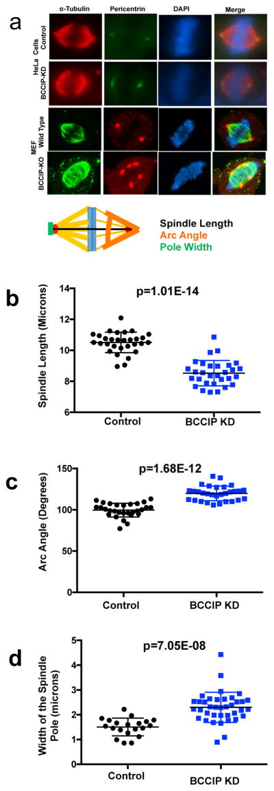
Figure 6. Abnormal spindle architecture in BCCIP deficient cells.

BCCIP knockdown HeLa or BCCIP knockout MEF cells were immunostained with α-tubulin and pericentrin and analyzed at metaphase. The distance between the paired centrosomes, the angles of spindle arcs and the width of the spindle poles were assessed. Representative image sets and a cartoon illustration of the assessed geometric measurements are shown in Panel-A. The distributions and averages of the spindle length, arc angles, and the width of the spindle poles are shown in panels B, C, and D respectively.
