Abstract
Purpose
A long natural history and a predominant osseous pattern of metastatic spread are impediments to the adoption of precision medicine in patients with prostate cancer. To establish the feasibility of clinical genomic profiling in the disease, we performed targeted deep sequencing of tumor and normal DNA from patients with locoregional, metastatic non-castrate, and metastatic castration-resistant prostate cancer (CRPC).
Methods
Patients consented to genomic analysis of their tumor and germline DNA. A hybridization capture-based clinical assay was employed to identify single nucleotide variations, small insertions and deletions, copy number alterations and structural rearrangements in over 300 cancer-related genes in tumors and matched normal blood.
Results
We successfully sequenced 504 tumors from 451 patients with prostate cancer. Potentially actionable alterations were identified in DNA damage repair (DDR), PI3K, and MAP kinase pathways. 27% of patients harbored a germline or a somatic alteration in a DDR gene that may predict for response to PARP inhibition. Profiling of matched tumors from individual patients revealed that somatic TP53 and BRCA2 alterations arose early in tumors from patients who eventually developed metastatic disease. In contrast, comparative analysis across disease states revealed that APC alterations were enriched in metastatic tumors, while ATM alterations were specifically enriched in CRPC.
Conclusion
Through genomic profiling of prostate tumors representing the disease clinical spectrum, we identified a high frequency of potentially actionable alterations and possible drivers of disease initiation, metastasis and castration-resistance. Our findings support the routine use of tumor and germline DNA profiling for patients with advanced prostate cancer, for the purpose of guiding enrollment in targeted clinical trials and counseling families at increased risk of malignancy.
Introduction
Prostate cancer is a disease characterized by distinct clinical states with highly variable outcomes1. Surgery and radiation therapy are potentially curative for patients with localized disease, whereas androgen deprivation therapy (ADT) is effective but palliative for patients who develop metastases with a testosterone level in the non-castrate range (metastatic non-castrate prostate cancer)2. Metastatic non-castrate prostate cancer inevitably evolves into castration-resistant prostate cancer (mCRPC), the lethal form of the disease.
Recent molecular profiling efforts, including the Stand Up to Cancer-PCF (SU2C-PCF) mCRPC project and The Cancer Genome Atlas (TCGA) primary localized prostate cancer study have identified distinct molecular subsets of prostate cancer and potentially targetable alterations that occur somatically as well as in the germline3–7. Notably, there is limited genomic data on metastatic non-castrate disease. While molecular profiling is not yet considered a standard-of-care for patients with the disease, new evidence points to enhanced treatment response in specific molecular contexts, paving the way for therapy selection based on tumor molecular characteristics. In particular, genomic alterations in genes involved in DNA damage repair by homologous recombination (HR) may predict for increased sensitivity to poly-ADP ribose polymerase (PARP) inhibitors and platinum-based therapy8,9. We sought to determine whether routine prospective genomic profiling in the clinical practice setting was feasible and informative for patients with prostate cancer, and to define the frequency of potential driver genomic alterations across the disease clinical spectrum.
Methods
Patients and Samples
Patients with prostate cancer consented to an institutional review board-approved protocol for tumor genomic profiling using the MSK-IMPACT targeted sequencing assay. Specific consent was required for analysis of germline variants in an identifiable manner. Following consent, either archival or new tumor samples were obtained, and blood was drawn for germline DNA. Archival tumor samples were formalin-fixed and paraffin embedded (FFPE). New biopsies were obtained under radiographic guidance and were formalin fixed. Bone biopsies were non-decalcified. All tumors were reviewed by pathologists specialized in genitourinary oncology for confirmation of malignant histology of prostatic origin.
Sequencing and Analysis
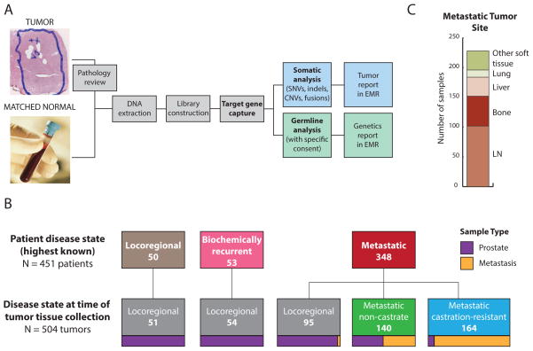
We employed the MSK-IMPACT assay as previously described14,15 (Fig. 1A). The assay is performed in a CLIA-certified laboratory, and designed to robustly identify single nucleotide variations (SNVs), small insertions and deletions (indels), copy number alterations (CNAs) and structural rearrangements in over 300 cancer-related genes in FFPE tumors and matched normal blood.
Figure 1. Clinical sequencing of tumors and germline for patients with prostate cancer.
A. MSK-IMPACT assay workflow. EMR = electronic medical record. B. 451 patients underwent tumor profiling in the clinic. Their last known disease state when seen in the clinic is represented at top of figure. Their disease state at the time of tissue collection for the 504 tumors that were profiled is represented at bottom. Tumors that were profiled represented all three prostate cancer clinical disease states: locoregional, metastatic non-castrate and metastatic castration-resistant. Locoregional disease indicates disease without distant clinical or pathological spread, including lymph node involvement in the pelvis only (TxN0/1). Sample type (prostate versus metastasis is represented at bottom. C. Site of disease for metastatic tumors successfully profiled by MSK-IMPACT (LN = lymph node).
Somatic variant analysis was performed as described14,15, with germline variants identified in matched blood samples filtered out in the somatic analysis process. Somatic findings were reported in the electronic medical record, as well as anonymized and uploaded to cBioPortal (http://www.cbioportal.org/study?id=prad_mskcc_2016) for visualization and analysis10,11. Clonality of mutations was estimated as cancer cell fraction (CCF)13, and implemented in the FACETS algorithm12.
Beginning in May 2015, patients were given the option to consent to analysis of germline variants identified through sequencing of normal blood samples. Germline analysis of 76 known cancer predisposing genes was performed as previously described14.
Supplementary methods are provided online.
Results
Targeted DNA sequencing of tumor-normal pairs from patients with prostate cancer
We successfully profiled 504 tumors from 451 prostate cancer patients who presented to the clinic using the MSK-IMPACT assay (Fig. 1A, Supplementary Table 1). In total, 348 patients (77%) had metastatic prostate cancer, 53 (12%) had biochemical recurrence after definitive therapy, and 50 (11%) had locoregional disease (Fig. 1B). The 504 tumors were either archival or newly acquired primary prostate or metastatic tumors of prostate origin, with 44 patients having more than one tumor profiled. Metastatic tumors successfully profiled were obtained from lymph node (45%), bone (22%), liver (14%), lung (5%) and other soft tissue sites (14%) (Fig. 1C). The disease state at the time of collection of the tumor is shown in Fig. 1B. Unlike the TCGA and SU2C-PCF studies, the tumors we profiled represented all three prostate cancer clinical states: locoregional, metastatic non-castrate and metastatic castration resistant.
We began with 746 biopsy/surgical samples to successfully sequence the 504 tumors reported above, with an overall success rate of 68% (Supplementary Fig. 1). The highest success rates were for prostate tumor samples obtained from diagnostic prostate needle biopsy, radical prostatectomy or transurethral resection of prostate performed for palliation. For metastatic samples, success rates of 69% and higher were seen for lymph node, liver and other soft tissues samples, whereas bone and lung samples were more challenging (42–52% success rate).
Somatic and germline alterations identified in the MSK-IMPACT dataset
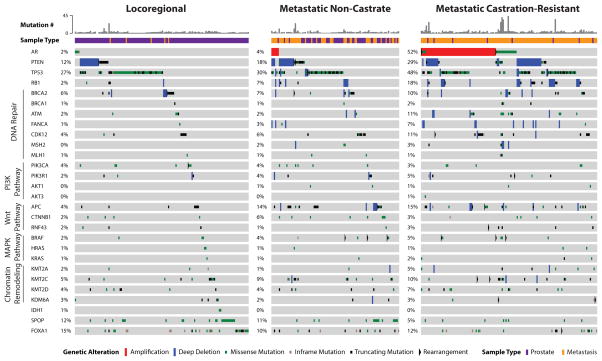
Somatic alterations in biologically-relevant genes in prostate cancer were identified in all disease states and are shown in Fig. 2, grouped in pathways that are potentially clinically actionable. Overall, the frequency of alterations in genes of interest in prostate cancer was similar for mCRPC tumors profiled with MSK-IMPACT and the SU2C-PCF mCRPC dataset4 (Supplementary Fig. 2), but demonstrated now in a clinical practice setting. However, notable differences were observed when comparing primary localized tumors in the MSK-IMPACT dataset to those profiled in the prospective TCGA study, including a higher frequency of alterations in TP53 and FOXA1 in the MSK-IMPACT tumors (Supplementary Fig. 3). This is most likely due to the more aggressive nature of the primary localized tumors in the MSK-IMPACT cohort, which were predominantly obtained from patients who subsequently developed biochemically recurrent and metastatic disease (Fig 1B), relative to the prospectively-acquired primary TCGA tumors (Supplementary Fig. 3).
Figure 2. Selected genomic alterations across disease states in the MSK-IMPACT dataset.
Each column represents an individual patient with prostate cancer whose tumor was acquired in the disease state indicated at top of figure. Total number of non-synonymous somatic mutations in the tumor is represented in histogram form. Sample type (prostate versus metastasis) is also represented. Alterations in commonly affected genes in prostate cancer (e.g. AR, PTEN, TP53, FOXA1) are shown, in addition to genes in potentially actionable or biologically-relevant pathways. The type of alteration (e.g. copy number variation, rearrangement, mutation) is indicated in the bottom row.
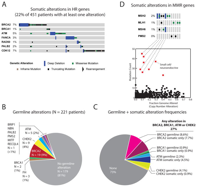
24% of patients carried somatic alterations in the PI3K/AKT pathway, including in PTEN, PIK3CA, PIK3CB, PIK3R1, AKT1 and AKT3 (Supplementary Fig. 4). The majority of point mutations in PIK3CA, AKT1 and AKT3 were known activating hotspot mutations in those genes15. Additionally, 5% of patients harbored somatic alterations in MAP kinase pathway genes (Supplementary Fig. 5), including hotspot mutations in BRAF, HRAS, KRAS and MAP2K1. 15% of patients carried somatic alterations in the Wnt-β catenin pathway, including in APC, CTNNB1 and RNF43 (Supplementary Fig. 6). Consistent with results of the SU2C-PCF study, 22% of patients harbored a somatic alteration in a gene involved in DNA damage repair (DDR) by homologous recombination, including BRCA2, BRCA1, ATM, FANCA, RAD50, PALB2 and CDK1216–20 (Fig. 3A, Supplementary Fig. 7).
Figure 3. Somatic and germline alterations in DNA damage repair genes.
A. Oncoprint of somatic alterations in genes involved in DNA repair by homologous recombination (HR) for 451 patients. 22% of patients harbor a tumor alteration in one of the listed genes. B. Germline alterations for patients who consented to germline analysis (N = 221). 19% of subjects were found to have a germline pathogenic alteration. C. Frequency of combined somatic and germline alterations in DNA repair genes BRCA2, BRCA1, ATM and CHEK2 for patients who consented to germline analysis. 27% of patients had either a germline or a somatic-only alteration in one of these genes. D. Oncoprint of somatic alterations in genes involved in DNA mismatch repair (MMR) (top). 3% of patients harbor a tumor alteration in one of these genes. Most tumors that display more than 20 somatic mutations (bottom) harbor a somatic alteration in a MMR gene.
A recent multi-institutional study identified a high frequency of DDR gene alterations in the germline of patients with advanced prostate cancer21. 221 patients in our dataset underwent formal germline analysis, the first 124 of whom were included in the previously-reported study21. Of these 221 patients, 42 (19% of total) had a known or likely pathogenic germline mutation in BRCA2 (9% of total), CHEK2 (4%), ATM (2%), BRCA1 (1%), FH (1 %), and PMS2, NBN, PALB2, and BRIP1 (<1% each) (Fig. 3B and Supplementary Table 2). While germline DDR gene alterations may predict for response to PARP inhibition or platinum agents, somatic-only alterations in these genes without a germline event may still predict for drug response9. Of the 221 patients who underwent germline analysis, 27% demonstrated alterations in BRCA2, BRCA1, ATM or CHEK2, either in the germline or somatically (Fig. 3C). Notably, germline analysis alone accounted for approximately half of these patients only, suggesting that both germline and somatic analysis should be performed to identify patients with DDR gene deficiency.
3% of patients had tumors with somatic alterations in mismatch repair (MMR) genes MSH2, MLH1, PMS2 or MSH6. These tumors accounted for the majority of samples with the highest mutation counts on MSK-IMPACT profiling (Fig. 3D), which were confirmed to be enriched for previously-described MMR and microsatellite instability (MSI) signatures22,23 (Supplementary Fig. 8). The identification of MMR-deficient prostate cancers may have immediate clinical applicability, given recent data suggesting sensitivity of such tumors to immune checkpoint blockade in colorectal cancer and other malignancies24–26.
In total, 36% of patients were found to have a potentially actionable alteration using the OncoKB annotation platform (Supplementary Fig. 9). This platform does not include non-BRCA/ATM germline alterations, missense alterations of unknown significance, and genes whose clinical significance is less clear, including CDK12 and FANCA. As genomic alterations undergo further characterization and new trials and drug targets emerge, the frequency of alterations defined as actionable may increase.
Comparative analysis of somatic alterations across disease states
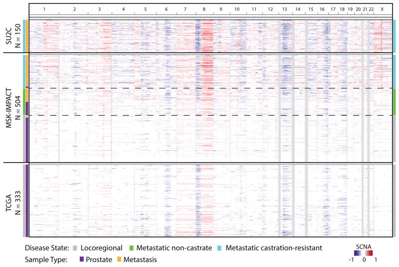
A unique aspect of this dataset is that it includes genetic profiles of tumors representing all three clinical states: locoregional, metastatic non-castrate and metastatic castration-resistant. The number of non-synonymous mutations per tumor increased significantly from tumors in the locoregional disease state to those in the mCRPC disease state (1.74 versus 4.02 mean mutations/megabase, p < 0.001), while tumors in the metastatic non-castration-resistant state had a mutation burden similar to locoregional tumors (2.08 mutations/megabase). Consistent with prior studies3,4, we identified recurrent areas of copy number loss involving chromosomes 6q, 8p, 13q and 16q, and areas of copy number gain involving chromosomes 1q, 3q, 7, 8q and X (Fig. 4). mCRPC tumors displayed the highest burden of copy number alterations, while those representing locoregional disease displayed the lowest (Fig. 4, Supplementary Fig. 10).
Figure 4. Cross-cohort comparative analysis of the pattern and degree of copy number alterations across the genome.
Represented here are regions of amplification (red) or deletion (blue) with chromosomes listed horizontally (top) in the MSK-IMPACT, SU2C-PCF (metastatic CRPC), and TCGA (primary localized prostate cancer) datasets. The MSK-IMPACT tumors are sorted by disease state at time of tissue acquisition, from locoregional (bottom, gray) to metastatic CRPC (top, blue). While sequencing platforms differ between studies, the degree and pattern of copy number alteration was similar for tumors acquired in the same disease state.
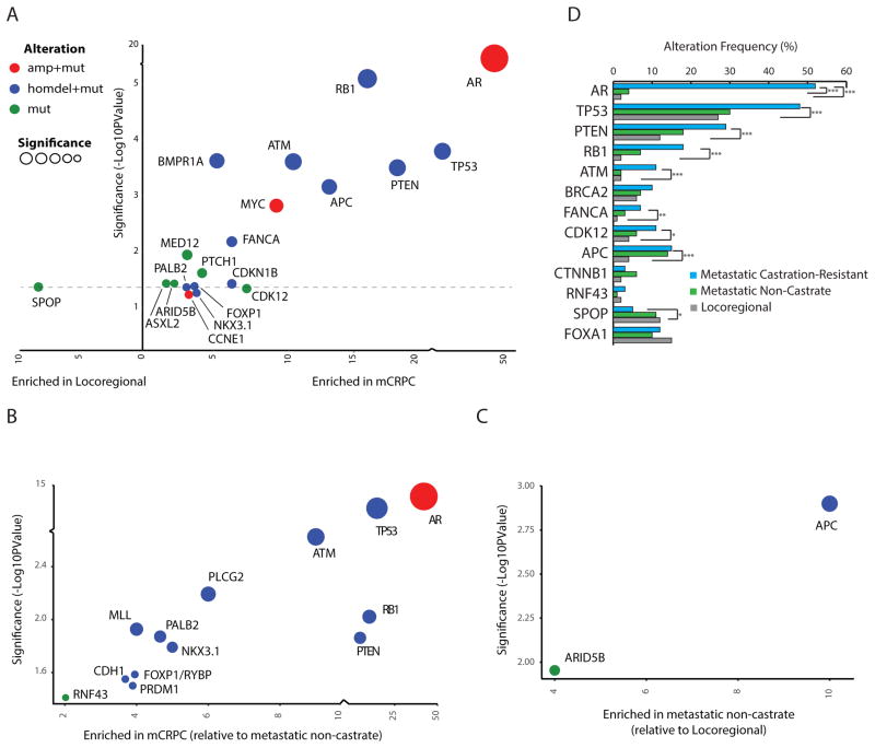
Aiming to identify possible genomic drivers of disease progression, we performed a selective enrichment analysis to identify genes that were more frequently altered in mCRPC compared with locoregional disease (Fig. 5A). AR amplification/mutation was the most enriched alteration in mCRPC, as shown in prior studies3,4. Other genes that were more commonly altered in mCRPC included TP53, RB1, PTEN, APC, ATM, FANCA and CDK12.
Figure 5. Enrichment of genomic alterations between disease states.
A. Enrichment of genomic alterations in tumors from patients with metastatic CRPC versus locoregional disease. The level of enrichment is represented as difference in frequency between the two indicated classes (x-axis) and its significance (p-value, y-axis). The type of alteration is represented by color (homdel = homozygous deletion; amp = amplification; mut = mutation). B. Enrichment of genomic alterations in tumors from patients with metastatic CRPC versus metastatic non-castrate disease. C. Enrichment of genomic alterations in tumors from patients with metastatic non-castrate disease versus locoregional disease. D. Frequencies of alterations in select genes across disease states in the MSK-IMPACT dataset. P-values are represented (Fisher’s exact test).
We performed a similar analysis comparing alterations in mCRPC versus metastatic non-castrate prostate cancer (Fig. 5B). AR was again the most enriched gene in this analysis. The high enrichment of alterations in AR in mCRPC relative to both locoregional and metastatic non-castrate disease serves as a positive control, consistent with the known role of AR as a driver of castration resistance27–30. Beyond frequent amplification of the gene, AR antiandrogen resistance mutations were identified in tumors from patients with mCRPC (Supplementary Fig. 11), including an F877L enzalutamide/ARN509 resistance mutation31,32 found in the tumor of a patient who progressed after 4 years of treatment on ARN509. Notably, a 4% alteration frequency in AR was identified in tumors from patients with metastatic non-castrate disease (Figs. 2, 5D). These were tumors that were exposed to ADT and were likely transitioning to a castration-resistant phenotype that had not yet manifested clinically. Four locoregional tumors were found to have mutations in AR, including an H875Y mutation known to confer resistance to flutamide28,33 in a prostatectomy sample from a patient who was treatment-naïve, but had received dutasteride for benign prostate enlargement.
In addition to AR, we again identified enrichment of alterations in TP53, RB1, PTEN and ATM in mCRPC compared with metastatic non-castrate disease. The enrichment of these genes in mCRPC relative to both earlier disease states implicates them in the development of castration-resistance, a finding that is of particular interest for ATM, a gene involved in DNA damage repair. FANCA and CDK12, two other DNA repair genes, did not show statistically-significant enrichment in mCRPC relative to metastatic non-castrate disease as they did relative to locoregional disease, though there is a trend that suggests a role in castration-resistance as well (Fig. 5D). When the analysis was limited to metastatic tumors or lymph nodes only, similar trends for enrichment were observed, although statistical significance was not always reached due to smaller sample size (Supplementary Fig. 12).
Only two genes were enriched in metastatic non-castrate versus locoregional disease: APC and to a lesser extent ARID5A (Fig. 5C). The enrichment of APC alterations in both metastatic states relative to locoregional disease (Fig. 5D) implicates this gene in metastasis. Conversely, alterations in SPOP7,34 were enriched in locoregional disease (and possibly metastatic non-castrate disease - though this does not meet statistical significance) relative to mCRPC (Figs. 5A, D), suggesting increased sensitivity of SPOP mutant tumors to ADT. These findings will require functional validation in the laboratory.
Matched samples identify clonal alterations in prostate cancer
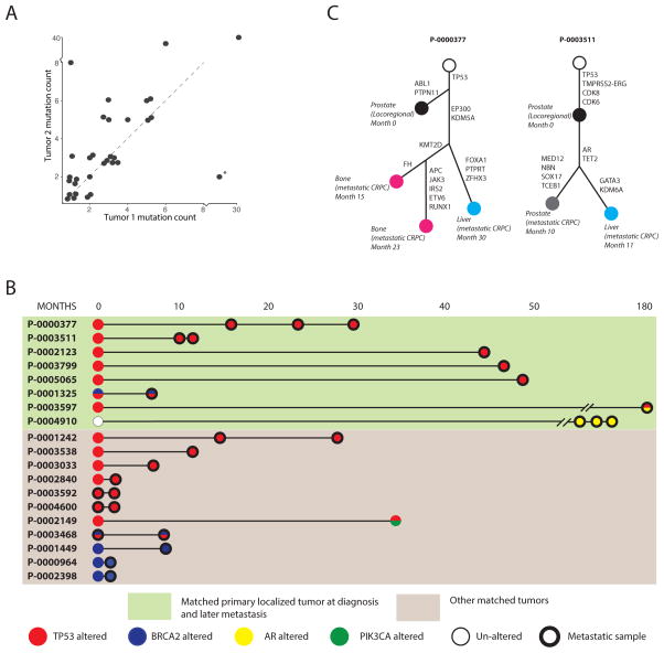
44 patients had more than one tumor site profiled by MSK-IMPACT, including 17 with a matched primary localized tumor and a subsequent metastatic tumor. Tumors from the same patient that were acquired at a later time point typically had a higher mutation count (Fig. 6A). Given the high frequency of TP53 alterations observed in primary localized tumors in this dataset, and prior reports of aggressive behavior of prostate tumors harboring TP53 alterations35,36, we sought to determine whether TP53 alterations were present in tumors early in their evolution, or whether they were acquired later at disease progression. As shown in Fig. 6B, TP53 alterations arose early in affected patients and were identified in all tumors from the same patient. TP53 mutations were clonal, including in cases where both a primary localized tumor and a later metastasis were available (cancer cell fraction ≥ 0.9 in the later metastasis). Likewise, we found that somatic alterations in BRCA2 were present in matched tumors, again suggesting that somatic BRCA2 loss of function alterations occur early in tumorigenesis for affected patients. Conversely, alterations in AR did not occur early in matched samples (Fig. 6B, patients P-0003597 and P-0004910), consistent with treatment-related changes that promote castration resistance. Of note, other potentially actionable alterations may arise later in disease evolution, as was the case for patient P-0002149 (Fig. 6B), who acquired an activating PIK3CA E545K mutation (Supplementary Fig. 4B) in a recurrent tumor nearly three years after his radical prostatectomy.
Figure 6. Somatic alterations identified in matched tumors from the same patients.
A. Somatic mutation count in pairs of matched tumors. The latter tumor (Tumor 2) tends to have a higher mutation burden. A notable exception (*) involves a bladder metastasis and a bone metastasis acquired 5 months later, where the patient had received salvage radiation to the pelvis, possibly explaining the higher mutation count in the earlier bladder tumor. B. Somatic alterations in TP53 (red), BRCA2 (blue), AR (yellow) and PIK3CA (green) in matched tumors in the dataset, including localized primaries and later metastases (green box) and other matched tumors from the same patients. C. Evolutionary analysis representing the acquisition of genomic alterations in sequential tumors obtained from 2 patients. Filled circles represent tumors sequenced by MSK-IMPACT, labeled with their sites, disease state and relative dates of acquisition.
To confirm the findings above, we performed phylogenetic analysis on cases where several matched tumors were available from the same patient (Fig. 6C). Both phylogenetic trees shown reveal the early truncal nature of TP53 alterations. For patient P-0000377, alterations in EP300 and KDM5A, genes involved in epigenetic modulation, occurred truncally for metastatic tumors. An alteration in KMT2D was identified subclonally in metachronous metastases from bone obtained from separate sites, but not in a liver metastasis. For patient P-0003511, AR amplification was truncal in castration-resistant tumors, consistent with its well-characterized role in castration resistance27. As the number of prostate cancer patients profiled longitudinally throughout their clinical care increases, such findings may provide insight into clonal driver events that promote disease progression and site-specific metastasis.
Discussion
Unlike prior prostate cancer genomic studies, we profiled tumors representing the clinical spectrum of the disease, from locoregional to metastatic non-castrate and metastatic castration-resistant disease, enabling comparisons of genomic landscape across disease states using a single assay. While locoregional tumors in this dataset typically represented more aggressive disease than TCGA, patients with such tumors are those in highest need of new treatment approaches and profiling of their tumors may have particular clinical relevance.
An increase in copy number alterations and mutation frequency was evident in mCRPC relative to earlier disease states, as was an increased frequency of alterations in AR, TP53, RB1 and PTEN. SPOP mutations, however, were more frequent in the earlier disease states, suggesting better outcomes for patients with SPOP-mutant tumors, possibly through increased sensitivity to ADT. Importantly, the ability to compare alteration frequencies across three prostate cancer disease states can provide insight into genes that promote metastasis versus castration resistance. In this analysis, APC and ATM emerged as candidate genes of interest that may independently contribute to metastasis and castration resistance respectively, pending functional validation in the laboratory. Other genes that emerged as being enriched in mCRPC, though not to the same extent, are FANCA and CDK12, which, like ATM, are involved in DNA damage repair, alluding to a possible role for DNA repair defects in the development of castration-resistance.
We also found that TP53 alterations are early clonal events in matched tumor samples from individual patients. This suggests that TP53 alterations in localized tumors may predict for an increased risk of progression to metastatic disease, consistent with recent reports of aggressive behavior of TP53-altered prostate cancers36–38. As long-term outcomes from prospective primary prostate cancer datasets become available, it will be possible to validate this finding, guiding more aggressive treatment approaches early on for these patients.
Overall, we identified potentially actionable alterations including hotspot activating alterations in genes that are known drug targets, consistent with the findings of the SU2C mCRPC study but this time in a prospective clinical practice setting. In allowing for separate somatic and germline analyses, our study showed that 27% of patients with advanced prostate cancer have a combination of either somatic or germline alterations in BRCA2, BRCA1, ATM or CHEK2, and that 3% harbor an alteration in an MMR gene. These findings have immediate therapeutic relevance, given the recently reported sensitivity of these tumors to PARP inhibition9 or immune checkpoint blockade24. The higher frequency of DNA repair alterations identified through integrative germline and somatic analysis strongly argues for performing both germline and somatic genomic analysis in all patients with advanced prostate cancer who will require systemic treatment, irrespective of screening based on family history.
In summary, this study shows that a large genomic dataset representing the clinical spectrum of prostate cancer can provide mechanistic insight into possible genomic drivers of disease initiation, metastasis and drug resistance. Our ability to profile metastatic tumors allowed us to detect the evolution of potential driver alterations in matched tumors from individual patients, identifying alterations in TP53 and BRCA2 as early events that may confer a more aggressive phenotype. Our study reveals that identifying actionable genomic alterations is feasible in the clinical practice setting for patients with prostate cancer, but several challenges remain. First, the availability of trials targeting these alterations remains a limitation. Trials of PARP inhibitors for prostate cancer patients with HR gene alterations are due to open shortly, and the findings of this study and others should prompt the development of multi-institutional molecularly-guided studies for the smaller subsets of patients with other molecular alterations. Second, a critical difficulty is in obtaining sufficient tumor material for sequencing, particularly for patients with disease that is restricted to bone. Circulating tumor DNA sequencing assays, currently under investigation in prostate cancer, may offer a solution to this problem. Overall, given the high frequency of potentially actionable alterations, early but compelling evidence of clinical benefit of targeted therapy for patients with DNA repair gene-deficient prostate cancers, and the implication of germline findings for family members, our data argue for the routine use of germline and somatic genomic profiling assays as standard practice for all patients with advanced prostate cancer.
Supplementary Material
Acknowledgments
Research Support:
NIH/NCI, Department of Defense, Prostate Cancer Foundation
We would like to thank Emily Waters, Vanessa Robinson, Amal Gulaid, Melanie Hullings, and Joseph Jang.
This work was supported by Prostate Cancer Foundation Challenge and Young Investigator Awards (W.A., H.S., N.A., B.S.T.), the National Institutes of Health (NIH/NCI Prostate SPORE P50-CA92629 and NIH/NCI Cancer Center Support Grant P30 CA008748), the Department of Defense Prostate Cancer Research Program (PC071610 and PC121111), the David H. Koch Fund for Prostate Cancer Research, the Marie-Josée and Henry R. Kravis Center for Molecular Oncology, the Robertson Foundation (N.S., B.S.T.), and an American Cancer Society Research Scholar Award (B.S.T.).
Footnotes
Study presented in part at:
ASCO Genitourinary Cancers Symposium 2015 (Prostate poster session), ASCO Annual Meeting 2015 (Genitourinary prostate poster session), AACR Annual Meeting 2016 (Late-breaking oral abstract minisymposium)
Disclaimers:
None
Authors’ disclosures of potential conflicts of interest
No conflicts of interest to declare.
References
- 1.Scher HI, Heller G. Clinical states in prostate cancer: toward a dynamic model of disease progression. Urology. 2000;55:323–7. doi: 10.1016/s0090-4295(99)00471-9. [DOI] [PubMed] [Google Scholar]
- 2.National Comprehensive Cancer Network. Prostate Cancer (Version 1.2015) [DOI] [PubMed] [Google Scholar]
- 3.Cancer Genome Atlas Research Network. The Molecular Taxonomy of Primary Prostate Cancer. Cell. 2015;163:1011–25. doi: 10.1016/j.cell.2015.10.025. [DOI] [PMC free article] [PubMed] [Google Scholar]
- 4.Robinson D, Van Allen EM, Wu YM, et al. Integrative clinical genomics of advanced prostate cancer. Cell. 2015;161:1215–28. doi: 10.1016/j.cell.2015.05.001. [DOI] [PMC free article] [PubMed] [Google Scholar]
- 5.Taylor BS, Schultz N, Hieronymus H, et al. Integrative genomic profiling of human prostate cancer. Cancer Cell. 2010;18:11–22. doi: 10.1016/j.ccr.2010.05.026. [DOI] [PMC free article] [PubMed] [Google Scholar]
- 6.Grasso CS, Wu YM, Robinson DR, et al. The mutational landscape of lethal castration-resistant prostate cancer. Nature. 2012;487:239–43. doi: 10.1038/nature11125. [DOI] [PMC free article] [PubMed] [Google Scholar]
- 7.Barbieri CE, Baca SC, Lawrence MS, et al. Exome sequencing identifies recurrent SPOP, FOXA1 and MED12 mutations in prostate cancer. Nat Genet. 2012;44:685–9. doi: 10.1038/ng.2279. [DOI] [PMC free article] [PubMed] [Google Scholar]
- 8.Cheng HH, Pritchard CC, Boyd T, et al. Biallelic Inactivation of BRCA2 in Platinum-sensitive Metastatic Castration-resistant Prostate Cancer. Eur Urol. 2015 doi: 10.1016/j.eururo.2015.11.022. [DOI] [PMC free article] [PubMed] [Google Scholar]
- 9.Mateo J, Carreira S, Sandhu S, et al. DNA-Repair Defects and Olaparib in Metastatic Prostate Cancer. N Engl J Med. 2015;373:1697–708. doi: 10.1056/NEJMoa1506859. [DOI] [PMC free article] [PubMed] [Google Scholar]
- 10.Cerami E, Gao J, Dogrusoz U, et al. The cBio cancer genomics portal: an open platform for exploring multidimensional cancer genomics data. Cancer Discov. 2012;2:401–4. doi: 10.1158/2159-8290.CD-12-0095. [DOI] [PMC free article] [PubMed] [Google Scholar]
- 11.Gao J, Aksoy BA, Dogrusoz U, et al. Integrative analysis of complex cancer genomics and clinical profiles using the cBioPortal. Sci Signal. 2013;6:pl1. doi: 10.1126/scisignal.2004088. [DOI] [PMC free article] [PubMed] [Google Scholar]
- 12.Shen R, Seshan VE. FACETS: allele-specific copy number and clonal heterogeneity analysis tool for high-throughput DNA sequencing. Nucleic Acids Res. 2016 doi: 10.1093/nar/gkw520. [DOI] [PMC free article] [PubMed] [Google Scholar]
- 13.McGranahan N, Favero F, de Bruin EC, et al. Clonal status of actionable driver events and the timing of mutational processes in cancer evolution. Sci Transl Med. 2015;7:283ra54. doi: 10.1126/scitranslmed.aaa1408. [DOI] [PMC free article] [PubMed] [Google Scholar]
- 14.Schrader KA, Cheng DT, Joseph V, et al. Germline Variants in Targeted Tumor Sequencing Using Matched Normal DNA. JAMA Oncol. 2016;2:104–11. doi: 10.1001/jamaoncol.2015.5208. [DOI] [PMC free article] [PubMed] [Google Scholar]
- 15.Chang MT, Asthana S, Gao SP, et al. Identifying recurrent mutations in cancer reveals widespread lineage diversity and mutational specificity. Nat Biotechnol. 2016;34:155–63. doi: 10.1038/nbt.3391. [DOI] [PMC free article] [PubMed] [Google Scholar]
- 16.Joshi PM, Sutor SL, Huntoon CJ, et al. Ovarian cancer-associated mutations disable catalytic activity of CDK12, a kinase that promotes homologous recombination repair and resistance to cisplatin and poly(ADP-ribose) polymerase inhibitors. J Biol Chem. 2014;289:9247–53. doi: 10.1074/jbc.M114.551143. [DOI] [PMC free article] [PubMed] [Google Scholar]
- 17.Bajrami I, Frankum JR, Konde A, et al. Genome-wide profiling of genetic synthetic lethality identifies CDK12 as a novel determinant of PARP1/2 inhibitor sensitivity. Cancer Res. 2014;74:287–97. doi: 10.1158/0008-5472.CAN-13-2541. [DOI] [PMC free article] [PubMed] [Google Scholar]
- 18.Buisson R, Dion-Cote AM, Coulombe Y, et al. Cooperation of breast cancer proteins PALB2 and piccolo BRCA2 in stimulating homologous recombination. Nat Struct Mol Biol. 2010;17:1247–54. doi: 10.1038/nsmb.1915. [DOI] [PMC free article] [PubMed] [Google Scholar]
- 19.McCabe N, Turner NC, Lord CJ, et al. Deficiency in the repair of DNA damage by homologous recombination and sensitivity to poly(ADP-ribose) polymerase inhibition. Cancer Res. 2006;66:8109–15. doi: 10.1158/0008-5472.CAN-06-0140. [DOI] [PubMed] [Google Scholar]
- 20.Cremona CA, Behrens A. ATM signalling and cancer. Oncogene. 2014;33:3351–60. doi: 10.1038/onc.2013.275. [DOI] [PubMed] [Google Scholar]
- 21.Pritchard CC, Mateo J, Walsh MF, et al. Inherited DNA-Repair Gene Mutations in Men with Metastatic Prostate Cancer. N Engl J Med. 2016 doi: 10.1056/NEJMoa1603144. [DOI] [PMC free article] [PubMed] [Google Scholar]
- 22.Alexandrov LB, Nik-Zainal S, Wedge DC, et al. Signatures of mutational processes in human cancer. Nature. 2013;500:415–21. doi: 10.1038/nature12477. [DOI] [PMC free article] [PubMed] [Google Scholar]
- 23.Alexandrov LB, Nik-Zainal S, Wedge DC, et al. Deciphering signatures of mutational processes operative in human cancer. Cell Rep. 2013;3:246–59. doi: 10.1016/j.celrep.2012.12.008. [DOI] [PMC free article] [PubMed] [Google Scholar]
- 24.Le DT, Uram JN, Wang H, et al. PD-1 Blockade in Tumors with Mismatch-Repair Deficiency. N Engl J Med. 2015;372:2509–20. doi: 10.1056/NEJMoa1500596. [DOI] [PMC free article] [PubMed] [Google Scholar]
- 25.Snyder A, Makarov V, Merghoub T, et al. Genetic basis for clinical response to CTLA-4 blockade in melanoma. N Engl J Med. 2014;371:2189–99. doi: 10.1056/NEJMoa1406498. [DOI] [PMC free article] [PubMed] [Google Scholar]
- 26.Van Allen EM, Miao D, Schilling B, et al. Genomic correlates of response to CTLA-4 blockade in metastatic melanoma. Science. 2015;350:207–11. doi: 10.1126/science.aad0095. [DOI] [PMC free article] [PubMed] [Google Scholar]
- 27.Chen CD, Welsbie DS, Tran C, et al. Molecular determinants of resistance to antiandrogen therapy. Nat Med. 2004;10:33–9. doi: 10.1038/nm972. [DOI] [PubMed] [Google Scholar]
- 28.Taplin ME, Bubley GJ, Shuster TD, et al. Mutation of the androgen-receptor gene in metastatic androgen-independent prostate cancer. N Engl J Med. 1995;332:1393–8. doi: 10.1056/NEJM199505253322101. [DOI] [PubMed] [Google Scholar]
- 29.Visakorpi T, Hyytinen E, Koivisto P, et al. In vivo amplification of the androgen receptor gene and progression of human prostate cancer. Nat Genet. 1995;9:401–6. doi: 10.1038/ng0495-401. [DOI] [PubMed] [Google Scholar]
- 30.Scher HI, Sawyers CL. Biology of progressive, castration-resistant prostate cancer: directed therapies targeting the androgen-receptor signaling axis. J Clin Oncol. 2005;23:8253–61. doi: 10.1200/JCO.2005.03.4777. [DOI] [PubMed] [Google Scholar]
- 31.Balbas MD, Evans MJ, Hosfield DJ, et al. Overcoming mutation-based resistance to antiandrogens with rational drug design. Elife. 2013;2:e00499. doi: 10.7554/eLife.00499. [DOI] [PMC free article] [PubMed] [Google Scholar]
- 32.Joseph JD, Lu N, Qian J, et al. A clinically relevant androgen receptor mutation confers resistance to second-generation antiandrogens enzalutamide and ARN-509. Cancer Discov. 2013;3:1020–9. doi: 10.1158/2159-8290.CD-13-0226. [DOI] [PubMed] [Google Scholar]
- 33.Duff J, McEwan IJ. Mutation of histidine 874 in the androgen receptor ligand-binding domain leads to promiscuous ligand activation and altered p160 coactivator interactions. Mol Endocrinol. 2005;19:2943–54. doi: 10.1210/me.2005-0231. [DOI] [PubMed] [Google Scholar]
- 34.Boysen G, Barbieri CE, Prandi D, et al. SPOP mutation leads to genomic instability in prostate cancer. Elife. 2015;4 doi: 10.7554/eLife.09207. [DOI] [PMC free article] [PubMed] [Google Scholar]
- 35.Aparicio AM, Shen L, Tapia EL, et al. Combined Tumor Suppressor Defects Characterize Clinically Defined Aggressive Variant Prostate Cancers. Clin Cancer Res. 2015 doi: 10.1158/1078-0432.CCR-15-1259. [DOI] [PMC free article] [PubMed] [Google Scholar]
- 36.Zhou Z, Flesken-Nikitin A, Corney DC, et al. Synergy of p53 and Rb deficiency in a conditional mouse model for metastatic prostate cancer. Cancer Res. 2006;66:7889–98. doi: 10.1158/0008-5472.CAN-06-0486. [DOI] [PubMed] [Google Scholar]
- 37.Chen Z, Trotman LC, Shaffer D, et al. Crucial role of p53-dependent cellular senescence in suppression of Pten-deficient tumorigenesis. Nature. 2005;436:725–30. doi: 10.1038/nature03918. [DOI] [PMC free article] [PubMed] [Google Scholar]
- 38.Hong MK, Macintyre G, Wedge DC, et al. Tracking the origins and drivers of subclonal metastatic expansion in prostate cancer. Nat Commun. 2015;6:6605. doi: 10.1038/ncomms7605. [DOI] [PMC free article] [PubMed] [Google Scholar]
Associated Data
This section collects any data citations, data availability statements, or supplementary materials included in this article.


