Abstract
Supramolecular materials composed of proteins and peptides have been receiving considerable attention towards a range of diseases and conditions from vaccines to drug delivery. Owing to the relative newness of this class of materials, the bulk of work to date has been preclinical. However, examples of approved treatments particularly in vaccines, dentistry, and hemostasis are demonstrating the translational potential of supramolecular polypeptides. Here we describe critical milestones in the clinical development of this class of materials and describe currently approved supramolecular polypeptide therapies. Additional examples of not-yet-approved materials that are steadily advancing towards clinical use are also featured. Spherical assemblies such as virus-like particles (VLPs), designed protein nanoparticles, and spherical peptide amphiphiles are highlighted, followed by fiber-forming systems such as fibrillizing peptides, fiber-forming peptide-amphiphiles, and filamentous bacteriophages.
Keywords: self-assembly, vaccine, translation, peptide, commercialization
Table of contents entry

Supramolecular Polypeptide Biomaterials are reaching clinical applications. This Progress Report describes self-assembled polypeptide biomaterials that have been translated into clinical applications, along with those making steady progress towards patients. Although the majority of new supramolecular polypeptide materials have yet to be translated, the regulatory approval and commercialization of several examples is encouraging for the field.
1. Introduction
Over the past few decades, researchers have built an expansive toolbox of self-assembling peptides and proteins that offer unique advantages over traditional small molecules and polymers for a range of biomedical applications. The first examples of synthetic self-assemblies were bioinspired derivatives of native proteins, but an increasing familiarity with the design rules governing supramolecular assembly has more recently facilitated the de novo design of this class of materials. The ability to create predictable and engineerable supramolecular structures has led to the implementation of several of these materials within biomedical applications. In this review, we highlight self-assembling polypeptide materials that have been clinically translated. We will also discuss the advantageous properties and features that have enabled their clinical implementation. For recent reviews on preclinical work relating to this class of materials including self-assembling immunomodulating materials,[1],[2] self-assembled materials for cell delivery,[3],[4] and self-assembled biomaterials as a whole,[5],[6] the reader is referred to the review articles indicated.
Properly designed polypeptides can self-assemble into a range of predictable structures including nanofibers, micelles, nanoparticles, and extended networks. These architectures are finding utilization in biomedical applications ranging between immunomodulation, drug delivery, tissue regeneration, defect repair, cell delivery, and combinations thereof. Many self-assembling systems have been investigated in preclinical research, but among these, relatively few have been successfully translated into approved devices and therapeutics. Those that are in current clinical use tend to possess the following features that have enabled their success:
-
-
Chemical Definition — Highly specified control over material composition, assembly properties, and bioactivity, in contrast with biologically sourced materials
-
-
Manufacturability — The extent to which the platform can be manufactured with relative ease at minimum cost and with maximum reproducibility
-
-
Tunability — The ability to adjust the amount of multiple selected active comopnents in a material with precision and reproducibility. Synthetic supramolecular systems tend to feature this property well beyond naturally derived materials.
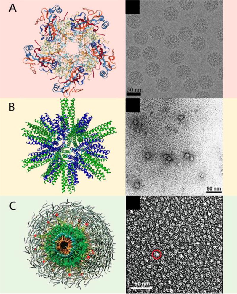
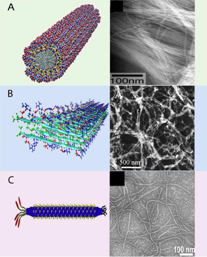
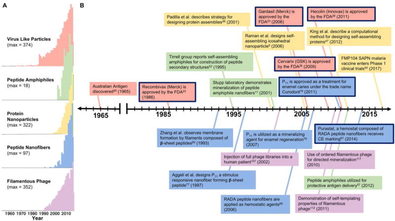
Selected platforms are discussed based on these features, because they have had a meaningful translation into clinical practice, or because they have had steady progress towards clinical translation. These include spherical assemblies such as virus-like particles, designed protein nanoparticles, and peptide amphiphiles (Figure 1); and elongated structures such as β-sheet nanofibers, fiber-forming peptide amphiphiles, and filamentous phage (Figure 2). A timeline of their development is shown in Figure 3.
Figure 1.
Compiled computer generated models and transmission electron microscopy images of spherical supramolecular assemblies (A) Virus-like particle, (B) Designed protein nanoparticle, and (C) Peptide amphiphile micelle. (A, left) adapted with permission.[7] 2007, ASBMB. (A, right) adapted with permission.[8] 2017, Elsevier. (B) adapted with permission.[9] 2006, Elsevier. (C) adapted with permission.[10] 2015, Wiley-VCH.
Figure 2.
Compiled computer generated models and transmission electron microscopy images of nanofibrillar supramolecular assemblies. (A) Peptide amphiphile nanofiber, (B) Beta sheet nanofiber, and (C) Filamentous phage. (A, left) adapted with permission.[11] 2001, AAAS. (A, right) adapted with permission.[12] 2002, PNAS. (B, left) adapted with permission.[13] 2013, American Chemical Society. (B, right) adapted with permission.[14] Wiley-VCH, 2008. (C, left) adapted with permission.[15] 2014, Elsevier. (C, right) adapted with permission.[16] 2014, Wiley-VCH.
Figure 3. Timeline of clinical development of supramolecular polypeptide materials.
(A) Citations per year of therapeutic materials in each of the listed classes of polypeptide-based supramolecular materials. (B) A timeline representing major events in the progression of self-assembling polypeptide therapeutics. Each family of materials is color-coded to match the citation data. Events in which a technology was approved for clinical use are outlined in black. Citation reports were generated using Web of Science and search terms indicating the therapeutic or clinical use of the relevant platforms.
2. Virus-like Particles
Some of the first engineered supramolecular structures to be translated effectively have been virus-like particles (VLPs), multiprotein constructs that self-assemble to mimic the organization, structure, and immunogenicity of native viruses but that lack infectious genetic materials (Figure 1A).[7,8] Their development has preceded other supramolecular materials discussed here (see timeline in Figure 3). VLPs are potent immunogens, able to stimulate B cell/antibody responses, CD4+ T-cell responses, and cytotoxic T-cell responses.[17,18] Owing to their lack of a viral genome, VLP-based vaccines circumvent some risks associated attenuated or inactivated live viruses, highlighting an advantage of supramolecular systems: they are more compositionally defined than the analogous biological structures, viruses. They also have advantages over subunit vaccines based on viral proteins or peptides conjugated to carrier proteins, which commonly require higher and more frequent dosing and adjuvants to be as effective as inactivated or attenuated viruses.[19] As alternatives to these previous platforms, VLPs were developed to display an array of epitopes that mimic the surface of native viruses more effectively than subunit or peptide vaccines, thus improving their immunogenic properties.[20] VLPs, like viruses, come in a range structures including those with a single capsid protein, multiple capsid proteins, or those without lipid envelopes.[21] VLPs consisting of multiple capsid proteins are expressed and assembled via subsequent processing or by co-expression of polycistronic genes within a cell.
Several additional aspects of VLPs have contributed to their clinical translation. Whereas the first translated VLP-based vaccines raised antibodies against the naturally occurring virus capsid proteins of the assembled structure, VLPs can also be used as vehicles to display heterologous antigens associated with other infectious diseases. This modularity of the platform is described below, in the discussion of chimeric and decorated VLPs. The manufacturability of VLPs depends upon the complexity of the platform. For example, GlaxoSmithKline’s (GSK’s) Cervarix™ (human papillomavirus) and Engerix™ (hepatitis B virus) and Merck & Co.’s Recombivax HB™ (hepatitis B virus) and Gardasil™ (human papillomavirus) are generated based on a single capsid protein—a feature that accelerated expression optimization and subsequent approval and commercialization. However, as platforms become increasingly complex, they become increasingly more challenging to manufacture at a large scale. Multi-subunit, chimeric, and other types of VLPs must overcome these challenges. Fortunately, a large assortment of expression systems (bacterial, yeast, insect, plant, mammalian, and cell-free) have been developed for the production of new VLP platform candidates.
The idea of using non-infectious virus particles to develop prophylactic human vaccines first became attractive when non-infectious VLPs composed of the surface antigen from the hepatitis B virus (HBsAg, also known as the Australia antigen), were discovered in human sera in 1965.[20] The first VLP-based human vaccine consisted of HBsAg VLPs derived from human plasma and was licensed in the United States in 1981 (Figure 3). Subsequently, after the advent of HIV/AIDS rendered plasma-derived products more challenging, the HBsAg particles were produced recombinantly in yeast, and the first recombinant human vaccine, Recombivax HB, was licensed by Merck in 1986.[22] Three years later in 1989, Engerix-B, a similar HBV vaccine, was licensed by GlaxoSmithKline. It took another 17 years before the next recombinant VLP-based vaccine was licensed for human use.[13]
Gardasil, licensed by Merck in 2006, is a vaccine against human papillomavirus (HPV) that protects against HPV-related diseases such as cervical cancer. In 1991, concurrently with several other laboratories, Frazer et al. observed that recombinantly expressed HPV L1 and L2 virus capsid proteins self-assembled into VLPs resembling the native virion structure.[23] In early 1992, based on this observation, Merck Research Laboratories (MRL) initiated an HPV vaccine program. At the time, a growing number of HPV genotypes were identified and associated with benign genital warts and or precursor lesions to cervical cancer. MRL devised a strategy to produce a quadrivalent HPV L1 VLP that would protect against cervical cancers (caused by HPV16 and 18) as well as genital warts (caused by HPV 6 and 11).
Several concurrent studies in the 1990s were additionally critical to the success of HPV vaccines. One important advancement was the development of an in vitro method of producing HPV virions. The HPV lifecycle is linked to human epithelial tissue differentiation which is difficult to achieve in vitro. Kreider et al. overcame this challenge by developing a complex xenograft model in which human foreskin epithelial tissue was infected with HPV 11 and grown under the renal capsule of athymic mice. This method, known as the Kreider method, allowed for the production of infectious HPV 11.[23] In 1995, animal challenge studies were published that continued to advance HPV vaccines. Two studies demonstrated measurable serum antibody responses to VLP immunization and subsequent protection in later challenges.[23] The third demonstrated the importance of maintaining the native VLP structure for optimal vaccine efficacy.[24] Another key contribution to the success of MRL’s HPV vaccine came from work developing a manufacturing process for HBsAg production in Saccharomyces cerevisiae. It provided a foundation for the development of methods to design, express, and purify large amounts of HPV VLPs.[23]
In 1997, HPV 11 L1 VLPs were evaluated for the first time in humans as a monovalent vaccine during a Phase I clinical trial to demonstrate safety and immunogenicity. The monovalent vaccine was chosen owing to the availability of robust models for the evaluation of immunogenicity, and because models for the other three HPV types had not yet been developed. The Phase 1 study outcomes were promising, with no reported significant adverse effects. A second study tested the ability of monovalent HPV 16 L1 VLP efficacy in preventing HPV infection, providing the first demonstration that vaccination with HPV VLPs could prevent disease. These favorable results supported the development of the multivalent vaccine and spurred the advancement of the program. In 2006, after impressive efficacy data and an acceptable safety profile, the first HPV VLP vaccine, Gardasil, was licensed (See timeline in Figure 3).[23,25]
Another HPV VLP human vaccine for the prevention of cervical cancer, Cervarix, was developed by GlaxoSmithKline and licensed in the United States in 2009. This vaccine includes HPV types 16 and 18 and has been produced using insect cells infected with recombinant baculovirus.[26] Cervarix demonstrated a 90.4% efficacy against cervical intraepithelial neoplasia lesions containing HPV 16 and 18 and indicated cross-protection with the HPV types 31 and 45, leading to the protection against 80% of cervical cancers.[27] Hecolin (Xiamen Innovax Biotech), a recombinant VLP-based vaccine for prophylactic use against hepatitis E virus infection, was licensed in China in 2011.[28] In the past decade, VLP vaccines have played a critical role in the improvement of human health and continue to be applied to new diseases. VLP-based vaccines are currently in clinical trials for the treatment of many infectious diseases: HBV, HIV, HPV, human parvovirus, influenza virus A, norwalk virus, ebola virus, and severe acute respiratory syndromerelated coronavirus (SARS-CoV). The inclusion of preclinical testing to this list broadens it even further, to diseases including chikungunya virus, west nile virus, and mumps virus.[29]
As mentioned in the introduction, one of the key strategic strengths of supramolecular systems is the modularity that they tend to possess. In VLPs, this modularity is exemplified by chimeric VLPs, in which epitopes of choice from various diseases are inserted into the particle-forming proteins. Provided that the new epitope can be inserted in a way that does not disrupt self-assembly, and that presents the epitope on the surface of the particle so that it is both antigenic and immunogenic, the modularity of the system is maintained.[20] By being able to insert epitopes of choice, the range of therapeutic applications becomes quite broad. For example, several clinical studies are currently evaluating chimeric VLP vaccines for the treatment of noninfectious diseases such as cancer (melanoma),[30] neurodegenerative diseases (Alzheimer’s disease),[31] autoimmune diseases (allergic rhinoconjunctivitis and asthmas),[32] and other disorders. Another approach for including chosen epitopes/antigens is separately expressing the VLP and target protein and conjugating them together. This approach is often preferable and necessary due to differences in optimal expression conditions for each component. Thus, postproduction methods have been developed to link the VLP and target antigen. This is achieved through genetic manipulation, coupling via supramolecular or covalent bonds, or by encapsulation of cargo by disassembling and reassembling purified VLPs in the presence of the desired molecule.[33] Coupling chemistries range from the use of bifunctional crosslinkers, click chemistry, sortase-mediated attachment, polyhistidine/NTA-Ni2+, or affinity-tag interactions to conjugate the VLP and target antigen[34] Notably, these postproduction-modified VLPs have led to several platforms currently being tested in clinical trials.[35,36,37]
Although a range of chemical cross-linking strategies have been developed for VLPs,[38,39] conjugation of target antigens with multiple reactive sites can lead to heterogenous coupling or unfavorable epitope display.[38] The Bachmann group addressed this challenge using the SpyTag-SpyCatcher system.[40] SpyTag is a peptide that forms a spontaneous and irreversible isopeptide bond with SpyCatcher, its protein partner.[40] The SpyCatcher-VLP platform is expressed in E. coli and mixed with SpyTag-Antigen to form “Plug-and-Display” decorated VLPs, another highly modular, tunable approach that allows for incorporation of a wide variety of antigens to the VLP surface. The group reported that SpyCatcher-VLPs decorated with the CIDR or Pfs25 antigens generated a robust antibody response are only a single immunization without requiring adjuvant. This platform shows promise in application such as drug delivery, enzyme scaffolds, biosensors, and cancer immunotherapy,[41] and it mitigates the need for complex chimeric protein expression or the incoroporation of unnatural amino acids, as would be necessary in copper-catalyzed azide-alkyne cycloaddition (click chemistry) or other chemoselective bioconjugation reactions.
VLPs, owing to their structural definition and flexibility in formulation, also make useful experimental tools for studying basic aspects of immunity. They usually range in diameter from 20–200 nm, an optimal size for drainage to lymph nodes and subsequent interactions with B cells. Whereas the differentiation of naïve B cells into memory B cells has been extensively studied at the cellular and molecular level, the fate of memory B cells upon antigen re-encounter has been comparatively less well-studied. The Bachmann group used a Qβ VLP as a model system and were able to track VLP-specific B cells via flow cytometry and histology to follow naïve and memory B cell responses.[42] They unexpectedly found that, during secondary B cell responses, secondary plasma cells are generated, whereas naïve B cells are recruited into a parallel primary B cell response. This phenomenon allows a plasticity of the memory B cell repertoire upon multiple antigenic exposures.
Shortcomings of VLPs include their requirement for a continuous and well-regulated cold chain, which negatively impacts their distribution to the developing world. To address this challenge, the Chackerian group has developed a VLP-based vaccine candidate that is compatible with spray drying, thus enhancing its stability over a broad range of temperatures.[43] This platform targets a highly conserved, broadly neutralizing epitope from the HPV minor capsid protein, L2. Not only does this vaccine elicit high-titer and long-lasting antibody immune responses,[44] but the spray dried VLPs were highly immunogenic in a mouse model after being stored for 14 months at either room temperature or 37°C. Other constraints that VLPs are subject to include the relatively small size of epitopes that can be accommodated within the particles. For example, large antigens such as HIV envelope and influenza hemagglutinin proteins are too large to be packaged within native VLPs. The practicality of VLP platforms is also limited by manufacturing considerations.[21] For example, VLPs derived from E. coli, the most widely used and most efficient expression system in the bio- technology industry, have a high degree of heterogeneity in their physical properties, including the shape and diameter of the particles. Such particles require post-purification disassembly and reassembly in optimized conditions. In addition, it may be possible to produce highly immunogenic VLP preparations, but the antigen might not be viable in the VLP context until stable formulations can be developed. Formulations must be resistant to aggregation upon exposure to low salt and protein concentration, as well as protection against surface adsorption and aggregation as a result of heat stress and physical agitation in order to achieve the multiple-year stability required for a marketed vaccine.[20] Newer expression systems in conjunction with innovative purification strategies could determine the pace for the next generation of VLP-based vaccine candidates. For example, yeast, insect, and mammalian expression systems have been used to circumvent the limitations of bacterial expression such as sub-optimal pH and lipid compositions within the bacteria. Additionally, the advancement of high-throughput biophysical and structural analyses of recombinant VLPs may play a key role in the assessment of VLP candidates. Electron microscopy, electrospray differential mobility analysis, atomic force microscopy, X-ray crystallography, and dynamic light scattering provide quantitative structural data for each vaccine candidate and play a valuable role in ensuring product robustness from the early clinical development stage and beyond.[20]
3. Designed Protein Nanoparticles
As an alternative to particles inspired by natural virus capsid proteins, fully designed protein nanoparticles have received considerable interest. Although “bottom-up” approaches for designing nanomaterials were popularized over 30 years ago,[45] the development of supramolecular polypeptide materials into successful medical technologies was initially slow, hindered by the sheer diversity of possible self-assembled structures and the lack of design rules. In 2001, the Yeates group developed an approach based on molecular symmetry to fabricate protein assemblies having a range of predictable architectures.[46] Their strategy was a breakthrough in rational self-assembling protein design.
In the past few years, the capacity to model and predict protein structures and energetics has increased along with computing power, leading to the computational design of de novo self-assembling protein nanoparticles.[47] The Baker group used naturally occurring oligomeric proteins as building blocks to design cage-like assemblies with accuracy. Recently, they generated a hyperstable 60-subunit protein icosahedron via symmetric modelling coupled with computational protein–protein interface design.[48] This structure is robust to genetic fusions, making it a notably modular platform that could be used in multivalent epitope display as well as drug delivery.
Taking inspiration from nature has also proven invaluable in the design of mechanically and chemically stable nanoparticles. In 2006, Raman et al. designed a self-assembling nanoparticle based on virus capsids via superposition of different protein oligomerization domains onto the symmetry axes of an icosahedron shown in Figure 1B. The monomer building block consisted of a protein chain made of two coiled coils connected by a short linker region. The association between the coiled coils caused the assembly of monomers into a roughly spherical nanoparticle.[9]
The self-assembling protein nanoparticle’s versatile and flexible design allow for optimization of biophysical and immunological properties making them a desirable vaccine platform. Whereas synthetic peptides are usually not sufficiently immunogenic and require adjuvants, the Burkhard group has developed a self-assembling protein nanoparticle platform that displays both B and T cell epitopes to produce a vaccine with self-adjuvanting qualities.[49] This platform is currently under development to improve a malaria vaccine, “RTS,S” that is based on the circumsporozoite protein of P. falciparum.[50] The self-assembled protein nanoparticles are able to stimulate high titer, high avidity antibodies and present CD8+ T-cell eptiopes to stimulate IL-2 and IFN-γ producing long-term memory T-cells in mouse models. The vaccine candidate FMP014, based on this platform, is currently undergoing phase 1 clinical trials.
A principal advantage of designed protein nanoparticles is their chemical definition. The ability to use computational methods to design nanoparticles of different geometries and sizes opens the door to applications such as drug delivery, in which the shape and size of the delivery vehicle are crucial. However, the manufacturability of these platforms, similar to VLPs, is a challenge due to their multi-subunit nature and the need to be recombinantly expressed.
4. Spherically Assembled Peptide Amphiphiles
Micellar nanocarriers composed of amphiphilic molecules have had particular success in the pharmaceutical realm as a tool to increase bioavailability, retention, and solubility of various drugs.[51] However, in this review, we shall be focusing on peptide amphiphiles as they relate to our theme of the clinical translation of peptide-based materials. Such structures self-assemble from molecules composed of a hydrophobic domain, usually an alkyl chain, and a hydrophilic peptide domain.[4,52] PA assembly is driven by hydrophobic-hydrophobic interactions in water, and bioactivity is programmed into the hydrophilic peptide head groups (see Figure 1C). For a comprehensive review on this topic, the reader is directed to Acar et al.[6]
Targeting, diagnostic, and theranostic platforms have been derived from peptide amphiphile micelles (PAMs). To increase the size of hydrophobic head groups and push systems toward a micellar packing morphology, the micelles consist of a biologically active peptide attached to a hydrophobic alkyl tail via a bulky PEG spacer. The PEG spacer allows for enhanced blood circulation times while retaining the packing parameters necessary for micelle formation.[6] These molecules are shown to circulate through the blood stream without causing blockage, and are cleared via the reticulo-endothelial system and renal system with 90% clearance and no toxicity after 7 days.[53]
PAMs are currently in preclinical development for both cancer and atherosclerosis diagnostic applications. For cancer applications, fluorescently-labeled PAs with the fibrin-binding peptide CREKA were used to target glioblastoma cells. Upon intravenous administration to GL261 glioma-bearing mice, non-targeting micelles passively accumulated at the fibrin deposits characteristic of tumor vasculature. These micelles displayed enhanced tumor homing as early as 1 h after administration without inducing cytotoxicity or tissue damage.[54] Towards a therapy for atherosclerosis, the CREKA targeting peptide, an antithrombin peptide called hirulog, and fluorescence molecules were assembled into theranostic micelles.[55] This fibrin-targeting micelle could also be functionalized by the addition of the gadolinium chelator diethylenetriaminepentaacetic acid, allowing for plaque localization and visualization using T1-weighted MRI imaging.[56] Similar PAMs have been designed for the targeting of monocytes as a strategy to diagnose atherosclerosis via the areas of heightened immunological activity characterized by the disease.[10]
Similarly to previously described vaccine nanoparticles, PA micelles are able to elicit either humoral or cell-mediated immunity without additional adjuvant. An antigen is simply conjugated to the tail domain of the molecule prior to self-assembly. Black et al. was able to assemble cylindrical micelles from monomers consisting of a dialkyl tail conjugated to a peptide containing the known cytotoxic T-cell epitope from the model tumor antigen ovalbumin.[57] These diC16-OVA micelles were able to stimulate OVA-specific T-cells, offering in vivo protection from tumors without any additional adjuvant. This observation spurred additional immunological studies to expand upon the potential of peptide amphiphile micelle vaccine platforms. In 2016, Barrett et. al used a Group A Streptococcus (GAS) B-cell antigen coupled to a diC16 tail which drove self-assembly of cylindrical micelles, to induce a micelle-mediated immune response (without adjuvant) that was stronger than seen with a conventional gold-standard vaccine formulation.[58] Although spherically assembled peptide amphiphiles have not yet reached regulatory approval for clinical applications, their versatility, modularity, and demonstrated success in preclinical work is encouraging.
5. Fiber-forming Platforms
Clinically translatable supramolecular materials are not limited to spherical morphologies. Extended structures with high aspect ratios have also seen considerable development towards a variety of medical technologies (Figure 2 A–C). Prominent among these have been fibrillar assemblies of peptides and their derivatives. These structures have been investigated over the past 20 years and have made progress towards therapies in hemostasis, dentistry, wound healing, and immunology.
While studying structural biology in Alexander Rich’s research group, Shuguang Zhang discovered a self-assembling β-sheet peptide based on the DNA-binding protein, zuotin.[59] The peptide formed amphiphilic tapes with two distinct surfaces, one hydrophobic and the other hydrophilic, and it also contained complementary charged residues that additionally favored this β-sheet folding (see Figure 2B). Following this discovery, a mimic of the native peptide was designed by mutating the charged and hydrophobic residues, but leaving the pattern intact.[60] This mimic demonstrated that self-assembly of peptides with these motifs was not a sequence-specific anomaly, but could be recreated in similar systems, forming the first steps towards design rules for fibrillizing peptides. The designer peptide was shown to also form macroscopic membranes and support the attachment of mammalian cells, demonstrating its utility as a biomaterial.[61] Subsequently, it was found that the original synthetic sequence RADA16-II (RARADADA)2 and its modified form RADA16-I (RADA)4 spontaneously formed hydrogels of entangled nanofibers in salt-containing solutions and cell culture media.[62] Mixtures of peptides bearing multiple bioactive groups could be incorporated into a single macroscopic gel by dosing in various amounts of monomeric peptides to the gel mixture. Additionally, the incorporation of ligands or epitopes within the materials simply required extension of the peptide at either terminus with the desired sequence, thus lowering the barrier of synthetic difficulty for biological researchers. These gels have been subsequently developed towards neuronal regeneration, cytokine delivery, as biotinylated scaffolds for versatile protein delivery, and other applications.[62–64]
The release of proteins and small molecules from RADA hydrogels largely depends on the size of the protein cargo regardless of charge and hydrophilic character, allowing a wide variety of biomolecules to be delivered in these gels.[65] Besides physical entrapment, a biotin-sandwich approach has been used to tether insulin-like growth factor 1 (IGF-1) to gels for long term localization of bioactive IGF-1 in the scaffold.[63] Biotinylation provides the advantage of modularity within the hydrogel delivery system, as any biotinylated protein can be immobilized to biotinylated fibers via a streptavidin linkage. Biotinylated fibers have been investigated for myocardial regeneration following infarction and showed success in rats when injected into infarcted hearts along with cardiac progenitor cells.[66]
Although RADA nanofibers have been investigated preclinically for a wide range of medical applications, they have had the most clinical success as a hemostatic agent. Marketed under the trade name Purastat™ by 3-D Matrix Ltd.,[67] the product is composed solely of peptide dissolved in sterile water, which forms fibers when in contact with biological fluids. This self-assembly provides a physical blockage in order to limit bleeding at the site of application. Although this peptide’s use as a hemostat is still being actively developed as a component of layer-by-layer wound dressings,[68] the original RADA16-I peptide has been useful as a surgical hemostat for multiple surgeries because of the dense mesh it creates at neutral pH. The RADA16-I peptide which became Purastat was first tested as a hemostatic agent in 2006 and demonstrated hemostasis in rats by stopping bleeding in under 15 seconds when applied as a 3–4% aqueous solution to wounds in skin, brain, spinal cord, femoral artery, and liver.[69] This rapid induction of hemostasis circumvented the need for components of the clotting cascade to activate the hemostat, it avoided the use of heat or pyrogenic substances, and it was effective in the presence of anti-coagulant therapies[70] without causing pronounced tissue responses.[71] Although the peptide is slightly acidic, it did not cause significant inflammation in any animal or clinical studies, even when applied to the brain.[72] Also named TDM-621, the RADA16-I peptide was first tested as a hemostat in human surgeries in Japan during cardiovascular procedures,[73] and later during endoscopic mucosal resection,[74] with no treatment-related adverse events in either trial. Purastat is limited to relatively low pressure hemorrhages in comparison to major arterial injuries,[75] but has general utility as a versatile, biodegradable hemostat. Clinical trials to monitor the post-market performance of Purastat for vascular surgery (NCT03103282), and to test the use of Purastat during endoscopic submucosal dissection are scheduled to begin soon (NCT02833558). Purastat has already achieved CE marking in Europe and has currently received medical device product registration approval in a number of coutries including Thailand, Mexico, and Indonesia, and Australia.
Initial studies of the RADA family of peptides inspired the development of general design principles for forming nanofibers, during which other peptides were shown to have similar self-assembling properties. Notably, peptide P11, developed by Amalia Aggeli and coworkers, would eventually lead to the development of the enamel regeneration product Curodont™. The first sequence designed by Aggeli et al. in 1997 was a mimic of a β-sheet transmembrane protein.[76,77] This peptide was designed de novo based on patterns found in the transmembrane protein and was shown to form high aspect-ratio fibrils that tangled to form hydrogels independently of pH in a manner similar to the protein-inspired peptide on which it was based.[77] Currently, this peptide is on the market in the EU and Switzerland in the form of a treatment for dental enamel caries, and it functions by creating a local environment that enhances enamel mineralization.[77] Upon injection, monomeric peptides assemble into nanotapes to create a 3D matrix that in some respects resembles the matrix environment necessary for enamel deposition.[78]
A major design improvement preceding the final Curodont™ peptide sequence was the introduction of pH-sensitivity. In 2003, Aggeli and coworkers rationally modified the original peptide sequence so that it was negatively charged at pH>8, thus creating electrostatic repulsion and allowing the peptides to remain in a monomeric, un-assembled state.[78] Upon switching to more acidic pH, however, the acidic residues became protonated and monomers assembled in to β-sheet tapes.[79] This pH-sensitivity allows for in situ assembly of peptides into nanofibers, which is hypothesized to improve the efficacy of Curodont™ as the material is able to completely fill demineralized defects of varying shapes and sizes. This pH-sensitive assembly was specifically explored in the context of enamel remineralization in 2007, where simulated intraoral conditions were employed to assess the performance of self-assembled scaffolds which were administered in a basic solution, allowed to fibriliize, and then incubated under cyclic pH.[78] These studies were completed on enamel lesions formed on extracted human teeth and indicated that the P11 scaffold caused hydroxyapatite mineralization where the peptide solution was applied.[78] Thus, positive and significant results were obtained without the need for specialized application methods or a poorly translatable animal model, a significant advantage for the development of polypeptide biomaterials towards dental applications. After the enamel restorative properties of P11 were developed in preclinical models, Credentis was founded in 2010, Curodont™ was launched as the company’s first product, and its safety and efficacy were verified in a clinical trial to treat dental caries.[78] In 2012 it received market approval (CE-label) for medical devices in the European Union. In keeping with the original peptide design, the final formulation of Curodont™ is composed of only a monomeric 11-amino acid peptide (P11) dissolved in water, with no additional bioactive agents.[80] Following a single application, the size and color of lesions are significantly improved, as observed one month after treatment.[80,81]
Peptide nanofibers have also increasingly received attention in immunological contexts, an area in which our group is active. However, because the focus here is on clinical development and these materials have not yet reached clinical trials, these applications will be only briefly described, and the reader is referred to other recent reviews for more expansive descriptions of this burgeoning field.[1,2,82,83] After the discovery that β-sheet fibrillized peptides can raise strong antibody responses without the requirement of supplemental adjuvant,[84] these materials have been investigated preclinically towards a range of diseases and conditions including malaria,[85] Staphylococus aureus infections,[86] influenza,[87] West Nile virus, cancer,[88] and cocaine abuse.[89] Immunogenic peptide nanofibers are produced by co-assembling fibrillizing peptides extended with specific epitopes or haptens along with other fibrillizing peptides bearing T-cell epitopes. They stimulate antibody/B-cell responses, CD4 T-cell responses, and CD8 T-cell responses[90] which can be raised in tunable magnitudes without causing inflammation.[85,86,89]
Other recent extensions of fibrillizing peptide technologies have included assemblies containing both peptides and other therapeutically active compounds. In these drug-peptide formulations, hydrophobic, π-π, and electrostatic interactions induce the assembly of the molecules into fibrillar networks much in the same way as pure peptide systems.[91,92] A recently described example is a strategy in which the FDA-approved anticancer drug Pemetrexed was conjugated to a four-amino acid peptide (FFEE) and used both as an MRI contrast agent and to form drug depots near tumor sites.[93] The dual function of the material (contrast and depot formation) was possible because the peptide-drug conjugate formed fibrillar hydrogels at high concentrations to form the drug depot, while lower concentrations in the circulation acted as the MRI contrast agent. A range of other strategies have likewise employed fibrillizing peptides to alter the delivery or pharmacokinetics of various drugs. For example, Hartgerink and coworkers showed that several different multivalent drug molecules such as clodronate, heparin, and suramin could be used to stabilize β-sheet fibrillar hydrogels by shielding the surface charges on the peptides that would otherwise inhibit gelation, thus forming drug depots.[94] Naphthalene-modified peptides have been explored to carry hydrophobic drugs such as Curcumin, which require carrier transport due to their low solubility.[95] These examples highlight considerable future potential for peptide nanofibers towards clinical applications.
6. Peptide Amphiphile Nanofibers
Alongside the use of β-sheet peptides, peptide amphiphiles composed of a peptide head group and an alkyl tail are a related class of materials that have seen considerable interest for creating supramolecular nanofibers (see Figure 2A). Although the self-assembly of peptide amphiphiles into spherical structures had been known for some time,[96] the landmark 2001 paper by Hartgerink, Stupp, and colleagues catalyzed much interest in the ability of this class of molecule to form nanofibrous materials for specific biomedical applications.[11] In this report, nanofibers spontaneously assembled from peptide amphiphiles into parallel bundles and promoted mineralization of hydroxyapatite.[11] Since then, fibrillar peptide amphiphile materials have been explored in a broad range of medical applications ranging between wound healing,[97] bone healing,[98] the delivery of proteins,[99] nervous tissue repair,[100] and others. Additional chemistries have been developed to stabilize the materials. For example, towards applications where mechanical integrity is necessary, adhesive groups have been incorporated into the hydrophilic heads to render the fibrous gels self-healing after they are strained mechanically.[101] A recent example of clinically directed peptide amphiphile nanofibers used them to deliver bioactive proteins such as bone morphogenic protein (BMP) to induce osteogenesis in an environment mimicking native bone growth.[98] Other examples have included nerve repair, which benefitted from the bioactivity and parallel alignment of fibrous scaffolds,[102] and burn injuries, where heparin-mimetic gels induced the formation of vascularized, collagen-rich tissues.[97] Peptide amphiphiles have also been used to deliver bioactive cargo outside of the regenerative context, including the electrostatic complexation of antisense oligonucleotides to a cationic peptide head to form a depot for sustained release of oligonucleotide.[103] Because networks of peptide-based fibers are morphologically similar to the extracellular matrix, they have also been highly useful as in vitro culture materials[104] and can be utilized as cell delivery vehicles, as demonstrated in the transplantation of islet cells[105] and bone marrow-derived pro-angiogenic cells (BMPACs) cultured prior to transplantation.[106] Although work with fibrous peptide amphiphiles has remained largely preclinical to date, is anticipated that the coming years will see many of these applications brought forward into approved therapeutics.
7. Nanofibers Formed from Filamentous Phage
Filamentous bacteriophages (see Figure 2C) represent an additional step in the progression of nanofiber-forming biomaterials. Although their production is considerably different than the chemical synthesis of PAs and short peptides, their elongated morphology and polyamino acid composition are similar in some respects, and can be used to endow phage-based materials with similar properties to the other fiber-forming platforms discussed above. Filamentous phages have highly engineerable coat proteins which allow the high density surface display of selected proteins. M13 phage are naturally filamentous, and they resemble peptide and peptide-amphiphile nanofibers in morphology as they are less than 10 nm in diameter yet almost 1 µm in length. Because M13 bacteriophages are unable to infect mammalian cells, there is negligible risk of virulent infection when using these viruses in medical applications. For these reasons, they have been historically used as antimicrobial agents and are currently approved for use in food products, but have had seen limited use in clinical trials as therapeutics. A few examples exist where the tissue-targeting and tumor-homing abilities of full phage libraries were tested in humans.[107,108]
Bacteriophages were initially investigated as nanomaterials when they were observed to form liquid crystals, and they proved useful for the templating of inorganic structures by incorporating metal binding peptides on the viral coat proteins.[109,110] Following the discovery of this functionality, their liquid crystalline behavior was utilized to form aligned matrices for neural cell culture by displaying RGD and IKVAV peptides on the phage surface.[111] This work included the use of standard phage display to select optimal 8-amino acid sequences for receptor binding, and the resultant filamentous phages were produced in E. coli, a relatively simple manufacturing process that is likely to be scalable.
In another recent example of preclinical materials development using filamentous phages, their self-templating properties proved useful for controlling the direction of osteoblast growth using the orientation of the phage-based substrate.[112] The use of both self-templated and fabricated directionality in phage substrates has allowed for the directional growth of human fibroblasts,[113] proliferation and elongation of neural progenitor cells,[114] and stimulated the differentiation of mesenchymal stem cells into osteoblasts when the phages displayed an osteogenic peptide on their surface.[115] As in vivo injectable materials, phages have been used preclinically as carriers for magnetic nanoparticles for targeted imaging of cancerous tumors by displaying a high density of targeting ligands and metal binding peptides on the phage surface.[116] A related study used a modified approach by conjugating a streptavidin-linked fluorophore to phages displaying tumor-targeting and streptavidin-binding peptides on their surface for targeted cancer imaging without the use of metal particles.[15] These studies demonstrated the importance of directional patterning in combination with the display of specific peptides on the phages. Moreover, the incorporation of multiple bioactive phage populations into a single material only requires adjustment of the mixture of various deposited phages, so in this way these materials feature the modularity characteristic of supramolecular systems.
Similar to other nanofibrous materials, filamentous phage materials have received significant attention for inducing osteogenesis because they can form collagen-mimetic bundles for the mineralization of hydroxyapatite similarly to PA and β-sheet peptide nanofibers. Display of anioinic peptides caused parallel assembly of individual phage in the presence of cations, followed by formation of oriented crystals when counterions were introduced owing to the local supersaturation of inorganic ions.[117] This approach closely resembled the demonstration of PA mineralization presented by Stupp and coworkers and illustrates conservation of biological processes across materials platforms.[11] Since the demonstration of phage assembly mineralization, osteogenesis studies have expanded to include presentation of a hydroxyapatite nucleating protein, Dentin Matrix Protein-1, as an alternative moiety for inducing crystal formation.[118] Tobacco Mosiac Virus (TMV), another rod-like virus was shown to cause differentiation of mesenchymal stem cells into bone cells as culture on the TMV coated substrate caused an upregulation of BMP-2, osteocalcin, and calcium sequestration, which are all markers of bone development.[119] Recently, RGD-bearing phages were 3D-printed into a ceramic scaffold to induce osteogenesis and angiogenesis concurrently without the addition of exogenous vascular endothelial growth factor.[16] Because of the scale at which phage can be produced, they may have manufacturing advantages over other designer materials.
8. Conclusions and Future Directions
Over the past fifty years, supramolecular assemblies of peptides and proteins have developed from single vaccines to a broad range of technologies that spans the breadth of biomaterials applications as drug delivery vehicles, scaffolds for tissue regeneration, and other therapeutics.[1,2,5,6] Currently, several have achieved regulatory approval for clinical use (Table 1), primarily in the vaccine space. The advancement of these platforms can be attributed to many factors. Peptides and proteins are more economical than ever to produce, and manufacturing efficiencies continue to be developed. The design rules for each subclass of materials has been significantly mapped in recent years. And strategies have been optimized for incorporating disease-specific ligands, epitopes, or other moieties within each platform. We expect the coming decades to witness the implementation of many new examples of supramolecular polypeptide therapies.
Table 1.
A collection of engineered recombinant and synthetic self-assembling protein and peptide biomaterials that have been clinically translated in the United States and Europe.
| Technology | Type | Disease Target | Manufacturer | Ref. |
|---|---|---|---|---|
| Recombivax-HB | VLP | Hepatitis B Virus | Merck | [120] |
| Engerix-B | VLP | Hepatitis B Virus | GlaxoSmithKline | [120] |
| GenHevac B | VLP | Hepatitis B Virus | Pasteur-Merieux Aventis | [120] |
| Hepavax-Gene | VLP | Hepatitus B Virus | Crucell | [120] |
| Gardasil | VLP | Human Papilloma Virus | Merck | [25] |
| Cervarix | VLP | Human Papilloma Virus | GlaxoSmithKline | [25] |
| Curodont | Betasheet Fiber | Enamel Regeneration | Credentis | [78] |
| Purastat | Betasheet Fiber | Hemostasis | 3D Matrix Medical Technology | [67] |
The immunogenic features of peptide assemblies are advantageous for the development of synthetic vaccines and other engineered immunotherapies, a topic which has been recently reviewed by our group and others[1,2,5,82,83], but it is also important to note for other assemblies containing high densities of protein or peptide ligands/epitopes that such multivalent displays may induce unwanted immune responses. It remains to be seen whether such responses can be tolerated in specific applications, or if they could even be turned in the favor of the material’s clinical performance. Interestingly, neither Curodont[78] nor Purastat[67] contain specific ligands/epitopes, possibly avoiding immunogenicity that may be observed in trials of other nanofibrous materials containing additional protein or peptide functional components.
Additional future work may focus on investigating the biodistribution and pharmacokinetics of preclinical self-assembling materials. Due to the dynamic nature of these materials, it is important to understand how the in vivo environment affects their long-term structural organization, retention, and clearance. In order to be clinically translated, these platforms must also be capable of large-scale production and stable storage. Unlike traditional small molecules, these materials must generally be kept in monomeric or otherwise stable states of assembly prior to administration. Each of these issues represents important considerations that have not yet been fully worked out for supramolecular materials.
With several self-assembling platforms being discovered and developed over the past 50 years, it is worth considering the cross-roads that lay ahead: should we focus on the development of the promising platforms discussed here, or should we continue searching for new, novel platforms? On the one hand, several of the aforementioned platforms have shown encouraging preclinical data and are being investigated in new disease models. On the other, the discovery of a new, more efficacious and versatile platforms could spur unforeseen therapies based on the self-assembling concept. Either way, we predict that self-assembling peptide and protein technologies will continue making strides in the preclinical and clinical realms.
Acknowledgments
Research on supramolecular peptide biomaterials in our group has been supported by the National Institutes of Health under grant numbers 5R01EB009701; 5R01AI118182; 5R21CA196434; and 7R21AR066244). This review’s contents are solely the responsibility of the authors and do not necessarily represent the official views of these agencies.
Biographies

Joel H. Collier, PhD is an Associate Professor at Duke University in the Biomedical Engineering Department. His research focuses on designing novel biomolecular materials for applications within immunotherapies, three-dimensional cell culture, and tissue repair. He received his BS in Materials Science from Rice University and his PhD in Biomedical Engineering from Northwestern University. In 2016 he moved from the Surgery Department at the University of Chicago to Duke Biomedical Engineering.

Chelsea N Fries is a doctoral student pursuing her PhD in Joel Collier’s group at Duke University in the Biomedical Engineering Department. Her research focuses on the molecular design of peptide-based materials for immunological applications. She received her BS in Biomedical Engineering from Northwestern Univerisity with a minor in Chemistry.

Kelly Hainline is a doctoral student in Joel Collier’s lab at Duke University. She received her BE in Biomedical Engineering with a minor in Materials Science and Engineering from Vanderbilt University in 2016. Her research focuses on the integration of functional proteins into supramolecular assemblies for 3D cell culture and immunotherapy applications.
References
- 1.Kelly SH, Shores LS, Votaw NL, Collier JH. Biomaterial strategies for generating therapeutic immune responses. Advanced Drug Delivery Reviews. 2017:1–16. doi: 10.1016/j.addr.2017.04.009. [DOI] [PMC free article] [PubMed] [Google Scholar]
- 2.Wen Y, Collier JH. Supramolecular peptide vaccines: tuning adaptive immunity. Current Opinion in Immunology. 2015;35:73–79. doi: 10.1016/j.coi.2015.06.007. [DOI] [PMC free article] [PubMed] [Google Scholar]
- 3.Branco MC, Schneider JP. Self-assembling materials for therapeutic delivery. Acta Biomaterialia. 2009;5(3):817–831. doi: 10.1016/j.actbio.2008.09.018. [DOI] [PMC free article] [PubMed] [Google Scholar]
- 4.Cui H, Webber MJ, Stupp SI. Self-assembly of peptide amphiphiles: From molecules to nanostructures to biomaterials. Biopolymers. 2010;94(1):1–18. doi: 10.1002/bip.21328. [DOI] [PMC free article] [PubMed] [Google Scholar]
- 5.Webber MJ, Appel EA, Meijer EW, Langer R. Supramolecular biomaterials. Nat Mater. 2016;15(1):13–26. doi: 10.1038/nmat4474. [DOI] [PubMed] [Google Scholar]
- 6.Acar H, Srivastava S, Chung EJ, Schnorenberg MR, Barrett JC, LaBelle JL, Tirrell M. Self-assembling peptide-based building blocks in medical applications. Advanced Drug Delivery Reviews. 2016 doi: 10.1016/j.addr.2016.08.006. [DOI] [PMC free article] [PubMed] [Google Scholar]
- 7.Bishop B, Dasgupta J, Klein M, Garcea RL, Christensen ND, Zhao R, Chen XS. Crystal Structures of Four Types of Human Papillomavirus L1 Capsid Proteins: Understanding the specificity of neutralizing monoclonal antibodies. The Journal of Biological Chemistry. 2007;282(43):31803–31811. doi: 10.1074/jbc.M706380200. [DOI] [PubMed] [Google Scholar]
- 8.Guan J, Bywaters SM, Brendle SA, Ashley RE, Makhov AM, Conway JF, Christensen ND, Hafenstein S. Cryoelectron Microscopy Maps of Human Papillomavirus 16 Reveal L2 Densities and Heparin Binding Site. Structure. 2017;25(2):253–263. doi: 10.1016/j.str.2016.12.001. [DOI] [PubMed] [Google Scholar]
- 9.Raman S, Machaidze G, Lustig A, Aebi U, Burkhard P. Structure-based design of peptides that self-assemble into regular polyhedral nanoparticles. Nanomedicine: Nanotechnology, Biology, and Medicine. 2006;2(2):95–102. doi: 10.1016/j.nano.2006.04.007. [DOI] [PubMed] [Google Scholar]
- 10.Chung EJ, Mlinar LB, Nord K, Sugimoto MJ, Wonder E, Alenghat FJ, Fang Y, Tirrell M. Monocyte-Targeting Supramolecular Micellar Assemblies: A Molecular Diagnostic Tool for Atherosclerosis. Advanced Healthcare Materials. 2015;4(3):367–376. doi: 10.1002/adhm.201400336. [DOI] [PMC free article] [PubMed] [Google Scholar]
- 11.Hartgerink JD, Benaish E, Stupp SI. Self-Assembly and Mineralization of Peptide-Amphiphile Nanofibers. Science. 2001;294:1684–1687. doi: 10.1126/science.1063187. [DOI] [PubMed] [Google Scholar]
- 12.Hartgerink JD, Beniash E, Stupp SI. Peptide-amphiphile nanofibers: A versatile scaffold for the preparation of self-assembling materials. Proc Natl Acad Sci USA. 2002;99(8):5133–5138. doi: 10.1073/pnas.072699999. [DOI] [PMC free article] [PubMed] [Google Scholar]
- 13.Cormier AR, Pang X, Zimmerman MI, Zhou H-X, Paravastu AK. Molecular Structure of RADA16-I Designer Self-Assembling Peptide Nanofibers. ACS Nano. 2013;7(9):7562–7572. doi: 10.1021/nn401562f. [DOI] [PMC free article] [PubMed] [Google Scholar]
- 14.Sieminski AL, Semino CE, Gong H, Kamm RD. Primary sequence of ionic self-assembling peptide gels affects endothelial cell adhesion and capillary morphogenesis. J. Biomed. Mater. Res. 2008;87A(2):494–504. doi: 10.1002/jbm.a.31785. [DOI] [PubMed] [Google Scholar]
- 15.Jin H-E, Farr R, Lee S-W. Collagen mimetic peptide engineered M13 bacteriophage for collagen targeting and imaging in cancer. Biomaterials. 2014;35(33):9236–9245. doi: 10.1016/j.biomaterials.2014.07.044. [DOI] [PubMed] [Google Scholar]
- 16.Wang J, Yang M, Zhu Y, Wang L, Tomsia AP, Mao C. Phage Nanofibers Induce Vascularized Osteogenesis in 3D Printed Bone Scaffolds. Adv. Mater. 2014;26(29):4961–4966. doi: 10.1002/adma.201400154. [DOI] [PMC free article] [PubMed] [Google Scholar]
- 17.Schirmbeck R, Böhm W, Reimann J. Virus-Like Particles Induce MHC Class I-Restricted T-Cell Responses. Intervirology. 1996;39(1–2):111–119. doi: 10.1159/000150482. [DOI] [PubMed] [Google Scholar]
- 18.Murata K, Lechmann M, Qiao M, Gunji T, Alter HJ, Liang TJ. Immunization with hepatitis C virus-like particles protects mice from recombinant hepatitis C virus-vaccinia infection. Proc Natl Acad Sci USA. 2003;100(11):6753–6758. doi: 10.1073/pnas.1131929100. [DOI] [PMC free article] [PubMed] [Google Scholar]
- 19.Noad R, Roy P. Virus-like particles as immunogens. Trends in Microbiology. 2003;11(9):438–444. doi: 10.1016/S0966-842X(03)00208-7. [DOI] [PubMed] [Google Scholar]
- 20.Zhao Q, Li S, Yu H, Xia N, Modis Y. Virus-like particle-based human vaccines: quality assessment based on structural and functional properties. Trends in Biotechnology. 2013;31(11):654–663. doi: 10.1016/j.tibtech.2013.09.002. [DOI] [PubMed] [Google Scholar]
- 21.Grgacic EVL, Anderson DA. Virus-like particles: Passport to immune recognition. Methods. 2006;40(1):60–65. doi: 10.1016/j.ymeth.2006.07.018. [DOI] [PMC free article] [PubMed] [Google Scholar]
- 22.McAleer WJ, Buynak EB, Maigetter RZ, Wampler DE, Miller WJ, Hilleman MR. Human hepatitis B vaccine from recombinant yeast. Nature. 1984;307(5947):178–180. doi: 10.1038/307178a0. [DOI] [PubMed] [Google Scholar]
- 23.Bryan JT, Buckland B, Hammond J, Jansen KU. Prevention of cervical cancer: journey to develop the first human papillomavirus virus-like particle vaccine and the next generation vaccine. Current Opinion in Chemical Biology. 2016;32:34–47. doi: 10.1016/j.cbpa.2016.03.001. [DOI] [PubMed] [Google Scholar]
- 24.Jansen KU, Rosolowsky M, Schultz LD, Markus HZ, Cook JC, Donnelly JJ, Martinez D, Ellis RW, Shaw AR. Vaccination with yeast-expressed cottontail rabbit papillomavirus (CRPV) virus-like particles protects rabbits from CRPV-induced papilloma formation. Vaccine. 1995;13(16):1509–1514. doi: 10.1016/0264-410X(95)00103-8. [DOI] [PubMed] [Google Scholar]
- 25.Shi L, Sings HL, Bryan JT, Wang B, Wang Y, Mach H, Kosinski M, Washabaugh MW, Sitrin R, Barr E. GARDASIL®: Prophylactic Human Papillomavirus Vaccine Development – From Bench Top to Bed-side. Clin Pharmacol Ther. 2007;81(2):259–264. doi: 10.1038/sj.clpt.6100055. [DOI] [PubMed] [Google Scholar]
- 26.Monie A, Hung C-F, Roden R, Wu T-C. Cervarix ™ : a vaccine for the prevention of HPV 16, 18-associated cervical cancer. Biologics Targets Therapy. 2008;2(1):107–113. [PMC free article] [PubMed] [Google Scholar]
- 27.Dubin G, Harper DM, Franco EL, Wheeler CM, Moscicki A-B, Romanowski B, Roteli-Martins CM, Jenkins D, Schuind A, Costa Clemens SA, Dubin G. Sustained efficacy up to 4·5 years of a bivalent L1 virus-like particle vaccine against human papillomavirus types 16 and 18: follow-up from a randomised control trial. The Lancet. 2006;367(9518):1247–1255. doi: 10.1016/S0140-6736(06)68439-0. [DOI] [PubMed] [Google Scholar]
- 28.Wu T, Li S-W, Zhang J, Ng M-H, Xia N-S, Zhao Q. Hepatitis E vaccine development. Human Vaccines & Immunotherapeutics. 2014;8(6):823–827. doi: 10.4161/hv.20042. [DOI] [PubMed] [Google Scholar]
- 29.Shirbaghaee Z, Bolhassani A. Different applications of virus-like particles in biology and medicine: Vaccination and delivery systems. Biopolymers. 2015;105(3):113–132. doi: 10.1002/bip.22759. [DOI] [PMC free article] [PubMed] [Google Scholar]
- 30.Braun M, Jandus C, Maurer P, Hammann-Haenni A, Schwarz K, Bachmann MF, Speiser DE, Romero P. Virus-like particles induce robust human T-helper cell responses. Eur. J. Immunol. 2011;42(2):330–340. doi: 10.1002/eji.201142064. [DOI] [PubMed] [Google Scholar]
- 31.Chackerian B, Rangel M, Hunter Z, Peabody DS. Virus and virus-like particle-based immunogens for Alzheimer's disease induce antibody responses against amyloid-β without concomitant T cell responses. Vaccine. 2006;24(37–39):6321–6331. doi: 10.1016/j.vaccine.2006.05.059. [DOI] [PubMed] [Google Scholar]
- 32.Senti G, Johansen P, Haug S, Bull C, Gottschaller C, Müller P, Pfister T, Maurer P, Bachmann MF, Graf N, Kündig TM. Use of A-type CpG oligodeoxynucleotides as an adjuvant in allergen-specific immunotherapy in humans: a phase I/IIa clinical trial. Clinical & Experimental Allergy. 2009;39(4):562–570. doi: 10.1111/j.1365-2222.2008.03191.x. [DOI] [PubMed] [Google Scholar]
- 33.Naskalska A, Pyrc K. Virus Like Particles as Immunogens and Universal Nanocarriers. Pol J Microbiol. 2015;64(1):3–13. [PubMed] [Google Scholar]
- 34.Frietze KM, Peabody DS, Chackerian B. Engineering virus-like particles as vaccine platforms. Current Opinion in Virology. 2016;18:44–49. doi: 10.1016/j.coviro.2016.03.001. [DOI] [PMC free article] [PubMed] [Google Scholar]
- 35.Klimek L, Willers J, Hammann-Haenni A, Pfaar O, Stocker H, Mueller P, Renner WA, Bachmann MF. Assessment of clinical efficacy of CYT003-QbG10 in patients with allergic rhinoconjunctivitis: a phase IIb study. Clinical & Experimental Allergy. 2011;41(9):1305–1312. doi: 10.1111/j.1365-2222.2011.03783.x. [DOI] [PubMed] [Google Scholar]
- 36.Ambühl PM, Tissot AC, Fulurija A, Maurer P, Nussberger J, Sabat R, Nief V, Schellekens C, Sladko K, Roubicek K, Pfister T, Rettenbacher M, Volk H-D, Wagner F, Müller P, Jennings GT, Bachmann MF. A vaccine for hypertension based on virus-like particles: preclinical efficacy and phase I safety and immunogenicity. Journal of Hypertension. 2007;25(1):63–72. doi: 10.1097/HJH.0b013e32800ff5d6. [DOI] [PubMed] [Google Scholar]
- 37.Tissot AC, Maurer P, Nussberger J, Sabat R, Pfister T, Ignatenko S, Volk H-D, Stocker H, Müller P, Jennings GT, Wagner F, Bachmann MF. Effect of immunisation against angiotensin II with CYT006-AngQb on ambulatory blood pressure: a double-blind, randomised, placebo-controlled phase IIa study. The Lancet. 2008;371:821–827. doi: 10.1016/S0140-6736(08)60381-5. [DOI] [PubMed] [Google Scholar]
- 38.Smith MT, Hawes AK, Bundy BC. Reengineering viruses and virus-like particles through chemical functionalization strategies. Current Opinion in Biotechnology. 2013;24(4):620–626. doi: 10.1016/j.copbio.2013.01.011. [DOI] [PubMed] [Google Scholar]
- 39.Sapsford KE, Algar WR, Berti L, Gemmill KB, Casey BJ, Oh E, Stewart MH, Medintz IL. Functionalizing Nanoparticles with Biological Molecules: Developing Chemistries that Facilitate Nanotechnology. Chem. Rev. 2013;113(3):1904–2074. doi: 10.1021/cr300143v. [DOI] [PubMed] [Google Scholar]
- 40.Zakeri B, Fierer JO, Celik E, Chittock EC, Schwarz-Linek U, Moy VT, Howarth M. Peptide tag forming a rapid covalent bond to a protein, through engineering a bacterial adhesin. Proc Natl Acad Sci USA. 2012;109(12):E690–E697. doi: 10.1073/pnas.1115485109. [DOI] [PMC free article] [PubMed] [Google Scholar]
- 41.Brune KD, Leneghan DB, Brian IJ, Ishizuka AS, Bachmann MF, Draper SJ, Biswas S, Howarth M. Plug-and-Display: decoration of Virus-Like Particles via isopeptide bonds for modular immunization. Sci Rep. 2016;6(1):1904. doi: 10.1038/srep19234. [DOI] [PMC free article] [PubMed] [Google Scholar]
- 42.Zabel F, Kündig TM, Bachmann MF. Virus-induced humoral immunity: on how B cell responses are initiated. Current Opinion in Virology. 2013;3(3):357–362. doi: 10.1016/j.coviro.2013.05.004. [DOI] [PubMed] [Google Scholar]
- 43.Saboo S, Tumban E, Peabody J, Wafula D, Peabody DS, Chackerian B, Muttil P. Optimized Formulation of a Thermostable Spray-Dried Virus-Like Particle Vaccine against Human Papillomavirus. Mol. Pharmaceutics. 2016;13(5):1646–1655. doi: 10.1021/acs.molpharmaceut.6b00072. [DOI] [PMC free article] [PubMed] [Google Scholar]
- 44.Tumban E, Muttil P, Escobar CAA, Peabody J, Wafula D, Peabody DS, Chackerian B. Preclinical refinements of a broadly protective VLP-based HPV vaccine targeting the minor capsid protein, L2. Vaccine. 2015;33(29):3346–3353. doi: 10.1016/j.vaccine.2015.05.016. [DOI] [PMC free article] [PubMed] [Google Scholar]
- 45.Drexler KE. Molecular engineering: An approach to the development of general capabilities for molecular manipulation. Proceedings of the National Academy of Sciences of the United States of America. 1981;78(9):5275–5278. doi: 10.1073/pnas.78.9.5275. [DOI] [PMC free article] [PubMed] [Google Scholar]
- 46.Padilla JE, Colovos C, Yeates TO. Nanohedra: Using symmetry to design self assembling protein cages, layers, crystals, and filaments. Proc Natl Acad Sci USA. 2001;98(5):2217–2221. doi: 10.1073/pnas.041614998. [DOI] [PMC free article] [PubMed] [Google Scholar]
- 47.King NP, Sheffler W, Sawaya MR, Vollmar BS, Sumida JP, Andre I, Gonen T, Yeates TO, Baker D. Computational design of self-assembling protein nanomaterials with atomic level accuracy. Science. 2012;336(6085):1171–1174. doi: 10.1126/science.1219364. [DOI] [PMC free article] [PubMed] [Google Scholar]
- 48.Hsia Y, Bale JB, Gonen S, Shi D, Sheffler W, Fong KK, Nattermann U, Xu C, Huang P-S, Ravichandran R, Yi S, Davis TN, Gonen T, King NP, Baker D. Design of a hyperstable 60-subunit protein icosahedron. Nature. 2016;535(7610):136–139. doi: 10.1038/nature18010. [DOI] [PMC free article] [PubMed] [Google Scholar]
- 49.Kaba SA, Brando C, Guo Q, Mittelholzer C, Raman S, Tropel D, Aebi U, Burkhard P, Lanar DE. A Nonadjuvanted Polypeptide Nanoparticle Vaccine Confers Long-Lasting Protection against Rodent Malaria. The Journal of Immunology. 2009;183(11):7268–7277. doi: 10.4049/jimmunol.0901957. [DOI] [PMC free article] [PubMed] [Google Scholar]
- 50.Heppner DG, Kester KE, Ockenhouse CF, Tornieporth N, Ofori O, Lyon JA, Stewart VA, Duboise P, Lanar DE, Krzych U, Moris P, Angov E, Cummings JF, Leach A, Hall BT, Dutta S, Schwenk R, Hillier C, Ogutu B, Ware LA, Nair L, Darko CA, Withers MR, Ogutu B, Polhemus ME, Fukuda M, Pichyangkul S, Gettyacamin M, Diggs C, Soisson L, Milman J, Dubois M-C, Garcon N, Tucker K, Wittes J, Plowe CV, Thera MA, Duombo OK, Pau MG, Goudsmit J, Ballou WR, Cohen J. Towards an RTS,S-based, multi-stage, multi-antigen vaccine against falciparum malaria: progress at the Walter Reed Army Institute of Research. Vaccine. 2005;23(17–18):2243–2250. doi: 10.1016/j.vaccine.2005.01.142. [DOI] [PubMed] [Google Scholar]
- 51.Torchilin VP. Micellar Nanocarriers: Pharmaceutical Perspectives. Pharm Res. 2006;24(1):1–16. doi: 10.1007/s11095-006-9132-0. [DOI] [PubMed] [Google Scholar]
- 52.Berndt P, Fields GB, Tirrell M. Synthetic Lipidation of Peptides and Amino Acids: Monolayer Structure and Properties. J. Am. Chem. Soc. 1995:9515–9522. [Google Scholar]
- 53.Chung EJ, Mlinar LB, Sugimoto MJ, Nord K, Roman BB, Tirrell M. In vivo biodistribution and clearance of peptide amphiphile micelles. Nanomedicine: Nanotechnology, Biology, and Medicine. 2015;11(2):479–487. doi: 10.1016/j.nano.2014.08.006. [DOI] [PubMed] [Google Scholar]
- 54.Chung EJ, Cheng Y, Morshed R, Nord K, Han Y, Wegscheid ML, Auffinger B, Wainwright DA, Lesniak MS, Tirrell MV. Fibrin-binding, peptide amphiphile micelles for targeting glioblastoma. Biomaterials. 2014;35(4):1249–1256. doi: 10.1016/j.biomaterials.2013.10.064. [DOI] [PMC free article] [PubMed] [Google Scholar]
- 55.Peters D, Kastantin M, Kotamraju VR, Karmali PP, Gujraty K, Tirrell M, Ruoslahti E. Targeting atherosclerosis by using modular, multifunctional micelles. Proc Natl Acad Sci USA. 2009;106(24):9815–9819. doi: 10.1073/pnas.0903369106. [DOI] [PMC free article] [PubMed] [Google Scholar]
- 56.Yoo SP, Pineda F, Barrett JC, Poon C, Tirrell M, Chung EJ. Gadolinium-Functionalized Peptide Amphiphile Micelles for Multimodal Imaging of Atherosclerotic Lesions. ACS Omega. 2016;1(5):996–1003. doi: 10.1021/acsomega.6b00210. [DOI] [PMC free article] [PubMed] [Google Scholar]
- 57.Black M, Trent A, Kostenko Y, Lee JS, Olive C, Tirrell M. Self-Assembled Peptide Amphiphile Micelles Containing a Cytotoxic T-Cell Epitope Promote a Protective Immune Response In Vivo. Adv. Mater. 2012;24(28):3845–3849. doi: 10.1002/adma.201200209. [DOI] [PubMed] [Google Scholar]
- 58.Barrett JC, Ulery BD, Trent A, Liang S, David NA, Tirrell MV. Modular Peptide Amphiphile Micelles Improving an Antibody-Mediated Immune Response to Group A Streptococcus. ACS Biomater. Sci. Eng. 2016;3(2):144–152. doi: 10.1021/acsbiomaterials.6b00422. [DOI] [PMC free article] [PubMed] [Google Scholar]
- 59.Zhang S, Lockshin C, Herbert A, Winter E, Rich A. Zuotin, A Putative Z-Dna Binding Protein in Saccharomyces Cerevisiae. The EMBO Journal. 1992;11(10):3787–3796. doi: 10.1002/j.1460-2075.1992.tb05464.x. [DOI] [PMC free article] [PubMed] [Google Scholar]
- 60.Zhang S, Holmes T, Lockshin C, Rich A. Spontaneous assembly of a self-complimentary oligopeptide to form a stable macroscopic membrane. Proceedings of the National Academy of the Sciences. 1993;90:3334–3338. doi: 10.1073/pnas.90.8.3334. [DOI] [PMC free article] [PubMed] [Google Scholar]
- 61.Zhang S, Holmes T, DiPersio M, Hynes R, Su X, Rich A. Self-complementary oligopeptide matrices support mammalian cell attachment. Biomaterials. 1995;16:1385–1393. doi: 10.1016/0142-9612(95)96874-y. [DOI] [PubMed] [Google Scholar]
- 62.Holmes T, de Lacalle S, Su X, Liu G, Rich A, Zhang S. Extensive neurite outgrowth and active synapse formation on self-assembling peptide scaffolds. Proceedings of the National Academy of the Sciences. 2000;97(12):6728–6733. doi: 10.1073/pnas.97.12.6728. [DOI] [PMC free article] [PubMed] [Google Scholar]
- 63.Davis ME, Hsieh PCH, Takahashi T, Song Q, Zhang S, Kamm RD, Grodzinsky A, Anversa P, Lee R. Local myocardial insulin-like growth factor 1 (IGF-1) delivery with biotinylated peptide nanofibers improves cell therapy for myocardial infarction. Proceedings of the National Academy of the Sciences. 2006;103(21):8155–8160. doi: 10.1073/pnas.0602877103. [DOI] [PMC free article] [PubMed] [Google Scholar]
- 64.Gelain F, Unsworth LD, Zhang S. Slow and sustained release of active cytokines from self-assembling peptide scaffolds. Journal of Controlled Release. 2010;145(3):231–239. doi: 10.1016/j.jconrel.2010.04.026. [DOI] [PubMed] [Google Scholar]
- 65.Koutsopoulos S, Unsworth LD, Nagai Y, Zhang S. Controlled release of functional proteins through designer self-assembling peptide nanofiber hydrogel scaffold. Proceedings of the National Academy of the Sciences. 2009;106(12):4623–4628. doi: 10.1073/pnas.0807506106. [DOI] [PMC free article] [PubMed] [Google Scholar]
- 66.Padin-Iruegas ME, Misao Y, Davis ME, Segers VFM, Esposito G, Tokunou T, Urbanek K, Hosoda T, Rota M, Anversa P, Leri A, Lee RT, Kajstura J. Cardiac Progenitor Cells and Biotinylated Insulin-Like Growth Factor-1 Nanofibers Improve Endogenous and Exogenous Myocardial Regeneration After Infarction. Circulation. 2009;120(10):876–887. doi: 10.1161/CIRCULATIONAHA.109.852285. [DOI] [PMC free article] [PubMed] [Google Scholar]
- 67.Pioche M, Camus M, Rivory JRM, Leblanc S, Lienhart I, Barret M, Chaussade S, Saurin J-C, Prat F, Ponchon T. A self-assembling matrix-forming gel can be easily and safely applied to prevent delayed bleeding after endoscopic resections. Endosc Int Open. 2016;04(04):E415–E419. doi: 10.1055/s-0042-102879. [DOI] [PMC free article] [PubMed] [Google Scholar]
- 68.Hsu BB, Conway W, Tschabrunn CM, Mehta M, Perez-Cuevas MB, Zhang S, Hammond PT. Clotting Mimicry from Robust Hemostatic Bandages Based on Self-Assembling Peptides. ACS Nano. 2015;9(9):9394–9406. doi: 10.1021/acsnano.5b02374. [DOI] [PMC free article] [PubMed] [Google Scholar]
- 69.G Ellis-Behnke R, Liang Y-X, Tay DKC, Kau PWF, Schneider GE, Zhang S, Wu W, So K-F. Nano hemostat solution: immediate hemostasis at the nanoscale. Nanomedicine: Nanotechnology, Biology, and Medicine. 2006;2(4):207–215. doi: 10.1016/j.nano.2006.08.001. [DOI] [PubMed] [Google Scholar]
- 70.Csukas D, Urbanics R, Moritz A, Ellis-Behnke R. AC5 Surgical Hemostat™ as an effective hemostatic agent in an anticoagulated rat liver punch biopsy model. Nanomedicine: Nanotechnology, Biology, and Medicine. 2015;11(8):2025–2031. doi: 10.1016/j.nano.2015.01.001. [DOI] [PubMed] [Google Scholar]
- 71.Song H, Zhang L, Zhao X. Hemostatic Efficacy of Biological Self-Assembling Peptide Nanofibers in a Rat Kidney Model. Macromol. Biosci. 2010;10(1):33–39. doi: 10.1002/mabi.200900129. [DOI] [PubMed] [Google Scholar]
- 72.Xu F-F, Wang Y-C, Sun S, Ho ASW, Lee D, Kiang KMY, Zhang X-Q, Lui W-M, Liu B-Y, Wu W-T, Leung GKK. Comparison between self-assembling peptide nanofiber scaffold (SAPNS) and fibrin sealant in neurosurgical hemostasis. Clinical And Translational Science. 2015;8(5):490–494. doi: 10.1111/cts.12299. [DOI] [PMC free article] [PubMed] [Google Scholar]
- 73.Masuhara H, Fujii T, Watanabe Y, Koyama N, Tokuhiro K. Novel Infectious Agent-Free Hemostatic Material (TDM-621) in Cardiovascular Surgery. Ann Thorac Cardiovasc Surg. 2012;18(5):444–451. doi: 10.5761/atcs.oa.12.01977. [DOI] [PubMed] [Google Scholar]
- 74.Yoshida M, Goto N, Kawaguchi M, Koyama H, Kuroda J, Kitahora T, Iwasaki H, Suzuki S, Kataoka M, Takashi F, Kitajima M. Initial clinical trial of a novel hemostat, TDM-621, in the endoscopic treatments of the gastric tumors. J Gastroenterol Hepatol. 2014;29:77–79. doi: 10.1111/jgh.12798. [DOI] [PubMed] [Google Scholar]
- 75.Jukes A, Murphy J, Vreugde S, Psaltis A, Wormald P. Nano-hemostats and a Pilot Study of Their Use in a Large Animal Model of Major Vessel Hemorrhage in Endoscopic Skull Base Surgery. J Neurol Surg B. 2016:1–7. doi: 10.1055/s-0036-1597277. [DOI] [PMC free article] [PubMed] [Google Scholar]
- 76.Aggeli A, Boden N, Cheng Y, Findlay J, Knowles PF, Kovatchev P, Turnbull P. Peptides Modeled on the Transmembrane Region of the Slow Voltage-Gated IsK Potassium Channel: Structural Characterization of Peptide Assemblies in the β-Strand Conformation. Biochemistry. 1996;35(50):16213–16221. doi: 10.1021/bi960891g. [DOI] [PubMed] [Google Scholar]
- 77.Aggeli A, Bell M, Boden N, Keen JN, Knowles PF, McLeish T, Pitkeathly M, Radford SE. Responsive gels formed by the spontaneous self-assembly of peptides into polymeric B-sheet tapes. Nature. 1997;386:259–262. doi: 10.1038/386259a0. [DOI] [PubMed] [Google Scholar]
- 78.Kirkham J, Firth A, Vernals D, Boden N, Robinson C, Shore RC, Brookes SJ, Aggeli A. Self-assembling Peptide Scaffolds Promote Enamel Remineralization. J Dent Res. 2007;86(5):426–430. doi: 10.1177/154405910708600507. [DOI] [PubMed] [Google Scholar]
- 79.Aggeli A, Bell M, Carrick LM, Fishwick CWG, Harding R, Mawer PJ, Radford SE, Strong AE, Boden N. pH as a Trigger of Peptide β-Sheet Self-Assembly and Reversible Switching between Nematic and Isotropic Phases. J. Am. Chem. Soc. 2003;125(32):9619–9628. doi: 10.1021/ja021047i. [DOI] [PubMed] [Google Scholar]
- 80.Brunton PA, Davies RPW, Burke JL, Smith A, Aggeli A, Brookes SJ, Kirkham J. Treatment of early caries lesions using biomimetic self-assembling peptides – a clinical safety trial. British Dental Journal. 2013;215(4):1–6. doi: 10.1038/sj.bdj.2013.741. [DOI] [PMC free article] [PubMed] [Google Scholar]
- 81.Jablonski-Momeni A, Heinzel-Gutenbrunner M. Efficacy of the self-assembling peptide P11-4 in construct- ing a remineralization scaffold on artificially-induced enamel lesions on smooth surfaces. J Orofac Orthop. 2014;75(3):175–190. doi: 10.1007/s00056-014-0211-2. [DOI] [PubMed] [Google Scholar]
- 82.Tostanoski LH, Jewell CM. Engineering self-assembled materials to study and direct immune function. Advanced Drug Delivery Reviews. 2017:1–19. doi: 10.1016/j.addr.2017.03.005. [DOI] [PMC free article] [PubMed] [Google Scholar]
- 83.Mora-Solano C, Collier JH. Engaging adaptive immunity with biomaterials. J. Mater. Chem. B. 2014;2(17):2409–2421. doi: 10.1039/C3TB21549K. [DOI] [PMC free article] [PubMed] [Google Scholar]
- 84.Rudra JS, Tian YF, Jung JP, Collier JH. A self-assembling peptide acting as an immune adjuvant. Proc Natl Acad Sci USA. 2010;107(2):622–627. doi: 10.1073/pnas.0912124107. [DOI] [PMC free article] [PubMed] [Google Scholar]
- 85.Rudra JS, Mishra S, Chong AS, Mitchell RA, Nardin EH, Nussenzweig V, Collier JH. Self-assembled peptide nanofibers raising durable antibody responses against a malaria epitope. Biomaterials. 2012;33(27):6476–6484. doi: 10.1016/j.biomaterials.2012.05.041. [DOI] [PMC free article] [PubMed] [Google Scholar]
- 86.Pompano RR, Chen J, Verbus EA, Han H, Fridman A, McNeely T, Collier JH, Chong AS. Titrating T-cell epitopes within self-assembled vaccines optimizes CD4+ helper T cell and antibody outputs. 2014;3(11):1898–1908. doi: 10.1002/adhm.201400137. [DOI] [PMC free article] [PubMed] [Google Scholar]
- 87.Chen J, Pompano RR, Santiago FW, Maillat L, Sciammas R, Sun T, Han H, Topham DJ, Chong AS, Collier JH. The use of self-adjuvanting nanofiber vaccines to elicit high-affinity B cell responses to peptide antigens without inflammation. Biomaterials. 2013;34(34):8776–8785. doi: 10.1016/j.biomaterials.2013.07.063. [DOI] [PMC free article] [PubMed] [Google Scholar]
- 88.Huang Z-H, Shi L, Ma J-W, Sun Z-Y, Cai H, Chen Y-X, Zhao Y-F, Li Y-M. a Totally Synthetic, Self-Assembling, Adjuvant-Free MUC1 Glycopeptide Vaccine for Cancer Therapy. J. Am. Chem. Soc. 2012;134(21):8730–8733. doi: 10.1021/ja211725s. [DOI] [PubMed] [Google Scholar]
- 89.Rudra JS, Ding Y, Neelakantan H, Ding C, Appavu R, Stutz S, Snook JD, Chen H, Cunningham KA, Zhou J. Suppression of Cocaine-Evoked Hyperactivity by Self-Adjuvanting and Multivalent Peptide Nanofiber Vaccines. ACS Chem. Neurosci. 2016;7(5):546–552. doi: 10.1021/acschemneuro.5b00345. [DOI] [PMC free article] [PubMed] [Google Scholar]
- 90.Chesson CB, Huelsmann EJ, Lacek AT, Kohlhapp FJ, Webb MF, Nabatiyan A, Zloza A, Rudra JS. Antigenic peptide nanofibers elicit adjuvant-free CD8+ T cell responses. Vaccine. 2014;32(10):1174–1180. doi: 10.1016/j.vaccine.2013.11.047. [DOI] [PubMed] [Google Scholar]
- 91.Caplan MR, Moore PN, Zhang S, Kamm RD, Lauffenburger DA. Self-Assembly of a β-Sheet Protein Governed by Relief of Electrostatic Repulsion Relative to van der Waals Attraction. Biomacromolecules. 2000;1(4):627–631. doi: 10.1021/bm005586w. [DOI] [PubMed] [Google Scholar]
- 92.Caplan MR, Schwartzfarb EM, Zhang S, Kamm RD, Lauffenburger DA. Control of self-assembling oligopeptide matrix formation through systematic variation of amino acid sequence. Biomaterials. 2002;23:219–227. doi: 10.1016/s0142-9612(01)00099-0. [DOI] [PubMed] [Google Scholar]
- 93.Lock LL, Li Y, Mao X, Chen H, Staedtke V, Bai R, Ma W, Lin R, Li Y, Liu G, Cui H. One-Component Supramolecular Filament Hydrogels as Theranostic Label-Free Magnetic Resonance Imaging Agents. ACS Nano. 2017;11(1):797–805. doi: 10.1021/acsnano.6b07196. [DOI] [PMC free article] [PubMed] [Google Scholar]
- 94.Kumar VA, Shi S, Wang BK, Li I-C, Jalan AA, Sarkar B, Wickremasinghe NC, Hartgerink JD. Drug-Triggered and Cross-Linked Self-Assembling Nanofibrous Hydrogels. J. Am. Chem. Soc. 2015;137(14):4823–4830. doi: 10.1021/jacs.5b01549. [DOI] [PMC free article] [PubMed] [Google Scholar]
- 95.Liu J, Liu J, Xu H, Zhang Y, Chu L, Liu Q, Song N, Yang C. Novel tumor-targeting, self-assembling peptide nanofiber as a carrier for effective curcumin delivery. IJN. 2013:197–11. doi: 10.2147/IJN.S55875. [DOI] [PMC free article] [PubMed] [Google Scholar]
- 96.Yu Y-C, Berndt P, Tirrell M, Fields GB. Self-Assembling Amphiphiles for Construction of Protein Molecular Architecture. J. Am. Chem. Soc. 1996;118:12515–12520. [Google Scholar]
- 97.Yergoz F, Hastar N, Cimenci CE, Ozkan AD, Tekinay T, Guler MO, Tekinay AB. Heparin mimetic peptide nanofiber gel promotes regeneration of full thickness burn injury. Biomaterials. 2017;134:117–127. doi: 10.1016/j.biomaterials.2017.04.040. [DOI] [PubMed] [Google Scholar]
- 98.Lee SS, Hsu EL, Mendoza M, Ghodasra J, Nickoli MS, Ashtekar A, Polavarapu M, Babu J, Riaz RM, Nicolas JD, Nelson D, Hashmi SZ, Kaltz SR, Earhart JS, Merk BR, McKee JS, Bairstow SF, Shah RN, Hsu WK, Stupp SI. Gel Scaffolds of BMP-2-Binding Peptide Amphiphile Nanofibers for Spinal Arthrodesis. 2014;4(1):131–141. doi: 10.1002/adhm.201400129. [DOI] [PMC free article] [PubMed] [Google Scholar]
- 99.Choe S, Veliceasa D, Bond CW, Harrington DA, Stupp SI, McVary KT, Podlasek CA. Sonic hedgehog delivery from self-assembled nanofiber hydrogels reduces the fibrotic response in models of erectile dysfunction. Acta Biomaterialia. 2016;32:89–99. doi: 10.1016/j.actbio.2016.01.014. [DOI] [PMC free article] [PubMed] [Google Scholar]
- 100.Li A, Hokugo A, Yalom A, Berns EJ, Stephanopoulos N, McClendon MT, Segovia LA, Spigelman I, Stupp SI, Jarrahy R. A bioengineered peripheral nerve construct using aligned peptide amphiphile nanofibers. Biomaterials. 2014;35(31):8780–8790. doi: 10.1016/j.biomaterials.2014.06.049. [DOI] [PMC free article] [PubMed] [Google Scholar]
- 101.Ceylan H, Urel M, Erkal TS, Tekinay AB, Dana A, Guler MO. Mussel Inspired Dynamic Cross-Linking of Self-Healing Peptide Nanofiber Network. Adv. Funct. Mater. 2012;23(16):2081–2090. doi: 10.1002/adfm.201202291. [DOI] [Google Scholar]
- 102.Zhan X, Gao M, Jiang Y, Zhang W, Wong WM, Yuan Q, Su H, Kan X, Dai X, Zhang W, Guo J, Wu W. Nanofiber scaffolds facilitate functional regeneration of peripheral nerve injury. Nanomedicine: Nanotechnology, Biology, and Medicine. 2013;9(3):305–315. doi: 10.1016/j.nano.2012.08.009. [DOI] [PubMed] [Google Scholar]
- 103.Bulut S, Erkal TS, Toksoz S, Tekinay AB, Tekinay T, Guler MO. Slow Release and Delivery of Antisense Oligonucleotide Drug by Self-Assembled Peptide Amphiphile Nanofibers. Biomacromolecules. 2011;12(8):3007–3014. doi: 10.1021/bm200641e. [DOI] [PubMed] [Google Scholar]
- 104.Koutsopoulos S, Zhang S. Long-term three-dimensional neural tissue cultures in functionalized self-assembling peptide hydrogels, Matrigel and Collagen I. Acta Biomaterialia. 2013;9(2):5162–5169. doi: 10.1016/j.actbio.2012.09.010. [DOI] [PubMed] [Google Scholar]
- 105.Uzunalli G, Tumtas Y, Delibasi T, Yasa O, Mercan S, Guler MO, Tekinay AB. Improving pancreatic islet in vitro functionality and transplantation efficiency by using heparin mimetic peptide nanofiber gels. Acta Biomaterialia. 2015;22:8–18. doi: 10.1016/j.actbio.2015.04.032. [DOI] [PubMed] [Google Scholar]
- 106.Tongers J, Webber MJ, Vaughan EE, Sleep E, Renault M-A, Roncalli JG, Klyachko E, Thorne T, Yu Y, Marquardt K-T, Kamide CE, Ito A, Misener S, Millay M, Liu T, Jujo K, Qin G, Losordo DW, Stupp SI, Kishore R. Enhanced potency of cell-based therapy for ischemic tissue repair using an injectable bioactive epitope presenting nanofiber support matrix. Journal of Molecular and Cellular Cardiology. 2014;74:231–239. doi: 10.1016/j.yjmcc.2014.05.017. [DOI] [PMC free article] [PubMed] [Google Scholar]
- 107.Arap W, Kolonin MG, Trepel M, Lahdenranta J, Cardo-Vila M, Giordano RJ, Mintz PJ, Ardelt PU, Yao VJ, Vidal CI, Chen L, Flamm A, Valtanen H, Weavind LM, Hicks ME, Pollock RE, Botz GH, Bucana CD, Koivunen E, Cahill D, Troncoso P, Baggerley KA, Pentz RD, Do K-A, Logothetis CJ, Pasqualini R. Steps toward mapping the human vasculature by phage display. Nat Med. 2002;8(2):121–127. doi: 10.1038/nm0202-121. [DOI] [PubMed] [Google Scholar]
- 108.Krag DN, Shukla G, Shen G-P, Pero S, Ashikaga T, Fuller S, Weaver DL, Burdette-Radoux S, Thomas C. Selection of Tumor-binding Ligands in Cancer Patients with Phage Display Libraries. Cancer Research. 2006;66(15):7724–7733. doi: 10.1158/0008-5472.CAN-05-4441. [DOI] [PubMed] [Google Scholar]
- 109.Lee S-W, Mao C, Flynn CE, Belcher AM. Ordering of Quantum Dots Using Genetically Engineered Viruses. Science. 2002;296:892–895. doi: 10.1126/science.1068054. [DOI] [PubMed] [Google Scholar]
- 110.Huang Y, Chiang C-Y, Lee SK, Gao Y, Hu EL, Yoreo JD, Belcher AM. Programmable Assembly of Nanoarchitectures Using Genetically Engineered Viruses. Nano Lett. 2005;5(7):1429–1434. doi: 10.1021/nl050795d. [DOI] [PubMed] [Google Scholar]
- 111.Merzlyak A, Indrakanti S, Lee S-W. Genetically Engineered Nanofiber-Like Viruses For Tissue Regenerating Materials. Nano Lett. 2009;9(2):846–852. doi: 10.1021/nl8036728. [DOI] [PubMed] [Google Scholar]
- 112.Chung W-J, Oh J-W, Kwak K, Lee BY, Meyer J, Wang E, Hexemer A, Lee S-W. Biomimetic self-templating supramolecular structures. Nature. 2011;478(7369):364–368. doi: 10.1038/nature10513. [DOI] [PubMed] [Google Scholar]
- 113.Yoo SY, Chung W-J, Kim TH, Le M, Lee S-W. Facile patterning of genetically engineered M13 bacteriophage for directional growth of human fibroblast cells. Soft Matter. 2011;7(2):363–368. doi: 10.1039/C0SM00879F. [DOI] [Google Scholar]
- 114.Chung W-J, Merzlyak A, Yoo SY, Lee S-W. Genetically Engineered Liquid Crystalline Viral Films for Directing Neural Cell Growth. Langmuir. 2010;26(12):9885–9890. doi: 10.1021/la100226u. [DOI] [PubMed] [Google Scholar]
- 115.Zhu H, Cao B, Zhen Z, Laxmi AA, Li D, Liu S, Mao C. Controlled growth and differentiation of MSCs on grooved films assembled from monodisperse biological nanofibers with genetically tunable surface chemistries. Biomaterials. 2011;32(21):4744–4752. doi: 10.1016/j.biomaterials.2011.03.030. [DOI] [PMC free article] [PubMed] [Google Scholar]
- 116.Ghosh D, Lee Y, Thomas S, Kohli AG, Yun DS, Belcher AM, Kelly KA. M13-templated magnetic nanoparticles for targeted in vivo imaging of prostate cancer. Nature Nanotech. 2012;7(10):677–682. doi: 10.1038/nnano.2012.146. [DOI] [PMC free article] [PubMed] [Google Scholar]
- 117.He T, Abbineni G, Cao B, Mao C. Nanofibrous Bio-inorganic Hybrid Structures Formed Through Self-Assembly and Oriented Mineralization of Genetically Engineered Phage Nanofibers. Small. 2010;6(20):2230–2235. doi: 10.1002/smll.201001108. [DOI] [PMC free article] [PubMed] [Google Scholar]
- 118.Xu H, Cao B, George A, Mao C. Self-Assembly and Mineralization of Genetically Modifiable Biological Nanofibers Driven by β-Structure Formation. Biomacromolecules. 2011;12(6):2193–2199. doi: 10.1021/bm200274r. [DOI] [PMC free article] [PubMed] [Google Scholar]
- 119.Lee LA, Muhammad SM, Nguyen QL, Sitasuwan P, Horvath G, Wang Q. Multivalent Ligand Displayed on Plant Virus Induces Rapid Onset of Bone Differentiation. Mol. Pharmaceutics. 2012;9(7):2121–2125. doi: 10.1021/mp300042t. [DOI] [PubMed] [Google Scholar]
- 120.Roldão A, Mellado MCM, Castiljo LR, Carrondo MJT, Alves PM. Virus-like particles in vaccine development. Expert Rev. Vaccines. 2010;9(10):1149–1176. doi: 10.1586/erv.10.115. [DOI] [PubMed] [Google Scholar]


