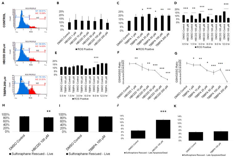
Figure 5. TBBPA Causes ROS Production in Spermatogenic Cells Derived from hESCs, while HBCDD and TBBPA Exposure Decrease GSH/GSSG Ratios.
(A) Flow cytometry-based analysis of DHE labeling. Blue indicates ROS−. Red indicates ROS+.
(B and C) Graphical representation showing that HBCDD does not affect ROS generation in hESCs differentiated in in vitro spermatogenic conditions (B), but TBBPA exposure causes overwhelming increase in ROS production (C).
(D and E) Graphical representation showing the generation of overwhelming ROS over a 12-hr period post-exposure to 100 μM HBCDD (D) and TBBPA (E).
(F and G) Graphical representation showing that HBCDD (F) and TBBPA (G) exposure decreases the GSH/GSSG ratio of hESCs differentiated in in vitro spermatogenic conditions.
(H–K) A 12-hr pre-treatment with 1 μM L-sulforaphane rescues 100 μM TBBPA-mediated cell death (I and K) but does not rescue cell death following 100 μM HBCDD exposure (H and J). A total of 5,000 events were analyzed, with three replications performed for each condition for (B)–(C) and (F)–(G). Three replications were performed for each condition for (D)–(E) and (H)–(K). Significant changes in ROS generation, GSH/GSSG ratio, and cell viability were determined using a 1-way analysis of variance (ANOVA) and validated via a Student’s t test, where * is p < 0.05, ** is p < 0.01, and *** is p < 0.001. Data are represented as mean ± SEM.
See also Figure S5.

