

Abstract
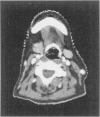
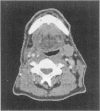
The success of radiation therapy depends critically on accurately delineating the target volume, which is the region of known or suspected disease in a patient. Methods that can compute a contour set defining a target volume on a set of patient images will contribute greatly to the success of radiation therapy and dramatically reduce the workload of radiation oncologists, who currently draw the target by hand on the images using simple computer drawing tools. The most challenging part of this process is to estimate where there is microscopic spread of disease. We are developing methods for automatically selecting and adapting standardized regions of tumor spread based on the location of lymph nodes in a standard or reference case, together with image registration techniques. The best available image registration techniques (deformable transformations computed using "mutual information" optimization) appear promising but will need to be supplemented by anatomic knowledge-based methods to achieve a clinically acceptable match.
Full text
PDF


Images in this article

Selected References
These references are in PubMed. This may not be the complete list of references from this article.
- Kalet I. J., Jacky J. P., Austin-Seymour M. M., Hummel S. M., Sullivan K. J., Unger J. M. Prism: a new approach to radiotherapy planning software. Int J Radiat Oncol Biol Phys. 1996 Sep 1;36(2):451–461. doi: 10.1016/s0360-3016(96)00322-7. [DOI] [PubMed] [Google Scholar]
- Maes F., Collignon A., Vandermeulen D., Marchal G., Suetens P. Multimodality image registration by maximization of mutual information. IEEE Trans Med Imaging. 1997 Apr;16(2):187–198. doi: 10.1109/42.563664. [DOI] [PubMed] [Google Scholar]
- Rosse C., Shapiro L. G., Brinkley J. F. The digital anatomist foundational model: principles for defining and structuring its concept domain. Proc AMIA Symp. 1998:820–824. [PMC free article] [PubMed] [Google Scholar]
- Som P. M., Curtin H. D., Mancuso A. A. An imaging-based classification for the cervical nodes designed as an adjunct to recent clinically based nodal classifications. Arch Otolaryngol Head Neck Surg. 1999 Apr;125(4):388–396. doi: 10.1001/archotol.125.4.388. [DOI] [PubMed] [Google Scholar]