

Abstract
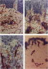
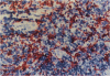
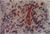
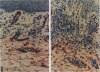
Dermal dendritic cells from eleven cases of mycosis fungoides (MF) (six patch and five plaque stage), two cases of pre-MF, and five specimens of normal human skin, were characterized immunohistochemically using a panel of antibodies including anti-human Thy-1, intercellular adhesion molecule-1 (ICAM-1; CD54), endothelial leukocyte adhesion molecule-1 (ELAM-1), vascular cell adhesion molecule-1 (VCAM-1), CD1a, CD2, CD14, CD18, CD34, MAC387, KP-1, EBM-11, factor XIIIa, factor XIIIs, and S100. Thy-1 expression in normal skin was limited to the microvascular endothelium and perivascular dendritic cells. An extensive interstitial network of Thy-1+ dendritic cells was seen in the papillary dermis of all cases of MF, whereas no epidermal cells were Thy-1+. The mean +/- standard deviation of interstitial Thy-1+ cells per high power field in the dermis was: normal skin, 2.86 +/- 0.34; pre-MF, 15; patch stage MF, 13.4 +/- 7.08; plaque stage MF, 49.96 +/- 21.29. Thy-1+ dendritic cells morphologically resembled the factor XIIIa+ "dermal dendrocyte" (DD) and shared their VCAM-1+, ICAM-1+, CD1a, CD2-, CD14+, CD18+, EMB11+, factor XIIIa+, factor XI-IIs-, S100-, MAC387- and KP-1-immunophenotype in MF. Double labeling studies revealed up to 50% of Thy-1+DD were also factor XIIIa+ in MF. Immediately beneath these cells was a similar network of CD34+, Thy-1-, factor XIIIa- dendritic cells limited to the reticular dermis. Strong microvascular endothelial cell expression of Thy-1 and VCAM-1, and focal vascular ELAM-1 expression were also seen in MF. Distinct cellular compartmentalization (papillary dermis versus reticular dermis versus epidermis) of dendritic cells is demonstrated by the differential expression of Thy-1, factor XIIIa, and CD34 antigens. The extensive number and prominent dermal dendritic network in the papillary dermis juxtaposed between epidermal keratinocytes (KC) and dermal/epidermal T cells, suggests an important pathophysiologic role for this newly recognized and immunophenotypically distinctive cell population in MF.
Full text
PDF


Images in this article

Selected References
These references are in PubMed. This may not be the complete list of references from this article.
- Amornsiripanitch S., Barnes L. M., Nordlund J. J., Trinkle L. S., Rheins L. A. Immune studies in the depigmenting C57BL/Ler-vit/vit mice. An apparent isolated loss of contact hypersensitivity. J Immunol. 1988 May 15;140(10):3438–3445. [PubMed] [Google Scholar]
- Basch R. S., Berman J. W. Thy-1 determinants are present on many murine hematopoietic cells other than T cells. Eur J Immunol. 1982 May;12(5):359–364. doi: 10.1002/eji.1830120502. [DOI] [PubMed] [Google Scholar]
- Bergstresser P. R., Sullivan S., Streilein J. W., Tigelaar R. E. Origin and function of Thy-1+ dendritic epidermal cells in mice. J Invest Dermatol. 1985 Jul;85(1 Suppl):85s–90s. doi: 10.1111/1523-1747.ep12275516. [DOI] [PubMed] [Google Scholar]
- Bonneville M., Itohara S., Krecko E. G., Mombaerts P., Ishida I., Katsuki M., Berns A., Farr A. G., Janeway C. A., Jr, Tonegawa S. Transgenic mice demonstrate that epithelial homing of gamma/delta T cells is determined by cell lineages independent of T cell receptor specificity. J Exp Med. 1990 Apr 1;171(4):1015–1026. doi: 10.1084/jem.171.4.1015. [DOI] [PMC free article] [PubMed] [Google Scholar]
- Caughman S. W., Breathnach S. M., Sharrow S. O., Stephany D. A., Katz S. I. Culture and characterization of murine dendritic Thy-1+ epidermal cells. J Invest Dermatol. 1986 Jun;86(6):615–624. doi: 10.1111/1523-1747.ep12275611. [DOI] [PubMed] [Google Scholar]
- Cerio R., Griffiths C. E., Cooper K. D., Nickoloff B. J., Headington J. T. Characterization of factor XIIIa positive dermal dendritic cells in normal and inflamed skin. Br J Dermatol. 1989 Oct;121(4):421–431. doi: 10.1111/j.1365-2133.1989.tb15509.x. [DOI] [PubMed] [Google Scholar]
- Cerio R., Spaull J., Oliver G. F., Jones W. E. A study of factor XIIIa and MAC 387 immunolabeling in normal and pathological skin. Am J Dermatopathol. 1990 Jun;12(3):221–233. doi: 10.1097/00000372-199006000-00002. [DOI] [PubMed] [Google Scholar]
- Chambers D. A. The Thy-1 epidermal cell: perspective and prospective. Br J Dermatol. 1985 Jul;113 (Suppl 28):24–33. doi: 10.1111/j.1365-2133.1985.tb15623.x. [DOI] [PubMed] [Google Scholar]
- Fivenson D. P., Rheins L. A., Nordlund J. J., Pomaranski M., Douglass M. C., Krull E. A. Thy-1 and T-cell receptor antigen expression in mycosis fungoides and benign inflammatory dermatoses. J Natl Cancer Inst. 1991 Aug 7;83(15):1088–1092. doi: 10.1093/jnci/83.15.1088. [DOI] [PubMed] [Google Scholar]
- Griffiths C. E., Nickoloff B. J. Keratinocyte intercellular adhesion molecule-1 (ICAM-1) expression precedes dermal T lymphocytic infiltration in allergic contact dermatitis (Rhus dermatitis). Am J Pathol. 1989 Dec;135(6):1045–1053. [PMC free article] [PubMed] [Google Scholar]
- Hansen E. R., Baadsgaard O., Lisby S., Cooper K. D., Thomsen K., Vejlsgaard G. L. Cutaneous T-cell lymphoma lesional epidermal cells activate autologous CD4+ T lymphocytes: involvement of both CD1+OKM5+ and CD1+OKM5- antigen-presenting cells. J Invest Dermatol. 1990 Apr;94(4):485–491. doi: 10.1111/1523-1747.ep12874650. [DOI] [PubMed] [Google Scholar]
- Haynes B. F., Scearce R. M., Lobach D. F., Hensley L. L. Phenotypic characterization and ontogeny of mesodermal-derived and endocrine epithelial components of the human thymic microenvironment. J Exp Med. 1984 Apr 1;159(4):1149–1168. doi: 10.1084/jem.159.4.1149. [DOI] [PMC free article] [PubMed] [Google Scholar]
- Knobler R. M., Edelson R. L. Cutaneous T cell lymphoma. Med Clin North Am. 1986 Jan;70(1):109–138. doi: 10.1016/s0025-7125(16)30972-5. [DOI] [PubMed] [Google Scholar]
- Lisby S., Baadsgaard O., Cooper K. D., Vejlsgaard G. L. Decreased number and function of antigen-presenting cells in the skin following application of irritant agents: relevance for skin cancer? J Invest Dermatol. 1989 Jun;92(6):842–847. doi: 10.1111/1523-1747.ep12696867. [DOI] [PubMed] [Google Scholar]
- McKenzie J. L., Fabre J. W. Human thy-1: unusual localization and possible functional significance in lymphoid tissues. J Immunol. 1981 Mar;126(3):843–850. [PubMed] [Google Scholar]
- Murphy G. F., Bronstein B. R., Knowles R. W., Bhan A. K. Ultrastructural documentation of M241 glycoprotein on dendritic and endothelial cells in normal human skin. Lab Invest. 1985 Mar;52(3):264–269. [PubMed] [Google Scholar]
- Nickoloff B. J. The human progenitor cell antigen (CD34) is localized on endothelial cells, dermal dendritic cells, and perifollicular cells in formalin-fixed normal skin, and on proliferating endothelial cells and stromal spindle-shaped cells in Kaposi's sarcoma. Arch Dermatol. 1991 Apr;127(4):523–529. [PubMed] [Google Scholar]
- Rice G. E., Munro J. M., Bevilacqua M. P. Inducible cell adhesion molecule 110 (INCAM-110) is an endothelial receptor for lymphocytes. A CD11/CD18-independent adhesion mechanism. J Exp Med. 1990 Apr 1;171(4):1369–1374. doi: 10.1084/jem.171.4.1369. [DOI] [PMC free article] [PubMed] [Google Scholar]
- Shiohara T., Moriya N., Gotoh C., Hayakawa J., Nagashima M., Saizawa K., Ishikawa H. Loss of epidermal integrity by T cell-mediated attack induces long-term local resistance to subsequent attack. I. Induction of resistance correlates with increases in Thy-1+ epidermal cell numbers. J Exp Med. 1990 Apr 1;171(4):1027–1041. doi: 10.1084/jem.171.4.1027. [DOI] [PMC free article] [PubMed] [Google Scholar]
- Singer K. H., Scearce R. M., Tuck D. T., Whichard L. P., Denning S. M., Haynes B. F. Removal of fibroblasts from human epithelial cell cultures with use of a complement fixing monoclonal antibody reactive with human fibroblasts and monocytes/macrophages. J Invest Dermatol. 1989 Feb;92(2):166–170. doi: 10.1111/1523-1747.ep12276685. [DOI] [PubMed] [Google Scholar]
- Sreilein J. W. Antigen-presenting cells in the induction of contact hypersensitivity in mice: evidence that Langerhans cells are sufficient but not required. J Invest Dermatol. 1989 Oct;93(4):443–448. doi: 10.1111/1523-1747.ep12284018. [DOI] [PubMed] [Google Scholar]
- Sullivan S., Bergstresser P. R., Tigelaar R. E., Streilein J. W. Induction and regulation of contact hypersensitivity by resident, bone marrow-derived, dendritic epidermal cells: Langerhans cells and Thy-1+ epidermal cells. J Immunol. 1986 Oct 15;137(8):2460–2467. [PubMed] [Google Scholar]