

Abstract
For the benefit of the first-year gross anatomy students, we digitized and published on a Web site images that had been collected over a 30-year period. We provided a CD-ROM (compact disk, read-only media) containing the image set in higher quality format to students and faculty. We supplemented basic images with hot topics such as CT angiography, virtual colonography, computer-aided diagnosis, and 3D post-processing. Full motion video and moving JPEG (Joint Photo Expert Group) animations were integrated into the atlas. On the post course questionnaire medical students reported that the images on CD-ROM were helpful during the course and for review prior to examinations. Faculty and medical students used the CD-ROM for problem-based learning sections and facilitator training. The images were clear and easily projected during review sessions and were useful for the small group sessions, where they served as examples of normal anatomy.
Keywords: Re-engineering, teaching file, Internet-based, CD-ROM
Our purpose was to teach radiologic anatomy using preserved images from the past and new methods such as 3D (three-dimensional) imaging and computer-aided diagnosis. To achieve our objective, we developed a digital teaching atlas of previously presented plain film examples of normal anatomy and basic pathology.
The transition from plain film to electronic media has repercussions beyond the Radiology Department. In this article the implementation and re-engineering of workflow for electronic instruction in a first-year gross anatomy course at a large teaching institution is discussed. In the past, images were available during the Gross Anatomy lab for only a few hours each week. Students lined up at view boxes before and after their cadaver dissections to look at the day’s radiographs. At the end of the day the films were taken down from the view boxes and replaced with new images. The films were not seen again unless they were presented on examinations. Some of the films demonstrated anatomy through techniques such as bronchograms that are no longer used in clinical practice. Over the years these films began to deteriorate from exposure to sunlight and the chemicals used in the anatomy lab.
The advantages of digital teaching files have been widely reported published.1,2,3,4,5 Publication and archiving using Internet servers and CD-ROMs (compact disk read only media) have also been described.5,6,7,8,9
Creation of a CD-ROM for storage and distribution of course images to over 250 medical students and faculty each year was the method chosen to be most cost effective. Each CD-ROM costs less than 10 cents to copy. In addition to the radiology images, the course syllabus, evaluation forms, normal laboratory values, and selected articles were included. The cost of duplicate printing of 200 images on film would total $342 per student (200 × $1.71 per film [price of one 14 × 17 inch film]). This cost does not include chemicals or technologist expense. This would be cost prohibitive for 250 people, even without the burden of the weight and the difficulty of sorting such a collection.
MATERIALS AND METHODS
Over the last 30 years, faculty members have collected plain film images emphasizing teaching points for basic anatomy. These images were the basis for teaching radiological anatomy to first-year medical students during their first 2 months of medical school. Some of the films were obtained with techniques no longer in use. CD-ROM and Internet–World Wide Web (WWW) format were the preferred methods of distribution. Most of the images were digitized using a UMAX Power Look 2100XL film scanner (UMAX, Dallas, TX) and a Microtek Scanmaker 9600XL film scanners (Microtek, Carson, CA).
Many of the films were faded and brittle, but they were firmly established tools in the course lectures and labs. A glass plate that predated film was also available. After there images were scanned, we used Photoshop (Adobe, San Jose CA) and Paint Shop Pro (Jasc Software, Inc Eden Prairie, MN) for cropping and post-processing. Many of the images contained patient identifiers, which were removed with the above programs for compliance with the Health Insurance Portability and Accountability Act of 1996.10 Purchase of a commercial Vidar Diagnostic Pro Plus film scanner (Vidar Pacific Grove, CA) allowed batch scanning of up to 25 films at a time. New images were easily added to the collection from our PACS (Picture Archiving and Communication System). Residents and faculty used a variety of DICOM (Digital Imaging and Communications in Medicine) viewers to view the PACS (Eastman Kodak, Rochester, NY) images and save them in new dynamic formats such as 3D animated GIF (CompuServe Graphics Interchange Format) files and video clips. The viewers contained either screen capture or export functions, enabling residents and faculty to save the window and level settings that best demonstrated the targeted anatomy. The export functions in Voxar 3D (Voxar Inc., Framingham, MA), eFilm (Merge eFilm, Milwaukee, WI), and Centricity Enterprise Web (GE Medical Systems, Milwaukee, WI) were especially easy to use. Voxar 3D and eFilm allowed saving in AVI (Audio Video Interleave) format. Radiologists worked closely with anatomy faculty and staff to label the anatomic detail displayed on the selected studies. The course graphics designer published the images in HTML format using Microsoft FrontPage (Microsoft, Redmond, WA) and Macromedia Authorware Version 6 (Macromedia Inc, San Francisco, CA) on a Web site dedicated to the first-year Gross Anatomy and Radiology course. The Web images were in compressed JPEG and GIF formats. The compressed format allowed students and faculty with network privileges to download the images rapidly at home or in the classroom as needed.
A CD-ROM containing the entire image set in higher quality image formats than those available online, uncompressed JPEG, GIF, and AVI, was provided to medical students and faculty at the beginning of the course. The AVI format was especially useful for displaying 3D and multi-planar images. The functionality had not been available to our medical students in the past. Voxar, eFilm, and Advantage Windows Version 4.1 (GE Medical Systems), running on a Unix-based Ultras ARC 2 (Sun Microsystems, Mountain View, CA), were used to generate shaded surface display (SSD), maximum intensity projection (MIP), and 3D medical images. Techniques such as “fly-through” virtual angiography, virtual colonography, and 3D post-processing supplemented basic images posted on the Web site. Full-motion video and moving GIF animations were integrated with image descriptions.
For the past 3 years, the School of Medicine has used the interactive CD-ROM as a teaching tool and resource. The CD-ROM is menu-driven; it shows imaging modalities used in radiologic practice with normal human organs and structures. The CD-ROM program is divided into four sections (Fig 1). The introduction includes definitions of terms used in radiology. The second section, human sectional imaging, is divided into six subsections that traverse the human body from the head to the lower limbs. The third section includes a complete body axial CT scan and 3D imaging. The final section contains a review of anatomy featuring a real patient with clinical problems. The CD-ROM has the features of the latest software releases such as search functions and bookmarks. The CD, titled Introduction to Radiological Imaging of Human Gross Anatomy, is available for purchase at http://www.interdoc1.com/
Figure 1.
CD-ROM introductory page and index.
RESULTS
Medical students gave the CD-ROM excellent reviews on the final course evaluations. Some 96% of the students reported that the CD-ROM was a useful addition to the hard-copy images displayed in the lab, and they preferred using the CD-ROM to view the week’s radiologic images rather than waiting in line to look at the films on the view boxes. Films were still used in the Gross Anatomy lab because the computers available for projection were not always working. This was judged to be a hardware problem and not a problem with the images.
On the post-course questionnaire, 91% of the students said they had no difficulty using the CD-ROM on their computers. A few requested a Macintosh version of the CD-ROM. In total, 93.7% reported the format of the images/text on the CD-ROM was user friendly, and 88.5% of the students used the radiological images on the CD-ROM while studying.
Faculty used the Internet images for problem-based learning sessions and facilitator training throughout the course. Images were copied from the CD-ROM and used to supplement faculty lectures. The course director reported to the Medical School Curriculum Committee that the Radiologic Lab Images on CD-ROM distributed to medical students, facilitators, and lecturers was a huge success.
The overall cost of printing 250 CD-ROMs was less than printing the 82-page syllabus alone. The project was so successful that the university funded publication and marketing for use in medical schools throughout the United States.
DISCUSSION
Most radiologists collect files of interesting cases. Our collection is unique, however, in that it is composed predominantly of normal structures captured with common imaging techniques. Because of the age of many of the films in this collection and its gradual integration into the Gross Anatomy course over 30 years, many films had become faded and brittle. Some of the films, such as barium bronchograms, are unique in that they demonstrate normal anatomy visualized in ways not used in today’s radiologic practice (Fig 2). Preservation and restoration of these images is worthwhile because the images are visually clear teaching aids. Digital publishing and access to the hospital PACS allows for the addition of images as new techniques become available.
Figure 2.
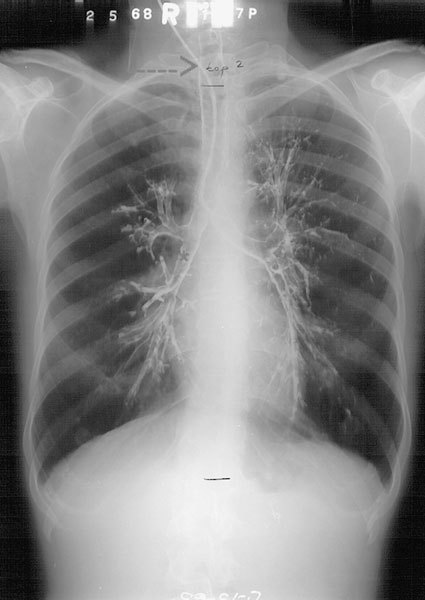
Barium bronchogram demonstrates the bronchial tree of the lung.
The CD-ROM provided faculty an opportunity to display some of the latest techniques in use by Radiology and allowed medical students to appreciate anatomy from multiple angles and views. The state-of-the-art techniques attracted and kept the interest of the students and may aid in recruiting the best students to Radiology residencies and research.
During the last few years there has been growing emphasis on correlating clinical applications with the traditional first two-year basic science courses. Recognition of anatomic structures and some clinical pathology is important for students of medicine as the basic science years of medical school and medical boards have become more clinically oriented.
The CD-ROM aids Radiology faculty in its relatively new participation in our first-year gross anatomy and core curriculum. This CD-ROM may also be useful to Physician Assistant, Nursing and Radiology Technologist students.
CONCLUSIONS
The use of radiologic images, including computed tomography, magnetic resonance imaging, 3-D imaging, ultrasound, and computer-aided diagnosis, has increased the understanding of human anatomy. Radiographic techniques that demonstrated unique anatomy, but are no longer in use can be preserved, digitized, and used for teaching. In our institution CD-ROM distribution was helpful to both medical students and faculty. The images were clear and easily projected during review sessions. They were also useful for the small group sessions, where they served as examples of normal anatomy.
Acknowledgments
We thank Annette Ayala, T. Nishino, Ph.D., and Victor Luciano for production and design of the CD-ROM.
References
- 1.Ernst R, Kawashima A, Tamm E, et al: A high-quality, low-cost, Internet/intranet-based digital imaging database. RSNA Electronic Journal page, RSNA Web site. Available at: http://ej.rsna.org/EJ_0_96/0056-97.fin/ . Accessed September 6, 2000
- 2.Bidgood Jr WD, Horii SC. Introduction to the ACR-NEMA DICOM standard. Radiographics. 1992;12:345–355. doi: 10.1148/radiographics.12.2.1561424. [DOI] [PubMed] [Google Scholar]
- 3.Siegel E, Reiner B. Electronic teaching files: seven-year experience using a commercial picture archiving and communication system. J Digit Imaging. 2001;14([suppl 1]):125S–127S. doi: 10.1007/BF03190314. [DOI] [PMC free article] [PubMed] [Google Scholar]
- 4.Ernst RD, Baumgartner BR, Tamm EP, et al. Development of a teaching file by using a DICOM database. Radiographics. 2002;22:217–221. doi: 10.1148/radiographics.22.1.g02ja10217. [DOI] [PubMed] [Google Scholar]
- 5.Zaidel M, Hopper K, Iyriboz T. Interactive Web-based radiology teaching file. J Digit Imaging. 1999;12:203–204. doi: 10.1007/BF03168803. [DOI] [PMC free article] [PubMed] [Google Scholar]
- 6.Tellis WM, Andriole KP, Avrin DE, et al. Web technology in the integration of a digital teaching file at the diagnostic workstation. J Digit Imaging. 1998;11:117–119. doi: 10.1007/BF03168277. [DOI] [PMC free article] [PubMed] [Google Scholar]
- 7.Weinberger E, Jakobovits R, Halsted M. MyPACS. net: A Web-Based Teaching File Authoring Tool. AJR Am J Roentgenol. 2002;179:579–582. doi: 10.2214/ajr.179.3.1790579. [DOI] [PubMed] [Google Scholar]
- 8.Goldberg DJ, DeMarco KJ, Parikh T. Internet-based interactive teaching file for neuroradiology. AJR Am J Roentgenol. 2000;175:1371–1373. doi: 10.2214/ajr.175.5.1751371. [DOI] [PubMed] [Google Scholar]
- 9.Tran TH, Roach NA, O’Kane PL, et al. Creating a digital radiographic teaching file and database using a PC and common software. AJR Am J Roentgenol. 2000;175:325–327. doi: 10.2214/ajr.175.2.1750325. [DOI] [PubMed] [Google Scholar]
- 10.Gostin LO. National Health Information Privacy: Regulations Under the Health Insurance Portability and Accountability Act. JAMA. 2001;285:3015–3021. doi: 10.1001/jama.285.23.3015. [DOI] [PubMed] [Google Scholar]