

Abstract
Nipah virus (NiV), a new member of the Paramyxoviridae, codes for a fusion (F) protein with five potential N-glycosylation sites. Because glycans are known to be important structural components affecting the conformation and function of viral glycoproteins, we analyzed the effect of the deletion of N-linked oligosaccharides on cell surface transport, proteolytic cleavage, and the biological activity of the NiV F protein. Each of the five potential glycosylation sites was removed either individually or in combination, revealing that four sites are actually utilized (g2 and g3 in the F2 subunit and g4 and g5 in the F1 subunit). While the removal of g2 and/or g3 had no or little effect on cleavage, surface transport, and fusion activity, the elimination of g4 or g5 reduced the surface expression by more than 80%. Similar to a mutant lacking all N-glycans, g4 deletion mutants in which the potential glycosylation site was destroyed by introducing a glycine residue were neither cleaved nor transported to the cell surface and consequently were not able to mediate cell-to-cell fusion. This finding indicates that in the absence of g4, the amino acid sequence around position 414 is important for folding and transport.
Nipah virus (NiV) together with the closely related Hendra virus forms the new Henipavirus genus within the Paramyxoviridae family (21). NiV codes for two surface glycoproteins, the receptor-binding G protein and the fusion (F) protein. The G protein mediates the attachment of virions to a cellular receptor which is not yet identified. The F protein functions in the penetration of the virus into host cells by initiating the pH-independent fusion of viral and cellular membranes. As in other paramyxoviruses, the NiV F protein is synthesized as an inactive precursor F0 that has to be cleaved into the two disulfide-linked subunits F1 and F2. Once the G/F1,2 complexes are expressed on the surfaces of infected cells, the hydrophobic fusion peptide located at the amino terminus of F1 mediates fusion with adjacent cells, resulting in multinucleated syncytia. Syncytium formation can also be observed with cells transfected with the genes encoding F and G (17). Since NiV causes a systemic infection in vivo and the majority of cell cultures tested so far supported NiV fusion, the cellular receptor appeared to be widely expressed and the viral F protein seemed to be ubiquitously activated (2, 5).
Despite high grades of similarity among the F proteins of Paramyxoviridae with respect to the size and location of hydrophobic domains and heptad repeats, the number, position, and functional importance of the attached glycans are varied (for a review, see reference 19). N-glycans not only determine the folding and the intracellular transport of viral glycoproteins (4, 15, 18) but also are known to modulate their antigenicity and their activity (1, 6, 7, 9, 13, 16, 20, 22). The NiV F protein contains five N-glycosylation consensus sites (N-X-S/T, in which X can be any amino acid except proline). However, which sites are actually used and how much N glycosylation affects the function of the protein remain to be elucidated. To address this question, we determined the number, location, and type of N-linked oligosaccharides in the F protein of NiV and analyzed their role in cell surface transport, proteolytic cleavage, and fusion activity. cDNA fragments spanning the F gene of the NiV genome (GenBank accession number AF212302) were cloned into a derivative of the replication-deficient murine leukemia virus vector pczCFG (8). To allow the detection of the protein with commercially available antibodies, a tagged version of the protein was established by replacing the 9 carboxy-terminal amino acids with amino acids 99 to 107 (YPYDVPDYA) of the human influenza virus hemagglutinin (HA) tag. The expression level, cleavage, and biological activity of the HA-tagged protein were unchanged from those of the parental F protein in transient transfection. The fusion activity of NiV F with and without the HA tag is shown in Fig. 1. All glycosylation mutants were based on the HA-tagged NiV F protein. The mutant F genes depicted in Fig. 2 were generated by introducing site-specific mutations into the double-stranded pczCFG5 plasmids. By using complementary mutagenic oligonucleotide primers, the third residue (S or T) of one or several of the five predicted glycosylation sites was changed to a glycine. The plasmids containing mutant F genes were transfected into MDCK cells by the use of Lipofectamine 2000 (Gibco BRL). To analyze the electrophoretic mobilities and proteolytic cleavage of the mutants, transfected cells were metabolically labeled at 24 h posttransfection by incubation with medium containing [35S]cysteine and [35S]methionine (Promix; Amersham) at a final concentration of 100 μCi/ml for 10 min. Subsequently, labeling medium was replaced by nonradioactive medium, and the cells were incubated at 37°C for 2 h. Radioimmunoprecipitation was essentially performed as described previously (11). Radiolabeled F proteins were precipitated with a polyclonal antiserum specific for HA-tagged proteins (Sigma) and subjected to sodium dodecyl sulfate-polyacrylamide gel electrophoresis (SDS-PAGE) under reducing conditions. Dried gels were exposed to Kodak BIOMAX films.
FIG. 1.

Fusion activities of F proteins with and without the HA tag. The NiV G gene was transfected either alone (A) or in combination with the gene encoding the NiV F protein with (B) or without (C) a C-terminal HA tag. At 24 h posttransfection, cell-to-cell fusion was visualized by Giemsa staining. Magnification, ×100.
FIG. 2.

Schematic diagram of the NiV F protein and amino acid sequences of mutated N-glycosylation sites. The two F protein subunits, F1 and F2, are indicated. Arrowheads point to the locations of the potential N-glycosylation sites. Numbers indicate the amino acid positions. Protein sequences are shown in single-letter code; boldface letters indicate exchanged amino acid residues. CD, cytoplasmic domain; FP, fusion peptide; LD, luminal domain; SP, signal peptide; TMD, transmembrane domain.
The expression of the standard F protein showed molecular masses of about 60 kDa for the precursor F0 and 49 and 19 kDa for the cleavage products F1 and F2, respectively (Fig. 3A, lane F). To visualize the electrophoretic profile of F protein lacking all N-glycans, immunoprecipitated standard F protein was treated with 500 U of N-glycosidase F (PNGase F; New England Biolabs) according to the instructions of the manufacturer before separation on an SDS gel. As shown in Fig. 3A, lane Fdeglyc, the removal of N-linked oligosaccharides by PNGase F resulted in faster migration of all F protein bands, indicating that N-glycans are attached to both the F1 and F2 subunits. Fg1 had the same profile as standard F, demonstrating that the very N-terminal glycosylation site (N64) is not used. In contrast, Fg2 and Fg3 had different migration patterns, with F0 and F2 products of about 58 and 17 kDa, respectively. Considering that an N-linked glycan residue has an average molecular mass of about 2 kDa, this shows that both N67 and N99 in the F2 subunit are glycosylated. The F2 product of Fg2 migrated slightly faster, indicating that the compositions of the N-glycans attached to N67 (g2) and N99 (g3) are not identical. As expected from the single mutants, the F2 subunit of the double mutant Fg2,3 comigrated with the PNGase F-digested F2 subunit of standard F protein. The two glycosylation sites in the F1 subunit are also utilized, because the F0 precursor of both Fg4 and Fg5 showed a reduced molecular weight compared to that of the precursor of standard F protein. The expected change in the electrophoretic mobility of F1 was detectable only in the Fg5 mutant, since Fg4 was not cleaved. Whereas Fg4 was cleavage deficient, the proteolytic processing of Fg1, Fg2, Fg3, Fg2,3, and Fg5 was as efficient as that of the fully glycosylated standard F protein.
FIG. 3.

Electrophoretic mobilities and endoglycosidase digestion analysis of the NiV F protein glycosylation mutants. (A) MDCK cells expressing either parental F (lane F) or glycosylation mutants (lanes Fg1, Fg2, Fg3, Fg4, Fg5, and Fg2,3) were radiolabeled with [35S]methionine (Promix) for 10 min and then incubated in chase medium for 2 h. F proteins were immunoprecipitated from cell lysates, separated on a 15% polyacrylamide gel under reducing conditions, and subjected to autoradiography. To demonstrate the migration pattern of F protein devoid of any N-glycans, the parental F protein was treated with PNGase F (lane Fdeglyc). (B) MDCK cells expressing parental F (lane F) or the glycosylation mutant Fg2, Fg3, Fg4, or Fg5 were radiolabeled as described above. Immunoprecipitated samples were divided into two aliquots and either digested with endo H (+) or kept untreated (−), followed by separation on a 15% polyacrylamide gel under reducing conditions.
To determine the type of N-glycans that are attached to the NiV F protein, high-mannose oligosaccharides were removed by digestion with endo-β-N-acetylglucosaminidase H (endo H; New England Biolabs). As shown in Fig. 3B, none of the F2 subunits contained endo H-sensitive glycans. This clearly demonstrates that the glycans attached to N67 (g2) and N99 (g3) are of the complex type. The fact that the F1 subunits of Fg2, Fg3, and standard F protein but not that of Fg5 showed a reduction in the apparent molecular mass after endo H treatment indicated that a high-mannose oligosaccharide is attached to N464 (g5).
The cell surface expression of the mutants was analyzed by using a biotinylation approach. Essentially as described earlier (12), the transfected cells were labeled twice for 20 min at 4°C with 2 mg of non-membrane-permeating sulfo-N-hydroxysuccinimidobiotin (Calbiochem) per ml. After biotinylation, cells were lysed and F proteins were immunoprecipitated by using the polyclonal antiserum directed against the HA tag. The precipitates were separated on an SDS gel under nonreducing conditions and transferred to nitrocellulose. Surface-expressed F protein was then visualized with a streptavidin-peroxidase complex (Amersham). Figure 4A shows that F, Fg1, Fg2, Fg3, Fg5, and Fg2,3 were expressed on the cell surface. Fg2, Fg3, standard F, and Fg1, which is identical to standard F because the very N-terminal glycosylation site is not utilized, were detected with signals of comparable intensities. The amount of surface-expressed Fg5 and Fg2,3 appeared to be reduced (17 and 36% of the amount in standard F protein, respectively), indicating that either the lack of g5 linked to N464 in the F1 subunit or the lack of both glycans of the F2 subunit downregulated the surface expression. Furthermore, Fig. 4A shows that no biotinylated Fg4 could be detected. This result suggests that not only cleavage but also surface transport of the NiV F protein essentially depends on the presence of the N-glycan at position N414 (g4) in the F1 subunit. In agreement with this, an immunofluorescence analysis revealed that the mutants lacking g4 accumulated in the endoplasmic reticulum (data not shown).
FIG. 4.
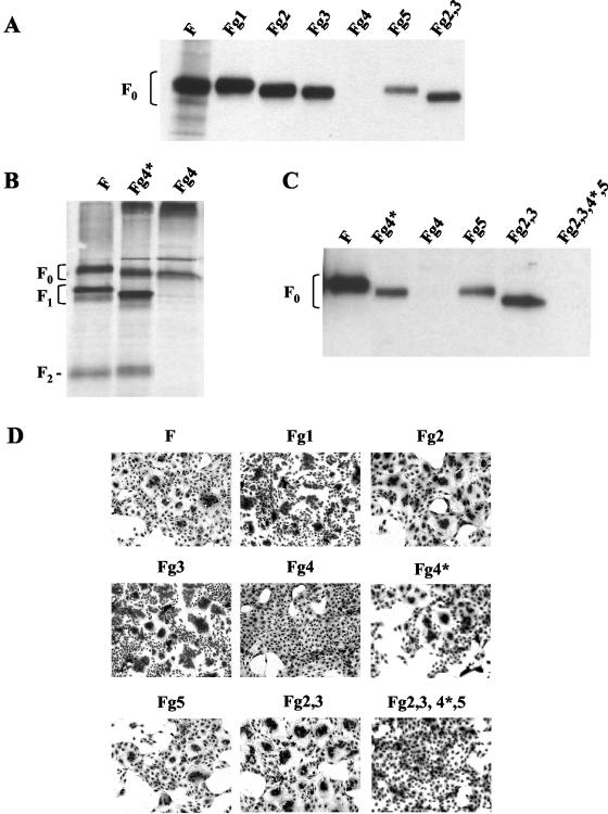
Cell surface expression, cleavage, and fusion activity of parental NiV F protein and glycosylation mutants. (A) At 24 h posttransfection, MDCK cells expressing either parental F or the glycosylation mutant Fg1, Fg2, Fg3, Fg4, Fg5, or Fg2,3 were surface labeled with biotin and lysed. Following immunoprecipitation, samples were subjected to SDS-PAGE under nonreducing conditions and blotted to nitrocellulose. Surface-labeled F proteins were visualized with streptavidin-peroxidase and chemiluminescence. (B) Cells were transfected with either parental F, Fg4*, or the Fg4 gene. At 24 h posttransfection, cells were radiolabeled, and F proteins were immunoprecipitated and subjected to SDS-PAGE and autoradiography as described for Fig. 3A. (C) Surface biotinylation of cells expressing either parental F or the glycosylation mutants Fg4*, Fg4, Fg5, Fg2,3, and Fg2,3,4*,5 was performed as described above. (D) The NiV G gene was cotransfected with either parental NiV F (subpanel F) or one of the glycosylation mutant genes (Fg1, Fg2, Fg3, Fg4, Fg4*, Fg5, Fg2,3, or Fg2,3,4*,5). At 24 h posttransfection, cell-to-cell fusion was visualized by Giemsa staining. Magnification, ×100.
To determine if the complete intracellular retention of Fg4 is due to the elimination of g4 rather than to the threonine-to-glycine mutation at position 416, we generated a mutant, Fg4*, in which asparagine at position 414 was replaced by glutamine. The mutation of N414 should also lead to the loss of g4. The mutant was analyzed by the immunoprecipitation of transfected cells after metabolic labeling and surface biotinylation. As with Fg4, Fg4* showed a mobility shift in comparison to the parental F protein. But in contrast to Fg4, Fg4* was found to be cleaved (Fig. 4B) and expressed on the cell surface (Fig. 4C). Even if the level of surface expression of Fg4* was lower than that of standard F or Fg2,3, it was comparable to the expression level of Fg5. In contrast to Fg4*, the quadruple mutant Fg2,3,4*,5 could not be detected on the cell surface, indicating that a NiV F protein lacking all N-glycans was no longer transported. Although surface transport of Fg4* was clearly downregulated (a reduction of more than 80% compared to the level in standard F protein), the different phenotypes of Fg4 and Fg4* suggest that intracellular transport is not only determined by the lack of the N-glycan at position 414 but also influenced by the amino acid sequence at this position. It appears that a glutamine at position 414 is a more favorable amino acid than a glycine at position 416 in terms of surface transport. The idea that a glycine at the potential glycosylation site from position 414 to position 416 negatively affects surface transport of the NiV F protein is further supported by the finding that another g4 deletion mutant with an asparagine-to-glycine mutation at position 414 (Fg4 N414G) displayed the same phenotype as Fg4 (neither cleavage nor surface expression; data not shown). Although the three-dimensional structure of the NiV F protein is not known, a comparison with the known structure of the Newcastle disease virus F protein (3) indicates that g4 linked to N414 is located in the head region. It must be assumed that both the glycan and the amino acid sequence at this position are critical for the conformational structure of the NiV F protein. Changes in the structure due to the loss of g4 and/or to unfavorable amino acid substitutions likely influence the binding to chaperones in the endoplasmic reticulum that affect protein folding and further transport.
To evaluate the importance of the N-glycans for biological activity, mutant F proteins were coexpressed with the NiV G protein, and syncytium formation was analyzed at 24 h posttransfection. Figure 4D shows that, as expected, the mutants that were neither cleaved nor surface expressed (Fg4 and Fg2,3,4*,5) were not able to induce cell-to-cell fusion. In contrast, Fg1, Fg2, Fg3, and also Fg2,3, Fg4*, and Fg5, the mutants with decreased surface expression levels, induced efficient syncytium formation. To evaluate the individual fusion capacities of the mutants, the percentage of cell fusion was quantitated as described previously (11). Standard F, Fg1, Fg2, Fg4*, and Fg5 induced almost similar levels of cell fusion (22, 28, 32, 29, and 25%, respectively), whereas Fg3 and Fg2,3 caused the fusion of 45 and 52% of the cells, respectively. Given their reduced surface expression levels, this result indicates an increased fusion capacity of Fg2,3, Fg4*, and Fg5.
The great variation in the number, location, and functional importance of N-glycans attached to paramyxoviral F proteins is well documented (1, 6, 9, 16, 22). There is no obvious evidence for the functional conservation of certain glycans. Even highly homologous F proteins such as those from bovine respiratory syncytial virus (RSV) and human RSV have different requirements with respect to N glycosylation (14, 22). Even if the spatial locations of different oligosaccharide chains could be partially predicted after the publication of the Newcastle disease virus F protein structure (3), the unpredictable implications of N-glycans in protein folding and intracellular transport make it necessary to analyze the individual roles of the N-glycans for each virus. For NiV, we found N glycosylation of N99, which is located only 10 amino acids upstream of the cleavage site (R109). Despite the short distance, the N-glycan at this position (g3) did not affect proteolytic activation. The cleavage of mutants lacking g3 was found to be as efficient as cleavage of the standard protein. Glycosylation in such proximity to a cleavage site is known only for the F protein of bovine RSV (23). Furthermore, our results show that in contrast to what has been proposed for the closely related Hendravirus F protein (10), both glycosylation sites in the F1 subunit of the NiV F protein are utilized. Thus, NiV, similar to the members of the Respirovirus, Rubulavirus, and Pneumovirus genera, contains N-linked glycans in both the F1 and F2 subunits. Both of the glycans g4 and g5 affected surface transport. Although the loss of g4 or g5 caused similar markedly reduced levels of surface expression, changes in amino acid composition at the glycosylation sites had different consequences. Whereas the g5 deletion mutant with a glycine at the third position of the glycosylation motif was proteolytically cleaved, partially transported to the cell surface, and therefore able to induce cell-to-cell fusion, g4 deletion mutants with a glycine at the first or the third position of the glycosylation motif no longer reached the cell surface. Similar to the mutant lacking all N-glycans, these mutants were neither cleaved nor transported to the cell surface and consequently were not able to mediate cell-to-cell fusion. In contrast to the glycans of the F1 subunit, none of the N-glycans of the F2 subunit was found to be of major importance for surface transport or functionality. This finding indicates that the individual N-linked oligosaccharides of the NiV F protein and the amino acid sequence at the potential glycosylation sites differ greatly in terms of their importance for protein folding and intracellular transport.
Acknowledgments
We thank Markus Czub for providing the cloned NiV F and G genes and Gert Zimmer and Georg Herrler for comments on the manuscript.
This work was supported by a grant from the German Research Foundation to A.M. (MA 1886/5-1).
REFERENCES
- 1.Bagai, S., and R. A. Lamb. 1995. Individual roles of N-linked oligosaccharide chains in intracellular transport of the paramyxovirus SV5 fusion protein. Virology 209:250-256. [DOI] [PubMed] [Google Scholar]
- 2.Bossart, K. N., L.-F. Wang, M. N. Flora, K. B. Chua, S. K. Lam, B. T. Eaton, and C. C. Broder. 2002. Membrane fusion tropism and heterotypic functional activities of the Nipah virus and Hendra virus envelope glycoproteins. J. Virol. 76:11186-11198. [DOI] [PMC free article] [PubMed] [Google Scholar]
- 3.Chen, L., J. J. Gorman, J. McKimm-Breschkin, L. J. Lawrence, P. A. Tulloch, B. J. Smith, P. M. Colman, and M. C. Lawrence. 2001. The structure of the fusion glycoprotein of Newcastle disease virus suggests a novel paradigm for the molecular mechanism of membrane fusion. Structure 9:255-266. [DOI] [PubMed] [Google Scholar]
- 4.Doms, R. W., R. A. Lamb, J. K. Rose, and A. Helenius. 1993. Folding and assembly of viral membrane proteins. Virology 193:545-562. [DOI] [PubMed] [Google Scholar]
- 5.Hooper, P., S. Zaki, P. Daniels, and D. Middleton. 2002. Comparative pathology of the diseases caused by Hendra and Nipah viruses. Microbes Infect. 3:315-322. [DOI] [PubMed] [Google Scholar]
- 6.Hu, A., T. Cathomen, R. Cattaneo, and E. Norrby. 1995. Influence of N-linked oligosaccharide chains on the processing, cell surface expression and function of the measles virus fusion protein. J. Gen. Virol. 76:705-710. [DOI] [PubMed] [Google Scholar]
- 7.Koch, M., M. Pancera, P. D. Kwong, P. Kolchinsky, C. Grundner, L. Wang, W. A. Hendrickson, J. Sodroski, and R. Wyatt. 2003. Structure-based, targeted deglycosylation of HIV-1 gp120 and effects on neutralization sensitivity and antibody recognition. Virology 313:387-400. [DOI] [PubMed] [Google Scholar]
- 8.Lindemann, D., T. Pietschmann, M. Picard-Maureau, A. Berg, M. Heinkelein, J. Thurow, P. Knaus, H. Zentgraf, and A. Rethwilm. 2001. A particle-associated glycoprotein signal peptide essential for virus maturation and infectivity. J. Virol. 75:5762-5771. [DOI] [PMC free article] [PubMed] [Google Scholar]
- 9.McGinnes, L., T. Sergel, J. Reitter, and T. Morrison. 2001. Carbohydrate modifications of the NDV fusion protein heptad repeat domains influence maturation and fusion activity. Virology 283:332-342. [DOI] [PubMed] [Google Scholar]
- 10.Michalski, W. P., G. Crameri, L. F. Wang, B. J. Shiell, and B. Eaton. 2000. The cleavage activation and sites of glycosylation in the fusion protein of Hendra virus. Virus Res. 69:83-93. [DOI] [PubMed] [Google Scholar]
- 11.Moll, M., H.-D. Klenk, and A. Maisner. 2002. Importance of the cytoplasmic tails of the measles virus glycoproteins for fusogenic activity and the generation of recombinant measles viruses. J. Virol. 76:7174-7186. [DOI] [PMC free article] [PubMed] [Google Scholar]
- 12.Moll, M., H.-D. Klenk, G. Herrler, and A. Maisner. 2001. A single amino acid change in the cytoplasmic domains of measles virus glycoproteins H and F alters targeting, endocytosis and fusion in polarized Madin-Darby canine kidney cells. J. Biol. Chem. 276:17887-17894. [DOI] [PubMed] [Google Scholar]
- 13.Munk, K., E. Pritzer, E. Kretzschmar, B. Gutte, W. Garten, and H.-D. Klenk. 1992. Carbohydrate masking of an antigenic epitope of influenza virus haemagglutinin independent of oligosaccharide size. Glycobiology 2:233-240. [DOI] [PubMed] [Google Scholar]
- 14.Pastey, M. K., and S. K. Samal. 1997. Role of individual N-linked oligosaccharide chains and different regions of bovine respiratory syncytial virus fusion protein in cell surface transport. Arch. Virol. 142:2309-2320. [DOI] [PubMed] [Google Scholar]
- 15.Roberts, P. C., W. Garten, and H.-D. Klenk. 1993. Role of conserved glycosylation sites in maturation and transport of influenza A virus hemagglutinin. J. Virol. 67:3048-3060. [DOI] [PMC free article] [PubMed] [Google Scholar]
- 16.Segawa, H., T. Yamashita, M. Kawakita, and H. Taira. 2000. Functional analysis of the individual oligosaccharide chains of Sendai virus fusion protein. J. Biochem. (Tokyo) 128:65-72. [DOI] [PubMed] [Google Scholar]
- 17.Tamin, A., B. H. Harcourt, T. G. Ksiazek, P. E. Rollin, W. J. Bellini, and P. A. Rota. 2002. Functional properties of the fusion and attachment glycoproteins of Nipah virus. Virology 296:190-200. [DOI] [PubMed] [Google Scholar]
- 18.Tamura, T., T. Yamashita, H. Segawa, and H. Taira. 2002. N-linked oligosaccharide chains of Sendai virus fusion protein determine the interaction with endoplasmic reticulum molecular chaperones. FEBS Lett. 513:153-158. [DOI] [PubMed] [Google Scholar]
- 19.von Messling, V., and R. Cattaneo. 2003. N-linked glycans with similar location in the fusion protein head modulate paramyxovirus fusion. J. Virol. 77:10202-10212. [DOI] [PMC free article] [PubMed] [Google Scholar]
- 20.Wagner, R., T. Wolff, A. Herwig, S. Pleschka, and H.-D. Klenk. 2000. Interdependence of hemagglutinin glycosylation and neuraminidase as regulators of influenza virus growth: a study by reverse genetics. J. Virol. 74:6316-6323. [DOI] [PMC free article] [PubMed] [Google Scholar]
- 21.Wang, L.-F., M. Yu, E. Hansson, L. I. Pritchard, B. Shiell, W. P. Michalski, and B. T. Eaton. 2000. The exceptionally large genome of Hendra virus: support for creation of a new genus within the family Paramyxoviridae. J. Virol. 74:9972-9979. [DOI] [PMC free article] [PubMed] [Google Scholar]
- 22.Zimmer, G., I. Trotz, and G. Herrler. 2001. N-glycans of F protein differentially affect fusion activity of human respiratory syncytial virus. J. Virol. 75:4744-4751. [DOI] [PMC free article] [PubMed] [Google Scholar]
- 23.Zimmer, G., M. Rohn, G. P. McGregor, M. Schemann, K. K. Conzelmann, and G. Herrler. 2003. Virokinin, a bioactive peptide of the tachykinin family, is released from the fusion protein of bovine respiratory syncytial virus. J. Biol. Chem. 278:46854-46861. [DOI] [PubMed] [Google Scholar]