

Abstract
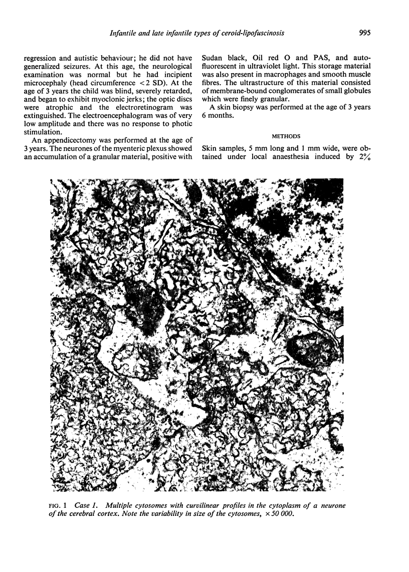
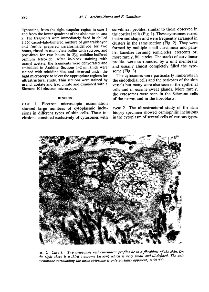
A skin biopsy was carried out in two children suffering from early infantile (Santavuori) and late infantile (Jansky-Bielschowsky) types of ceroid-lipofuscinosis. In both cases cytoplasmic inclusions, identical with those found in neurones, were present in the skin. Skin biopsy thus appears as a simple technique for the diagnosis of the ceroid-lipofuscinoses.
Full text
PDF


Images in this article


Selected References
These references are in PubMed. This may not be the complete list of references from this article.
- Carpenter S., Karpati G., Andermann F. Specific involvement of muscle, nerve, and skin in late infantile and juvenile amaurotic idiocy. Neurology. 1972 Feb;22(2):170–186. doi: 10.1212/wnl.22.2.170. [DOI] [PubMed] [Google Scholar]
- Carpenter S., Karpati G., Wolfe L. S., Andermann F. A type of juvenile cerebromacular degeneration characterized by granular osmiophilic deposits. J Neurol Sci. 1973 Jan;18(1):67–87. doi: 10.1016/0022-510x(73)90021-x. [DOI] [PubMed] [Google Scholar]
- Fawcett J. S., Andermann F., Wiglesworth F. W., Smith D. L. On the natural history of late infantile cerebromacular degeneration. Neurology. 1966 Nov;16(11):1130–1134. doi: 10.1212/wnl.16.11.1130. [DOI] [PubMed] [Google Scholar]
- Haltia M., Rapola J., Santavuori P. Infantile type of so-called neuronal ceroid-lipofuscinosis. Histological and electron microscopic studies. Acta Neuropathol. 1973 Oct 11;26(2):157–170. doi: 10.1007/BF00697751. [DOI] [PubMed] [Google Scholar]
- Joosten E., Gabreels F., Stadhouders A., Bolmers D., Gabreels-Festen A. Involvement of sural nerve in neuronal ceroid-lipofuscinoses: report of two cases. Neuropadiatrie. 1973 Jan;4(1):98–110. doi: 10.1055/s-0028-1091731. [DOI] [PubMed] [Google Scholar]
- Martin J. J., Jacobs K. Skin biopsy as a contribution to diagnosis in late infantile amaurotic idiocy with curvilinear bodies. Eur Neurol. 1973;10(5):281–291. doi: 10.1159/000114283. [DOI] [PubMed] [Google Scholar]
- Rapola J., Haltia M. Cytoplasmic inclusions in the vermiform appendix and skeletal muscle in two types of so-called neuronal ceroid-lipofuscinosis. Brain. 1973 Dec;96(4):833–840. doi: 10.1093/brain/96.4.833. [DOI] [PubMed] [Google Scholar]
- Santavuori P., Haltia M., Rapola J., Raitta C. Infantile type of so-called neuronal ceroid-lipofuscinosis. 1. A clinical study of 15 patients. J Neurol Sci. 1973 Mar;18(3):257–267. doi: 10.1016/0022-510x(73)90075-0. [DOI] [PubMed] [Google Scholar]
- Witzleben C. L., Smith K., Nelson J. S., Monteleone P. L., Livingston D. Ultrastructural studies in late-onset amaurotic idiocy: lymphocyte inclusions as a diagnostic marker. J Pediatr. 1971 Aug;79(2):285–293. doi: 10.1016/s0022-3476(71)80115-4. [DOI] [PubMed] [Google Scholar]
- van Haelst U. J., Gabreëls F. J. The electron microscopic study of the appendix as early diagnostic means in Batten-Spielmeyer-Vogt disease. Acta Neuropathol. 1972;21(2):169–175. doi: 10.1007/BF00687571. [DOI] [PubMed] [Google Scholar]