Abstract
The heptauridine tract at each gene end and intergenic region (IGR) at the gene junctions of vesicular stomatitis virus (VSV) have effects on synthesis of the downstream mRNA, independent of their respective roles in termination of the upstream mRNA. To investigate the role of the U tract and the IGR in downstream gene transcription, we altered the N/P gene junction of infectious VSV such that transcription levels would be affected and result in altered molar ratios of the N and P proteins, which are critical for optimal viral RNA replication. The changes included extended IGRs between the N and P genes and shortening the length of the heptauridine tract upstream of the P gene start. Viruses having various combinations of these changes were recovered from cDNA and selective pressure for efficient viral replication was applied by sequential passage in cell culture. The replicative ability and sequence at the altered intergenic junctions were monitored throughout the passages to compare the effects of the changes at the IGR and U tract. VSV variants with wild type U tracts upstream of the P gene replicated to levels similar to wt VSV. Variants with shortened U tracts were reduced in their ability to replicate. With passage, populations emerged that replicated to higher levels. Sequence analysis revealed that mutations had been selected for in these populations that increased the length of the U tract. This correlated with an increase in abundance of P mRNA and protein to provide improved N:P protein molar ratios. Extended IGRs resulted in decreased downstream transcription but the effect was not as extensive as that caused by shortened U tracts. Extended IGRs were not selected against in 5 passages. Our results indicate that the size of the upstream gene-end U tract is an important determinant of efficient downstream gene transcription in infectious virus.
INTRODUCTION
Vesicular stomatitis virus (VSV) is the prototype of the Rhabdoviruses in the order Mononegavirales, the nonsegmented negative strand (NNS) RNA viruses. The VSV genome contains 11.2 kb of negative sense RNA with five genes that are flanked by 3’ leader (le) and 5’ trailer (tr) regions in the order 3’-N-P-M-G-L-5’ (Rose and Schubert, 1987). VSV transcription is obligatorily sequential (Abraham and Banerjee, 1976; Ball and White, 1976). There is a single 3' entry site for the RNA dependant RNA polymerase (RdRp) and transcription of each downstream gene depends on the prior transcription and proper termination of the gene immediately upstream (Abraham and Banerjee, 1976; Ball and White, 1976; Barr, Whelan, and Wertz, 1997a; Barr, Whelan, and Wertz, 1997b; Barr, Whelan, and Wertz, 2002). In addition, VSV transcription is polar, with monocistronic mRNA transcript abundance decreasing with increasing distance from the genome 3’ end (Ball et al., 1999; Iverson and Rose, 1981; Villarreal, Breindl, and Holland, 1976; Wertz, Perepelitsa, and Ball, 1998). This results from the poorly understood phenomenon of transcription attenuation, causing an approximate 30% drop in transcript abundance as the RdRp crosses each gene junction (Iverson and Rose, 1981). Transcription of VSV mRNAs is regulated by cis-acting signals located within the 3' leader region and at the beginning and end of each gene as well as the 2 nucleotides that lie between each gene (Barr and Wertz, 2001; Barr, Whelan, and Wertz, 1997a; Barr, Whelan, and Wertz, 1997b; Barr, Whelan, and Wertz, 2002; Hwang, Englund, and Pattnaik, 1998; Li and Pattnaik, 1999; Stillman and Whitt, 1997; Stillman and Whitt, 1998; Stillman and Whitt, 1999; Wertz et al., 1994; Whelan and Wertz, 1999). Together, the sequences that comprise the upstream gene end, the intergenic dinucleotide and the downstream gene start are referred to as the gene junction. The gene junctions comprise 23 nucleotides that include a strictly conserved 11 nucleotide sequence at the end of each upstream gene, a two nucleotide intergenic region (IGR) and a conserved sequence at the start of the downstream gene (Barr, Whelan, and Wertz, 2002). These sequences direct the VSV RdRp to terminate and polyadenylate the upstream mRNA, and to initiate and cap the downstream mRNA (Barr and Wertz, 2001; Barr, Whelan, and Wertz, 1997a; Barr, Whelan, and Wertz, 1997b; Barr, Whelan, and Wertz, 2002; Hwang, Englund, and Pattnaik, 1998; Stillman and Whitt, 1997; Stillman and Whitt, 1998; Stillman and Whitt, 1999; Wang, McElvain, and Whelan, 2007).
Termination of mRNA transcription is a critical step in the control of gene expression for the NNS viruses. For VSV, mRNA polyadenylation and termination are signaled by the gene end sequence 3’-AUACUUUUUUU-5’ and the first nucleotide of the IGR (Barr and Wertz, 2001; Barr, Whelan, and Wertz, 1997a; Barr, Whelan, and Wertz, 1997b; Barr, Whelan, and Wertz, 2002; Hwang, Englund, and Pattnaik, 1998; Stillman and Whitt, 1997). The IGR, the sequence between the end of the upstream gene end sequence and the beginning of the downstream gene start sequence, is conserved as the dinucleotide 3’-GA-5’ at every VSV gene junction except the P/M gene junction, which is 3’-CA-5’. Limited extensions of the IGR up to 6 nt have shown that the RdRp responds to alterations in both the length and sequence of the IGR, suggesting that the sequence of the IGR affects both termination and initiation at the gene junction (Barr, Whelan, and Wertz, 1997b; Stillman and Whitt, 1997; Stillman and Whitt, 1998). It has been proposed that the RdRp scans the IGR for the appropriate downstream gene start sequence (Stillman and Whitt, 1998), and this hypothesis is supported by the observation that transcript initiation occurs at a low frequency from non-consensus gene start sequences located within an extended IGR (Hinzman, Barr, and Wertz, 2002; Stillman and Whitt, 1998).
Initiation of VSV mRNA was previously shown to be signaled by the gene start sequence, 3’-UUGUCnnUAG-5’ and influenced by the 5’ nucleotide of the intergenic region (Stillman and Whitt, 1997; Stillman and Whitt, 1999). The first three nucleotides in the gene start sequence were also found to play a role in capping of the nascent mRNA, a designation that was recently confirmed by individually mutating each of these conserved nucleotides and analyzing the resulting effects on transcript initiation and capping in infectious viruses (Wang, McElvain, and Whelan, 2007).
Additionally, our laboratory showed previously that nucleotides within the conserved gene end also play a critical role in signaling downstream gene transcription initiation (Hinzman, Barr, and Wertz, 2002). Since termination of the upstream gene, which is signaled by the gene end sequence, is required for initiation of the downstream gene, these studies were carried out by inserting an additional functional gene end sequence upstream of the existing gene end at the gene junction in subgenomic replicons. This separated the added gene end sequence from the downstream initiation signal by 88 nt. Because of the sequential nature of transcription, the RdRp terminated transcription at the first gene end sequence encountered (the added gene end), then initiated transcription at the downstream gene start sequence. The original gene end sequence immediately upstream of the gene start sequence was thus made redundant in terms of termination of the upstream mRNA, and could be altered without disrupting sequential transcription. Using this approach, mutagenesis of the upstream gene end sequences was carried out. We found that the size and sequence of the gene end U tract was important for efficient transcription of the downstream gene, independent of the role of the U tract in termination (Hinzman, Barr, and Wertz, 2002). A U tract size of 7 residues was optimal in signaling efficient downstream mRNA transcription, whereas either incrementally reducing or increasing the size of the U7 tract resulted in a corresponding reduction in the abundance of the downstream mRNA. Mutation of the 3’-AUAC-5’ tetranucleotide of the gene end sequence did not affect downstream transcription (Hinzman, Barr, and Wertz, 2002).
A consequence of duplicating the gene end sequence was that it resulted in an extension of the IGR. Recently, we investigated the effect of IGR length on transcription of VSV mRNAs by incrementally increasing the size of the IGR from the wt 2 nt up to 320 nt. We found that increasing the length of the IGR in a stepwise manner resulted in corresponding incremental decreases in the abundance of the downstream mRNA (Barr et al., 2008). In other words, increasing the length of the IGR resulted in a corresponding increase in transcriptional attenuation at that gene junction. As the IGRs of VSV are conserved in sequence and length, it is unclear what effect extended IGRs might have in the context of an infectious virus.
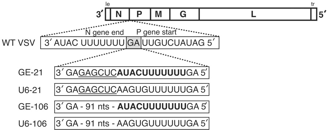
Having shown that efficient downstream transcription was affected by both the length and sequence of the U tract and the length of the IGR in experiments using VSV subgenomic replicons, in the present work we extend these studies to investigate the effect of these alterations on VSV gene expression and overall viral replication during the viral life cycle. Previous work has established that the N:P protein molar ratio is important for efficient viral replication (Ball et al., 1999; Howard and Wertz, 1989; La Ferla and Peluso, 1989; Peluso and Moyer, 1988) and that the virus population can rapidly adapt to relieve detrimental alterations in the N:P ratio (Wertz, Moudy, and Ball, 2002). We reasoned that selective pressure for efficient viral replication could be used to test the effect of extended IGRs at the N/P gene junction. We therefore engineered recombinant VSVs that contained extended IGRs and these were made also to have the length of U tract immediately upstream of the P gene start sequence between the N and P genes either shortened or left as the wild type heptauridine tract (Fig. 1). The viruses were repeatedly passaged in comparison to wt VSV. Thus we simultaneously tested the effect that extended IGRs and altered U tract length had on downstream transcription compared to wt VSV. The effects of these alterations on viral replication and gene expression at both the mRNA and protein level with successive passage was examined. We found that replication of VSV variants with shorter U tracts was compromised compared with wt VSV, regardless of the length of the IGR. With passage, mutations were selected that altered the length of the U tract upstream of the P gene start sequence, and correspondingly, an increase in expression of the P gene was observed. Our results show that absence of a U7 tract as a component of the initiation signal provides a greater selection pressure than an extended intergenic region.
FIG. 1.
Schematic diagram of wt VSV and variant viruses engineered to contain extended IGRs and U tract alterations at the N/P gene junction. The indicated sequences were inserted between the N gene end sequence and the P gene start sequence of the cDNA clone of VSV, and virus was rescued as described in Materials and Methods. The inserted gene end sequence is shown in bold, and the Xho I restriction site underlined. Abbreviations: le, leader; tr, trailer; WT, wild type; nts, nucleotides.
MATERIALS AND METHODS
Cells and Viruses
Baby hamster kidney (BHK-21) cells were used for transfections, passage of virus, growth of viral stocks, viral single-step growth analysis, and analysis of viral RNA and protein synthesis, as described previously (Ball et al., 1999; Flanagan, Ball, and Wertz, 2000; Whelan et al., 1995). African green monkey kidney (Vero-76) cells were used to determine viral titers and plaque morphology (Ball et al., 1999; Flanagan, Ball, and Wertz, 2000). The plasmid pVSV1(+), described previously (Whelan et al., 1995), was used to construct variant VSV cDNAs that contained extended intergenic regions of 21 nt (GE-21 and U6-21, see Fig 1) between the N gene end sequence and the P gene start sequence using standard cloning techniques. The GE virus contained a wild type gene end sequence, 3’-AUACUUUUUUUGA-5’, upstream of the P gene start sequence (Fig. 1). The U6 virus contained the sequence 3’-AAGUGUUUUUUGA-5’ upstream of the P gene start sequence. For the GE-106 and U6-106 viruses, additional sequence derived from VSV cDNA (corresponding to nt 1296 – 1374 on the VSV genome) was amplified by PCR and inserted at the XhoI site of either GE-21 or U6-21, respectively. Variant and wild-type VSV was recovered from the supernatant of vTF7-3 (Fuerst et al., 1986) infected cells transfected with the viral cDNA and plasmids encoding the N, P, and L proteins of VSV. Supernatant from the primary transfection was filtered once through a 0.22 µm filter and a fractional volume was passed onto fresh BHK-21 cells in the presence of 1-β-D-arabinofuranosylcytosine (AraC, 25 µg/mL), to inhibit vTF7-3 replication, for 36 hr at 37C. The supernatant was harvested, titered, and designated as the passage 1 stock for each virus. Virus was passaged at a multiplicity of infection (MOI) of 0.01 in BHK-21 cells at 37C for 20 h then harvested and titered.
Sequence Analysis
The N/P junction sequences were analyzed at indicated passages by cDNA sequence analysis of viral RNA purified from virions. Genomic RNA was purified from virus harvested from the supernatant of infected BHK-21 cells by phenol/chloroform extraction and ethanol precipitation. RT-PCR was performed using primers that flanked the N/P gene junction, 9121 (5’-TATCAGATCTGATATGATGCAGTATGCG-3’) and S&S (5’-GATTCTGTGTCAGAATCATCTGC-3’). The cDNA product from the RT-PCR reaction was isolated by gel purification and the sequence was determined using primer 9121. Bulk RT-PCR trace sequence chromatograms were visualized using the 4Peaks sequence analysis software (http://www.mekentosj.com/4peaks/). Clonal sequence analysis was done by subcloning of the cDNA products from two independent RT-PCR reactions and sequence determination of individual clones.
Plaque Morphology and Single Step Growth Analysis
Viral plaque diameters were determined by direct measurement to the nearest 0.5 mm of at least 30 individual viral plaques from Vero-76 monolayers, normalized to the wt VSV plaque diameter, and a 95% confidence interval was calculated for each virus at the indicated passage. Single step growth analysis was conducted in BHK-21 cells infected at an MOI of 5. After 1 hr of adsorption, the inoculum was removed, the monolayer was washed twice, fresh medium was added, and the cultures were incubated at 37C. Supernatant fluids were harvested at the indicated time intervals up to 24 hr and the viral titers were determined by plaque assay on Vero-76 cells.
RNA and Protein Analysis
Viral RNA synthesis was detected in BHK-21 cells infected at an MOI of 5 with the indicated virus by metabolic labeling with [3H]uridine (33 µCi/mL) in the presence of actinomycin D (ActD, 10 µg/mL) at 37C for 3 hrs starting 2 hrs post-adsorption. Total cytoplasmic RNA was harvested by phenol/chloroform extraction followed by ethanol precipitation, as described previously (Pattnaik et al., 1992; Wertz et al., 1994). A fraction of the RNA sample was electrophoresed through a 1.75% agarose-urea gel and the radiolabeled products were visualized by fluorography and autoradiography (Pattnaik et al., 1992; Wertz et al., 1994).
Viral protein and RNA synthesis were analyzed by infection of BHK-21 cells with the indicated virus at a MOI of 50 at 37°C in the presence of ActD (10 µg/mL). At 2 hr post-adsorption, the cells were washed twice and incubated in methionine-free medium with ActD (10 µg/mL) for 30 min. Cells were then incubated with [35S]methionine (50 µCi/mL) in the presence of ActD (10 µg/mL) for 30 min. Cytoplasmic extracts were prepared and divided into two fractions. Viral protein synthesis was analyzed from one fraction by protein separation on a denaturing 10% low bis polyacrylamide gel. Total RNA was harvested from the second fraction by phenol/chloroform extraction followed by ethanol precipitation and RNAs were quantitated by primer extension analysis. Two oligonucleotide primers were used in each reaction: the first primer, N–Pext (5’-ATTGATGTAAAGAGGAATCTCC-3’), corresponded to positions 192–171 of the VSV genome (annealing to the N mRNA); the second primer, P–Pext (5’-AATTTCTGGTTCAGATTCTGTG-3’), corresponded to positions 1599–1578 (annealing to the P mRNA). The primers (5 µM) were separately end labeled in the presence of [33P]-ATP (0.5 µCi/mL) and T4 polynucleotide kinase (0.4 U/µL, Invitrogen) for 30 min at 37°C and purified using a nucleotide removal kit (Qiagen). The primers were annealed in excess and extended with Superscript II (20 U/µl, Invitrogen) for 50 min at 50°C using reaction conditions recommended by the manufacturer. Radiolabeled cDNA products were separated on a denaturing 6% polyacrylamide gel, visualized by autoradiography, and quantitated by densitometry.
Quantitation
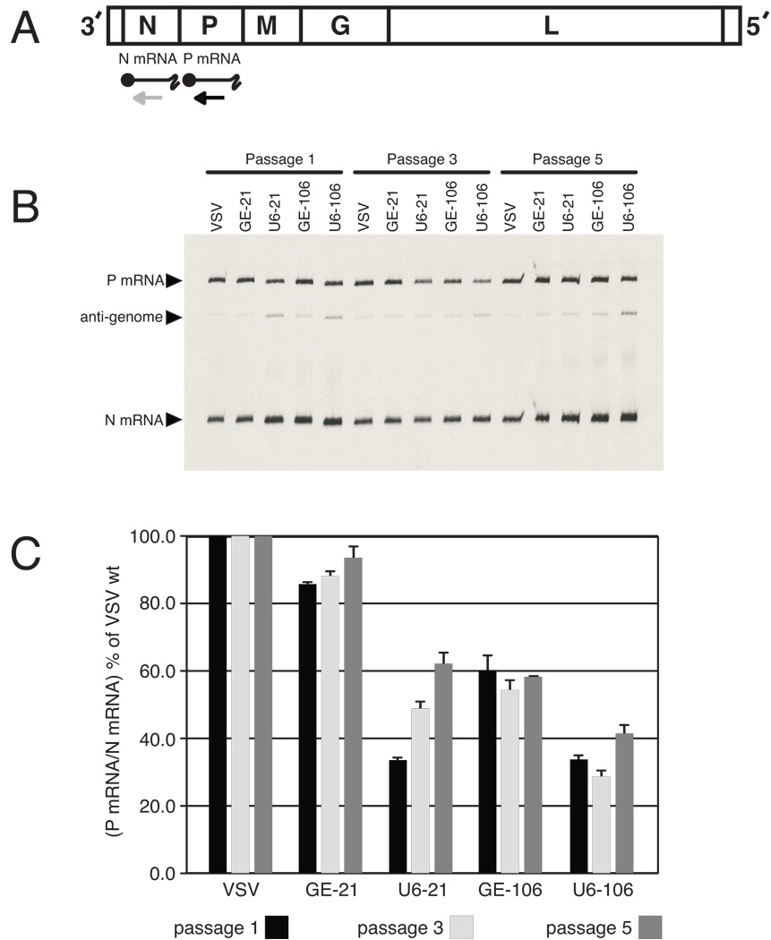
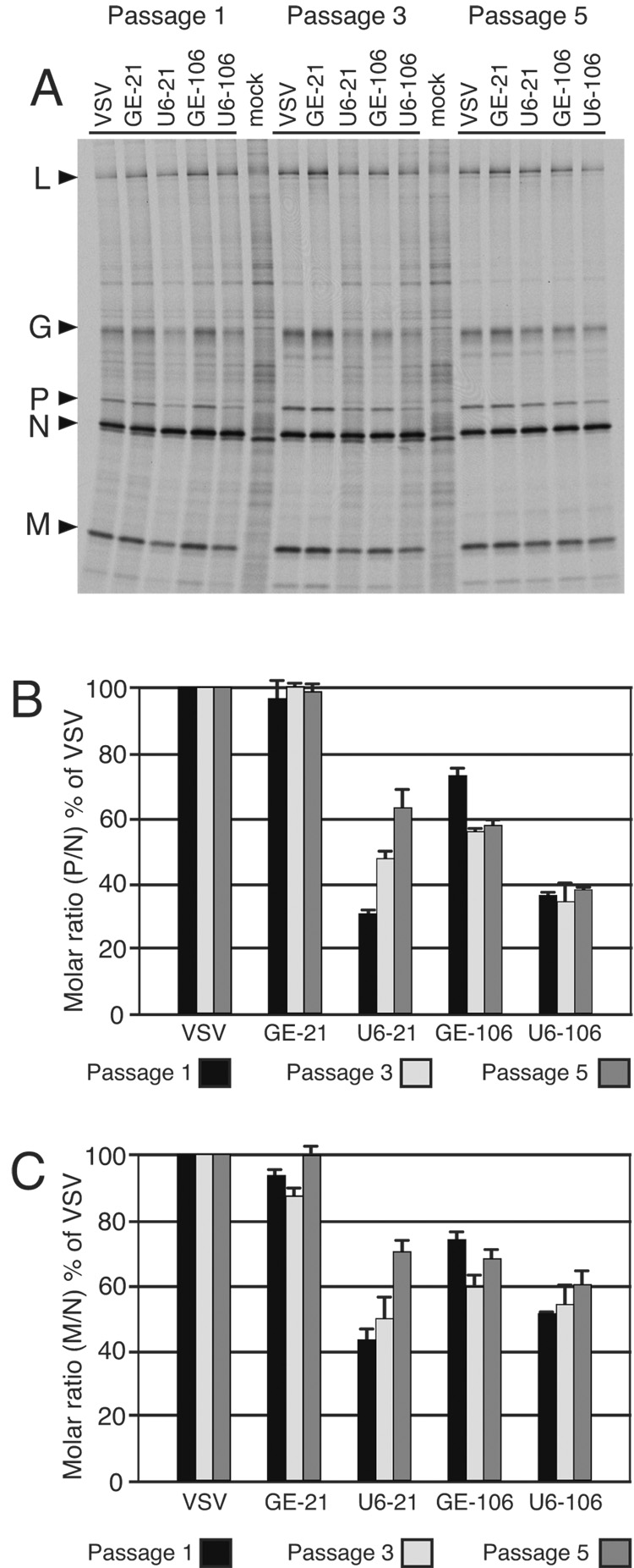
The quantities of VSV-specific primer extension products and metabolically labeled protein products were determined by densitometry using a Bio-Rad model GS-800 densitometer and Quantity One software. The cDNA corresponding to the P mRNA was compared with the cDNA corresponding to the N mRNA and expressed as a percentage of the P:N mRNA ratio from wt VSV (Fig 5c). Protein quantitation was adjusted for methionine content and expressed as a molar ratio of P or M protein to N protein, relative to wt VSV (Fig 6).
FIG. 5.
Comparison of the P:N mRNA ratios for wt VSV and variant viruses at passages 1, 3, and 5. (A) Schematic diagram of the VSV genome, the N and P mRNAs, and the relative positions of primers N-Pext and P-Pext that anneal to the N and P mRNA, respectively, and also to corresponding positions within the anti-genome. (B) Primer extension analysis of the N and P mRNAs from the indicated viruses at the indicated passage using end-labeled primers N-Pext and P-Pext as described in Materials and Methods. The position of the products corresponding to the antigenome, the N mRNA, and the P mRNA are indicated at the left of the figure. (C) Quantitation of primer extension products from three independent experiments, including that shown in panel B. The P:N mRNA ratio was determined, normalized to the wt VSV P: N mRNA ratio at that passage, and expressed as the mean percentage relative to wt VSV. Error bars represent the standard error of the mean. Where error bars are not visible, the standard error was negligible.
FIG. 6.
Comparison of the N/P protein molar ratios for wt VSV and variant viruses at passage 1, 3, and 5. (A) Viral proteins synthesized in BHK-21 cells infected with the indicated viruses at the specified passage. Protein was labeled with [35S]methionine as described in Materials and Methods, resolved by low bis PAGE, and detected by autoradiography. The virus and passage number are indicated at the top and the viral proteins are identified at the left of the figure. Mock 1 and 2 are uninfected cells labeled as described above. (B) P – N protein molar ratios from three independent experiments, including that shown in panel A. Molar ratios were calculated after normalizing for methionine content of the P and N proteins and expressed as a mean percentage of the wt VSV P:N ratio for the given passage. Error bars represent the standard error of the mean. (C) M:N protein molar ratios, determined and plotted as described in the legend for panel B.
RESULTS
Recovery of variant VSVs with altered intergenic regions and U tracts at the N/P gene junction
To examine in infectious virus the effect of alterations to the length of the IGR and, concurrently, alterations to the length of the U tract upstream of a gene start sequence, we made changes to the N/P gene junction of VSV. We took advantage of the high error rate of the VSV RdRp and the lack of a proofreading mechanism, as well as the observed selective pressure to maintain an optimal N:P protein molar ratio to test the relative importance of the IGR length versus the U tract length regarding P gene expression. Extended IGRs of either 21- or 106-nt were engineered between the N gene end sequence and the P gene start sequence of the infectious cDNA clone of VSV, pVSV1(+), as described in Materials and Methods. Two clones were constructed for each size of IGR: One set contained a duplicated gene end sequence at the end of the IGR, which included a wild-type U7 tract just upstream of the P gene start sequence (GE-21 and GE-106). The other set had a shortened U6 tract upstream of the P gene start sequence (U6-21 and U6-106) (Fig. 1). The sequence of the inserted IGRs was identical for the respective size except for a 4 nt region, which lies 8 nt upstream of the P gene start sequence (Fig. 1, UACU vs AGUG).
The two sets of viruses were designed to test the response to selective pressure in the following ways. The first set was designed to have inserted either a short (GE-21) or long (GE-106) IGR extension followed by a second functional gene end sequence, 3’-AUACU7-5’. For these viruses a single nucleotide change in the upstream N gene end sequence (either at the conserved C residue, or by deletion or alteration of a single U residue within the U tract) would abrogate termination at the upstream N gene end and result in termination occurring at the second, inserted gene end, thereby eliminating with a single nucleotide change the extended IGR.
The second set of viruses was designed to have inserted either a short (U6-21) or long (U6-106) IGR followed by a second inserted gene end, 3’-AGUGU6-5’ that was both nonfunctional for termination and also suboptimal for downstream transcription. These viruses were designed such that reversion to eliminate the extended intergenic region would be difficult, requiring both a change at the original upstream N gene end to eliminate its function, coupled with at least two changes to restore termination signaling at the second inserted gene end (3’-AGUGU6-5’; at least a C at position 4 and addition of another U to form a functional U7 tract). In contrast, reversion of the U6 tract to give the optimal U7 tract for downstream initiation could be achieved through a single nucleotide change. Thus the two sets of viruses were designed to test for the relative importance of a short intergenic versus a U7 tract for downstream transcriptional initiation - and in either case a single nucleotide change could accomplish this. We postulated that, with passage, if the length of the IGR was important for P gene expression, viral populations within GE-21 and GE-106 infected cells would be selected during passage that had corrected the IGR length to that suitable for efficient P mRNA transcription and viral replication. Similarly, if the length of the U tract immediately upstream of the P gene was important for efficient P gene expression, we anticipated that viral populations within U6-21 and U6-106 infected cells would be selected during passage that had altered the length of the U tract to that suitable for P gene expression.
Recovery, Passage, and Replication of Viruses with an Extended Intergenic Region
Viruses were recovered from each of the engineered cDNAs by transfection of the appropriately engineered plasmid into BHK-21 cells along with plasmids encoding the VSV N, P, and L proteins. Wt VSV was recovered in addition to the variant viruses and included as a control in the experiments described in this study. An initial virus stock was prepared from the primary transfection (see Materials and Methods) for each virus and this stock was designated as passage 1. The sequence at the N/P gene junction for passage 1 was examined and found to match the input cDNA sequence by RT-PCR and sequence analysis. Each virus was then passaged five successive times, in triplicate, at a MOI of 0.01 with propogation for 20 h at 37°C in BHK-21 cells. After each passage, the virus stock was titered on Vero-76 cells. The plaque diameter for each virus was measured and normalized to the wt VSV plaque diameter at that passage and a 95% confidence interval was calculated to estimate the mean plaque diameter for the indicated viral population (Fig. 2).
FIG. 2.
Mean plaque diameters for wt VSV and variant viruses following sequential passage. Virus from passage 1 (A), 3 (B), and 5 (C) were analyzed. Viral plaque diameters were measured directly from plaques formed on Vero-76 cell monolayers. The mean plaque diameter for each virus was determined, normalized to wt VSV at each passage, and expressed as the mean plaque diameter with a 95% confidence interval.
At passage 1, no difference was detected between the plaque diameter of the wt VSV population and those of the GE-21 or GE-106 populations, referred to collectively as the GE viruses (Fig. 2A). In contrast, the U6-21 and U6-106 populations (referred to collectively as the U6 viruses) were markedly reduced in their plaque diameter relative to wt VSV. This agreed with the observation that the titers from the U6 viruses were generally 10 fold lower than those of VSV and the GE viruses (data not shown). By passage 3, a significant increase in the plaque size for the U6 viruses was observed, relative to their passage 1 plaque size (Fig. 2B), suggesting that these viral populations were improving in their replicative ability. At pass 3, the GE virus populations remained indistinguishable from wt VSV, although the GE-21 plaque size had increased relative to its passage 1 diameter. By passage 5 (Fig. 2C), the U6-21 virus plaque diameter had increased such that no difference could be detected between its plaque size and that of wt VSV. The U6-106 plaque diameter at passage 5 had not changed from its passage 3 size. Concurrent with the increase in plaque diameter for the U6-21 viruses, the titer of U6-21 virus after 5 passages was similar to the wt VSV titer (data not shown).
Single Step Growth Analysis of Passaged Virus
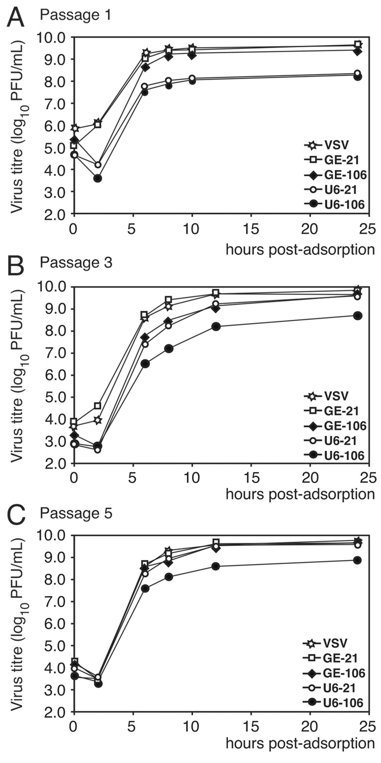
Replication of the engineered viruses was compared with that of wild type by single step growth assays at passages 1, 3, and 5 in BHK-21 cells infected with an MOI of 5 (Fig. 3). Supernatant fluids were harvested at the indicated times and titers were determined in Vero-76 cells, as described in Materials and Methods. As observed with the plaque diameter measurements at passage 1, GE-21 and GE-106 replicated to levels indistinguishable from wt VSV. In contrast, U6-21 and U6-106 replicated to titers approximately 10-fold lower than wt VSV (Fig. 3A). At passage 3, however, U6-21 replication had improved to wt levels at the 24 h time point, whereas U6-106 remained approximately 10 fold lower in titer (Fig. 3B). However, both U6-21 and GE-106 replicated to an intermediate level between wt VSV and U6-106 at the middle time points (Fig. 3B, time points 6, 8, and 12 hrs). By passage 5, U6-21 replicated to titers indistinguishable from wt VSV at all time points, while the U6-106 virus was reduced in replication by less than 10-fold (Fig. 3C). These data correlated with the plaque measurement data and suggest that by passage 5, virus subpopulation(s) within the U6-21 population had been selected for efficient viral replication.
FIG. 3.
Single step growth analysis of wt VSV and variant viruses. BHK-21 cells infected with indicated viruses at an MOI of 5 at 37C. Virus from passage 1 (A), 3 (B), and 5 (C) were analyzed. Samples of the supernatant medium were harvested at the indicated time points and titrated in duplicate by plaque assay on Vero-76 cells.
Sequence Determination of the N/P Junction of Passaged Virus
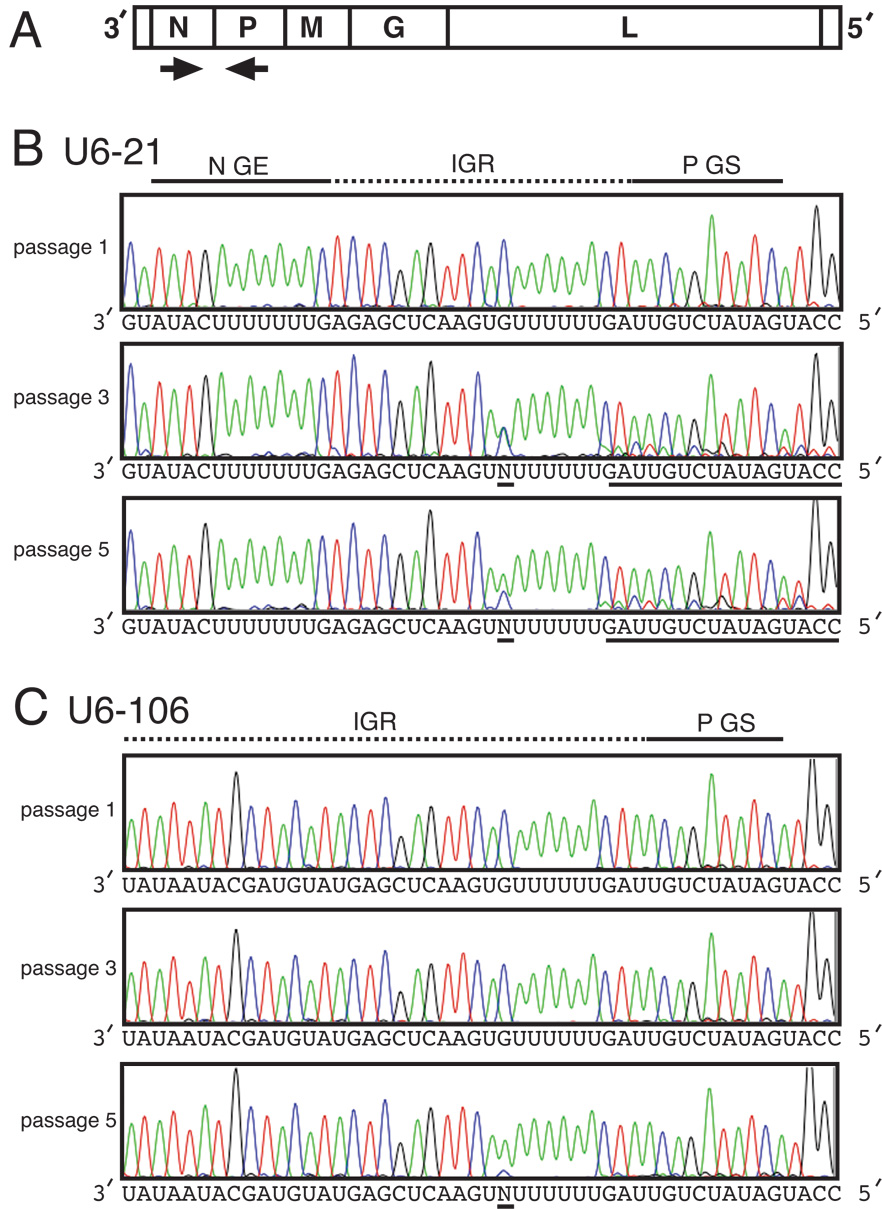
To investigate the possibility that mutations had been selected at the engineered N/P junction of the passaged viruses, we used RT-PCR to determine the genomic sequence at this junction for each of the viruses at passages 1, 3, and 5. The N/P junction was amplified using flanking primers (Figure 4A) and the bulk cDNA product was purified by gel extraction, and sequenced (see Materials and Methods). No alterations at the N/P junction were detected in the wt VSV, GE-21, or GE-106 populations at passages 1, 3, or 5 (data not shown). In contrast, the N/P junction sequence of the U6-21 virus population was heterogeneous at the region immediately upstream of the P gene start at both passage 3 and 5 (Fig. 4B, underlined sequence). Heterogeneity was observed at two points in the sequence, suggesting that at least two different subpopulations had been selected. One point of heterogeneity started directly after the U6 tract, where at least two sequence traces were evident, indicating that changes (insertions or deletions) had occurred in the genome at this point. The other point of change was at the residue immediately upstream of the U6 tract, where a subpopulation appeared by passage 3 that had incorporated a U residue in place of the G residue present at passage 1. The bulk N/P junction sequence of the U6-106 virus population appeared homogenous at passage 3, but it appeared that changes were beginning to be selected at passage 5 (Fig. 4C). As with the U6-21 population, a subpopulation of the U6-106 population had incorporated a U residue in place of the G residue upstream of the U6 tract. These results indicated that subpopulations within the U6-21 and U6-106 populations had been selected for during passage.
FIG. 4.
Sequence analysis of the N/P gene junction of the indicated viruses at passage 1, 3, and 5. (A) Diagram of the VSV genome and relative positions of primers used in RT – PCR analysis. (B and C) Sequence chromatogram traces of bulk RT – PCR products from U6-21 (B) and U6-106 (C) genomes at the indicated passage. The N gene end sequence (N GE), intergenic region (IGR), and P gene start sequence (P GS) are indicated above the chromatograms. Underlined sequence indicates potential change(s) from the passage 1 sequence.
To determine the relative proportions of viral subpopulations within the passage 5 population observed in Fig. 4, two independent RT-PCRs of the total RNA were done to amplify the N/P junction as described above. The bulk cDNA products were purified and subcloned for sequence determination of individual clones. At least 23 clones for each virus were selected for sequencing. Wt VSV was included as a control. The wt VSV N/P gene junction was unchanged in 96% of the clones, the sole exception being the extension of the U7 tract in the N gene end sequence by one nucleotide to U8 in 1/23 clones examined (Table 1). This mutation was not predicted to affect termination of the N mRNA according to previous work, and was likely not significant. No alteration was observed in the N gene end sequence, which preceded the IGR, for both the GE or U6 viruses (data not shown).
Table 1.
Partial intergenic junction sequences of pass 1 and pass 5 cloned cDNA*
| IGR – P Junction Sequence (3′ to 5′) | ||||
|---|---|---|---|---|
| Pass 1 | Pass 5 | Effective change | No. of clones | |
| VSVa | AUACUUUUUUUGAUUGUC | AUACUUUUUUUGAUUGUC | none | 22/23 |
| AUACUUUUUUUUGAUUGUC | U7 → U8 | 1/23 | ||
| GE-21 | AUACUUUUUUUGAUUGUC | AUACUUUUUUUGAUUGUC | none | 25/27 |
| AUACUUUUUUUUGAUUGUC | U7 → U8 | 2/27 | ||
| U6-21 | AAGUGUUUUUUGAUUGUC | AAGUGUUUUUUUUGAUUGUC | U6 → U8 | 12/33 |
| AAGUGUUUUUUUGAUUGUC | U6 → U7 | 2/33 | ||
| AAGUUUUUUUUGAUUGUC | U6 → U8 | 18/33 | ||
| AAGU21GAUUGUC | U6 → U21 | 1/33 | ||
| GE-106 | AUACUUUUUUUGAUUGUC | AUACUUUUUUUGAUUGUC | none | 22/23 |
| AUACUUUUUUUUGAUUGUC | U7 → U8 | 1/23 | ||
| U6-106 | AAGUGUUUUUUGAUUGUC | AAGUGUUUUUUGAUUGUC | none | 10/32 |
| AAGUUUUUUUUGAUUGUC | U6 → U8 | 20/32 | ||
| AAGUGUUUUUUUGAUUGUC | U6 → U7 | 2/32 | ||
IGR, intergenic region;
wild-type N – P gene junction
For the GE viruses, the intergenic region sequences at passage 5 remained identical to the passage 1 sequence, except for the addition of a single U residue at the U7 tract to increase the U tract to U8 (Table 1). The passage 1 sequence was observed in 93 and 95% of the passage 5 clones examined for GE-21 (25/27) and GE-106 (22/23), respectively. This is the same alteration identified with wt VSV, described above (table 1).
For the U6 viruses, however, substantial heterogeneity was observed at passage 5 in the U tract upstream of the P gene start sequence. This is in agreement with the sequence of the bulk population as shown in Figure 4. Various changes resulted in an increase in the length of the U tract (Table 1). The majority (91%; 30/33 clones examined) of the alterations observed in the U6-21 clones of passage 5 were a lengthening of the U6 tract, present at passage 1, to U8. Two other mutations observed were a U7 tract (2/33 clones) and a U21 tract (1/33 clones). None of the passage 5 viruses had maintained the original U6-21 sequence.
In the U6-106 population 31% of clones (10/32) at passage 5 had maintained the passage 1 sequence. Alterations were observed just upstream of the P gene in 69% of the U6-106 passage 5 clones, resulting in increased U tract lengths of either U8 (20/32 clones) or U7 (2/32 clones) (table 1). These data suggested that mutations were selected for in the upstream U tract that conferred an advantage for replication of U6-21 and U6-106. The increase in the size of the upstream U tract with passage correlated with the observed increase in the replicative ability of U6-21 (Fig. 2 and 3).
Analysis of the N to P mRNA and Protein Ratios of Passaged Viruses
To quantitate the ratio of N to P mRNA and protein as a function of passage, BHK-21 cells were infected at an MOI of 50 with viruses at passage 1, 3, and 5. Cytoplasmic extracts were prepared, divided into two fractions, and viral RNA and protein synthesis were analyzed (Fig. 5 and 6, respectively). The RNA was analyzed and quantitated by primer extension analysis using two primers in the same reaction: primer N–Pext annealed to the N mRNA and primer P–Pext annealed to the P mRNA (Fig. 5A). After extension by reverse transcriptase, the labeled cDNA products were electrophoresed (Fig. 5B), quantitated, and the ratio of the P mRNA to the N mRNA was compared for the viruses relative to wt VSV at the indicated passage (Fig. 5C). At passage 1, U6-21, GE-106, and U6-106 had reduced P:N ratios that were 33, 59, and 35% relative to wild-type, respectively. The P:N ratio of GE-21 was reduced to 86% of wt VSV. At passage 3, an increase in the U6-21 P:N mRNA ratio from passage 1 was observed (33 to 49% of wt VSV), whereas the P:N mRNA ratios for the other viruses remained similar to passage 1 levels. The P:N mRNA ratio for U6-21 was observed to increase again (62% of wt VSV) at passage 5. The observed increase in the U6-21 P:N mRNA ratio with passage correlated with the increase in viral replication detected over the same passages (Fig. 2 and 3). A modest increase in the U6-106 virus P:N ratio relative to wt VSV was detected at passage 5 compared to the passage 1 ratio (35 to 43% of wt VSV).
Protein synthesis was detected by metabolic labeling with [35S]methionine as described in Materials and Methods. Labeled protein extracts were resolved by SDS-PAGE (Fig. 6A), the molar ratios of the N and P proteins were quantitated, and the P:N ratio was calculated relative to wt VSV at each passage (Fig. 6B). In agreement with the primer extension data (Fig. 5), each virus maintained similar P:N protein molar ratios over passage except for the U6-21 virus, in which a steady increase in the P:N molar ratio was observed during passage (Fig. 6B). The P:N molar ratio of U6-21 increased from 31% of wt VSV at passage 1 to 63% of wt VSV at passage 5. Similar data were found for protein expression from genes downstream of the N/P junction as shown by comparison of the M:N protein molar ratio during passage (Fig. 6C) for the viruses. This indicated that genes downstream of the U6-21 N/P junction were similarly increased in expression concurrent with the increase in P protein expression, as has been shown previously and would be expected with obligatory sequential transcription(Wertz, Moudy, and Ball, 2002).
DISCUSSION
Transcriptional attenuation in non-segmented negative stranded RNA viruses is a critical determinant of viral gene expression, yet the underlying mechanism that controls this process is poorly understood. Our recent results have shown that both the length of the IGR and the presence of an upstream U tract are important determinants of the level of attenuation. In this report, we have investigated the relative importance of these two elements on downstream transcription. We engineered VSV variants to have duplicated gene end sequences, which extended the IGRs at the N/P gene junction to either 21- or 106- nt. Additionally, alterations were made such that the original gene end U tract immediately upstream of the P gene either remained as the wild-type U7 tract, or was shortened to a U6 tract. The N/P gene junction was chosen because of the observed selective pressure to maintain a N:P protein molar ratio suitable for efficient viral replication (Howard and Wertz, 1989; Pattnaik and Wertz, 1990; Peluso and Moyer, 1988; Wertz, Moudy, and Ball, 2002). The engineered viruses were recovered and those with a U6 tract were found to have a replicative disadvantage. All viruses were passaged separately in cell culture at low MOI to allow selection for viral subpopulations that replicated more efficiently by increasing expression of the P gene.
Our results show that the U6-21 virus that contained a U6 tract upstream of the P gene start sequence exhibited a reduced replication phenotype. However, upon passage, viral replication improved rapidly. Changes that resulted in an increase in U tract length were observed to accumulate in the U tract immediately upstream of the P gene. While selection for mutations in regions of the genome outside the N/P gene junction cannot be ruled out, we found that the increase in U tract size correlated with an increase in the P:N molar ratio to a level that permitted efficient viral replication. Increasing the number of passages or altering the passage conditions may select for additional mutations that permit an even greater increase in the abundance of the P gene products. Selection in the U6-106 population was slower than observed for the U6-21 population. The U6-106 population replicated less well than wt VSV at all passages, although a modest increase in U6-106 replication was detected. Sequence analysis showed that selection for longer U tracts upstream of the P gene was also occurring in the U6-106 population. The reason for the slower kinetics of selection in the U6-106 population relative to the U6-21 population is unknown, although one possibility is that the slow growth of U6-106 correspondingly reduced the number of genomes copied per passage.
These results confirm our previous findings that the U tract in the gene end immediately upstream of the downstream gene start sequence is an important part of the gene start signal (Barr et al., 2008; Hinzman, Barr, and Wertz, 2002). In addition, our results show that the U7 tract is important for initiation regardless of the size of the upstream IGR. For the U6-21 virus, the observed increase in the size of the U6 tract at the IGR/P junction by two nucleotides to U8 with passage resulted in a two-fold increase in the abundance of the P mRNA. The assays used in this study do not allow us to determine at which step of the transcriptive process the U tract exerts its effect. The U tract may, as proposed (Barr et al., 2008; Hinzman, Barr, and Wertz, 2002), simply serve as a functional part of the initiation sequence, reminiscent of how DNA-dependant RNA polymerases recognize and respond to sequences upstream and downstream of the initiating nucleotide of certain promoters. Investigation of the means by which the U tract regulates downstream transcription is underway.
The intergenic regions of VSV are highly conserved as the sequence 3’-G/CA-5’. The conservation of the dinucleotide IGR in VSV suggests that this length and sequence is optimal for efficient recognition of the various gene junction signals by the RdRp, including termination of the upstream transcript and initiation of the downstream mRNA. In VSV subgenomic replicons, the RdRp responds to certain alterations in either the sequence or length of the IGR, thereby affecting termination and/or initiation (Barr, Whelan, and Wertz, 1997b; Stillman and Whitt, 1998), but it was not clear how the presence of extended IGRs would affect the RdRp in the context of full-length VSV mRNA transcription. One exception to the conservation of the dinucleotide length IGR is the G/L IGR of the New Jersey serotype of VSV, which is 21 nt in length (Luk et al., 1987). This extended IGR was initially proposed to downregulate transcription of the L gene due to the size of the IGR (Luk et al., 1987; Stillman and Whitt, 1998). Alternatively, recent data suggest that the downregulation of the L mRNA may be the result of the separation of the U tract from the gene start sequence as the U tract of the upstream gene end sequence was shown to play a role in transcription of the downstream mRNA (Hinzman, Barr, and Wertz, 2002). The observed downregulation of the L mRNA could be the result of a combination of the length of the IGR and the separation of the U tract from the L gene start.
The GE-21 and GE-106 virus populations maintained the full-length of their respective extended IGRs for 5 passages under the conditions used in this study. No alteration in the N gene end sequence of individual clones was detected at passage 5 (data not shown). The VSV RdRp has been estimated to terminate mRNA transcription with an efficiency of 97 – 99% at the wt gene end sequence but single point mutations in this region have the potential to abrogate transcriptional termination (Barr, Whelan, and Wertz, 1997a). Accumulation of mutations in the upstream N gene end of the GE viruses would be expected to lead to transcriptional readthrough of the extended IGR and subsequent termination at the duplicated downstream gene end immediately upstream of the P gene to restore the wt 2 – nt IGR (see Fig. 1). The size of the IGR also affected the abundance of the downstream transcript (Fig. 5, compare GE-21 with GE-106 and U6-21 with U6-106), but not to a level that resulted in selection against the IGR length in the 5 passages carried out. It is possible that the selective pressure applied in the conditions used in this study was not strong enough or long enough to select against the IGR of the GE viruses, as these viruses replicated to levels similar to wt VSV. Nevertheless, these results suggest that the VSV RdRp can tolerate extended IGRs of up to 106 nt without a detectable reduction in viral replication. The N:P mRNA and protein molar ratios, however, were reduced in both GE-21 and GE-106 viruses relative to wt VSV, but not to the level observed in the U6 viruses and therefore viral replication may not have been debilitated enough to select against the extended IGR. The observed reduction of N:P mRNA and protein molar ratios in both GE-21 and GE-106 correlated with the size of the IGR, suggesting that downstream transcription is affected by the distance between the gene end and gene start signals. This observation is supported by further work showing a strong correlation between the length of the VSV IGR and a reduction in the abundance of downstream transcription (Barr et al., 2008).
The inverse relationship between IGR length and initiation ability may not hold true for all NNS viruses. The paramyxovirus human respiratory syncytial virus (HRSV) contains non-conserved IGRs of varying lengths and sequence (Collins et al., 1986). There is no apparent necessity for a U tract immediately upstream of the gene start sequence, although U-rich sequences are present in all but one of the longer IGRs of natural isolates (Moudy, Sullender, and Wertz, 2004). Extended IGRs of up to 160 nt in length have been placed between the M and G genes of a recombinant HRSV lacking the SH gene (Bukreyev, Murphy, and Collins, 2000). No effect of IGR length on transcription was detected, suggesting that at least for that particular gene junction in HRSV, the distance between the gene end and gene start sequences was not an important regulatory element or that the molar ratios of the upstream and downstream gene products relative to one another were not critical. An effect on HRSV replication, as assayed by plaque diameter measurement and single step growth analysis, was detected however. This seems to indicate that the HRSV IGR extension did have a detrimental effect, although was not apparent with the transcription analysis used (Bukreyev, Murphy, and Collins, 2000). A more recent study that analyzed the effect of increasing the F/HN and HN/L IRGs of infectious Newcastle disease virus (NDV) showed that all IGR changes affected pathogenesis in chickens. Examination of the effect of these IGR changes on gene expression also showed a decrease in downstream gene expression with increased IGR length (Yan and Samal, 2008). Interestingly, reduction in IGR length also resulted in decreased pathogenesis, and the authors suggested that the variable NDV IGRs may have been selected to insure molar ratios of expressed proteins that were favorable for viral replication.
The contrast between the results obtained with HRSV and those of the present study with VSV may reflect a difference in the way that gene expression of various NNS RNA viruses is controlled. Alternatively, these results could reflect the choice of placement of the extended IGR in the genome between genes where the relative molar ratios are important or a more fundamental difference in the response of the respective RdRps to the cis-acting signals. Experiments conducted in subgenomic replicons using IGRs derived from clinical isolates of HRSV showed that in some cases, the sequence of the IGR was an important determinant of local transcriptional activity, suggesting that the IGR may have a role in transcriptional regulation at certain gene junctions (Moudy et al., 2003; Moudy, Sullender, and Wertz, 2004).
In summary, we have used selective pressure for efficient viral replication to show that the length of the U tract upstream of the gene start sequence is important for efficient gene expression in VSV. We have also determined that the size of the IGR affects the abundance of downstream mRNA transcripts during viral transcription.
ACKNOWLEDGEMENTS
We thank the members of the G. W. Wertz and L. A. Ball laboratories for helpful discussions during the preparation of the manuscript. We acknowledge Drs. E. Brian Flanagan and Isidoro Martinez for expert technical advice. We acknowledge the DNA Sequencing Facility, Center for AIDS Research, UAB, for sequence determination.
This work was supported by Grant R37-AI12464 from the NIAID of National Institutes of Health to G. W. W.
Footnotes
Publisher's Disclaimer: This is a PDF file of an unedited manuscript that has been accepted for publication. As a service to our customers we are providing this early version of the manuscript. The manuscript will undergo copyediting, typesetting, and review of the resulting proof before it is published in its final citable form. Please note that during the production process errors may be discovered which could affect the content, and all legal disclaimers that apply to the journal pertain.
REFERENCES
- Abraham G, Banerjee AK. Sequential transcription of the genes of vesicular stomatitis virus. Proc Natl Acad Sci U S A. 1976;73(5):1504–1508. doi: 10.1073/pnas.73.5.1504. [DOI] [PMC free article] [PubMed] [Google Scholar]
- Arnheiter H, Davis NL, Wertz G, Schubert M, Lazzarini RA. Role of the nucleocapsid protein in regulating vesicular stomatitis virus RNA synthesis. Cell. 1985;41(1):259–267. doi: 10.1016/0092-8674(85)90079-0. [DOI] [PubMed] [Google Scholar]
- Ball LA, Pringle CR, Flanagan B, Perepelitsa VP, Wertz GW. Phenotypic consequences of rearranging the P, M, and G genes of vesicular stomatitis virus. J Virol. 1999;73(6):4705–4712. doi: 10.1128/jvi.73.6.4705-4712.1999. [DOI] [PMC free article] [PubMed] [Google Scholar]
- Ball LA, White CN. Order of transcription of genes of vesicular stomatitis virus. Proc Natl Acad Sci U S A. 1976;73(2):442–446. doi: 10.1073/pnas.73.2.442. [DOI] [PMC free article] [PubMed] [Google Scholar]
- Barr JN, Tang X, Hinzman E, Shen R, Wertz GW. The VSV polymerase can initiate at mRNA start sites located either up or downstream of a transcription termination signal but size of the intervening intergenic region affects efficiency of initiation. Virology. 2008 doi: 10.1016/j.virol.2007.12.023. [DOI] [PMC free article] [PubMed] [Google Scholar]
- Barr JN, Wertz GW. Polymerase slippage at vesicular stomatitis virus gene junctions to generate poly(A) is regulated by the upstream 3′-AUAC-5′ tetranucleotide: implications for the mechanism of transcription termination. J Virol. 2001;75(15):6901–6913. doi: 10.1128/JVI.75.15.6901-6913.2001. [DOI] [PMC free article] [PubMed] [Google Scholar]
- Barr JN, Whelan SP, Wertz GW. cis-Acting signals involved in termination of vesicular stomatitis virus mRNA synthesis include the conserved AUAC and the U7 signal for polyadenylation. J Virol. 1997a;71(11):8718–8725. doi: 10.1128/jvi.71.11.8718-8725.1997. [DOI] [PMC free article] [PubMed] [Google Scholar]
- Barr JN, Whelan SP, Wertz GW. Role of the intergenic dinucleotide in vesicular stomatitis virus RNA transcription. J Virol. 1997b;71(3):1794–1801. doi: 10.1128/jvi.71.3.1794-1801.1997. [DOI] [PMC free article] [PubMed] [Google Scholar]
- Barr JN, Whelan SP, Wertz GW. Transcriptional control of the RNA-dependent RNA polymerase of vesicular stomatitis virus. Biochim Biophys Acta. 2002;1577(2):337–353. doi: 10.1016/s0167-4781(02)00462-1. [DOI] [PubMed] [Google Scholar]
- Bukreyev A, Murphy BR, Collins PL. Respiratory syncytial virus can tolerate an intergenic sequence of at least 160 nucleotides with little effect on transcription or replication in vitro and in vivo. J Virol. 2000;74(23):11017–11026. doi: 10.1128/jvi.74.23.11017-11026.2000. [DOI] [PMC free article] [PubMed] [Google Scholar]
- Collins PL, Dickens LE, Buckler-White A, Olmsted RA, Spriggs MK, Camargo E, Coelingh KV. Nucleotide sequences for the gene junctions of human respiratory syncytial virus reveal distinctive features of intergenic structure and gene order. Proc Natl Acad Sci U S A. 1986;83(13):4594–4598. doi: 10.1073/pnas.83.13.4594. [DOI] [PMC free article] [PubMed] [Google Scholar]
- Davis NL, Arnheiter H, Wertz GW. Vesicular stomatitis virus N and NS proteins form multiple complexes. J Virol. 1986;59(3):751–754. doi: 10.1128/jvi.59.3.751-754.1986. [DOI] [PMC free article] [PubMed] [Google Scholar]
- Flanagan EB, Ball LA, Wertz GW. Moving the glycoprotein gene of vesicular stomatitis virus to promoter-proximal positions accelerates and enhances the protective immune response. J Virol. 2000;74(17):7895–7902. doi: 10.1128/jvi.74.17.7895-7902.2000. [DOI] [PMC free article] [PubMed] [Google Scholar]
- Fuerst TR, Niles EG, Studier FW, Moss B. Eukaryotic transient-expression system based on recombinant vaccinia virus that synthesizes bacteriophage T7 RNA polymerase. Proc Natl Acad Sci U S A. 1986;83(21):8122–8126. doi: 10.1073/pnas.83.21.8122. [DOI] [PMC free article] [PubMed] [Google Scholar]
- Hinzman EE, Barr JN, Wertz GW. Identification of an upstream sequence element required for vesicular stomatitis virus mRNA transcription. J Virol. 2002;76(15):7632–7641. doi: 10.1128/JVI.76.15.7632-7641.2002. [DOI] [PMC free article] [PubMed] [Google Scholar]
- Howard M, Wertz G. Vesicular stomatitis virus RNA replication: a role for the NS protein. J Gen Virol. 1989;70(Pt 10):2683–2694. doi: 10.1099/0022-1317-70-10-2683. [DOI] [PubMed] [Google Scholar]
- Hwang LN, Englund N, Pattnaik AK. Polyadenylation of vesicular stomatitis virus mRNA dictates efficient transcription termination at the intercistronic gene junctions. J Virol. 1998;72(3):1805–1813. doi: 10.1128/jvi.72.3.1805-1813.1998. [DOI] [PMC free article] [PubMed] [Google Scholar]
- Iverson LE, Rose JK. Localized attenuation and discontinuous synthesis during vesicular stomatitis virus transcription. Cell. 1981;23(2):477–484. doi: 10.1016/0092-8674(81)90143-4. [DOI] [PubMed] [Google Scholar]
- La Ferla FM, Peluso RW. The 1:1 N-NS protein complex of vesicular stomatitis virus is essential for efficient genome replication. J Virol. 1989;63(9):3852–3857. doi: 10.1128/jvi.63.9.3852-3857.1989. [DOI] [PMC free article] [PubMed] [Google Scholar]
- Li T, Pattnaik AK. Overlapping signals for transcription and replication at the 3′ terminus of the vesicular stomatitis virus genome. J Virol. 1999;73(1):444–452. doi: 10.1128/jvi.73.1.444-452.1999. [DOI] [PMC free article] [PubMed] [Google Scholar]
- Luk D, Masters PS, Gill DS, Banerjee AK. Intergenic sequences of the vesicular stomatitis virus genome (New Jersey serotype): evidence for two transcription initiation sites within the L gene. Virology. 1987;160(1):88–94. doi: 10.1016/0042-6822(87)90048-1. [DOI] [PubMed] [Google Scholar]
- Moudy RM, Harmon SB, Sullender WM, Wertz GW. Variations in transcription termination signals of human respiratory syncytial virus clinical isolates affect gene expression. Virology. 2003;313(1):250–260. doi: 10.1016/s0042-6822(03)00299-x. [DOI] [PubMed] [Google Scholar]
- Moudy RM, Sullender WM, Wertz GW. Variations in intergenic region sequences of Human respiratory syncytial virus clinical isolates: analysis of effects on transcriptional regulation. Virology. 2004;327(1):121–133. doi: 10.1016/j.virol.2004.06.013. [DOI] [PubMed] [Google Scholar]
- Pattnaik AK, Ball LA, LeGrone AW, Wertz GW. Infectious defective interfering particles of VSV from transcripts of a cDNA clone. Cell. 1992;69(6):1011–1020. doi: 10.1016/0092-8674(92)90619-n. [DOI] [PubMed] [Google Scholar]
- Pattnaik AK, Wertz GW. Replication and amplification of defective interfering particle RNAs of vesicular stomatitis virus in cells expressing viral proteins from vectors containing cloned cDNAs. J Virol. 1990;64(6):2948–2957. doi: 10.1128/jvi.64.6.2948-2957.1990. [DOI] [PMC free article] [PubMed] [Google Scholar]
- Peluso RW, Moyer SA. Viral proteins required for the in vitro replication of vesicular stomatitis virus defective interfering particle genome RNA. Virology. 1988;162(2):369–376. doi: 10.1016/0042-6822(88)90477-1. [DOI] [PubMed] [Google Scholar]
- Rose JK, Schubert M. Rhabdovirus genomes and their products. In: Wagner RR, editor. The Rhabdoviruses. New York, N. Y: Plenum Press; 1987. pp. 127–166. [Google Scholar]
- Stillman EA, Whitt MA. Mutational analyses of the intergenic dinucleotide and the transcriptional start sequence of vesicular stomatitis virus (VSV) define sequences required for efficient termination and initiation of VSV transcripts. J Virol. 1997;71(3):2127–2137. doi: 10.1128/jvi.71.3.2127-2137.1997. [DOI] [PMC free article] [PubMed] [Google Scholar]
- Stillman EA, Whitt MA. The length and sequence composition of vesicular stomatitis virus intergenic regions affect mRNA levels and the site of transcript initiation. J Virol. 1998;72(7):5565–5572. doi: 10.1128/jvi.72.7.5565-5572.1998. [DOI] [PMC free article] [PubMed] [Google Scholar]
- Stillman EA, Whitt MA. Transcript initiation and 5′-end modifications are separable events during vesicular stomatitis virus transcription. J Virol. 1999;73(9):7199–7209. doi: 10.1128/jvi.73.9.7199-7209.1999. [DOI] [PMC free article] [PubMed] [Google Scholar]
- Villarreal LP, Breindl M, Holland JJ. Determination of molar ratios of vesicular stomatitis virus induced RNA species in BHK21 cells. Biochemistry. 1976;15(8):1663–1667. doi: 10.1021/bi00653a012. [DOI] [PubMed] [Google Scholar]
- Wang JT, McElvain LE, Whelan SP. Vesicular stomatitis virus mRNA capping machinery requires specific cis-acting signals in the RNA. J Virol. 2007;81(20):11499–11506. doi: 10.1128/JVI.01057-07. [DOI] [PMC free article] [PubMed] [Google Scholar]
- Wertz GW, Moudy R, Ball LA. Adding genes to the RNA genome of vesicular stomatitis virus: positional effects on stability of expression. J Virol. 2002;76(15):7642–7650. doi: 10.1128/JVI.76.15.7642-7650.2002. [DOI] [PMC free article] [PubMed] [Google Scholar]
- Wertz GW, Perepelitsa VP, Ball LA. Gene rearrangement attenuates expression and lethality of a nonsegmented negative strand RNA virus. Proc Natl Acad Sci U S A. 1998;95(7):3501–3506. doi: 10.1073/pnas.95.7.3501. [DOI] [PMC free article] [PubMed] [Google Scholar]
- Wertz GW, Whelan S, LeGrone A, Ball LA. Extent of terminal complementarity modulates the balance between transcription and replication of vesicular stomatitis virus RNA. Proc Natl Acad Sci U S A. 1994;91(18):8587–8591. doi: 10.1073/pnas.91.18.8587. [DOI] [PMC free article] [PubMed] [Google Scholar]
- Whelan SP, Ball LA, Barr JN, Wertz GT. Efficient recovery of infectious vesicular stomatitis virus entirely from cDNA clones. Proc Natl Acad Sci U S A. 1995;92(18):8388–8392. doi: 10.1073/pnas.92.18.8388. [DOI] [PMC free article] [PubMed] [Google Scholar]
- Whelan SP, Wertz GW. Regulation of RNA synthesis by the genomic termini of vesicular stomatitis virus: identification of distinct sequences essential for transcription but not replication. J Virol. 1999;73(1):297–306. doi: 10.1128/jvi.73.1.297-306.1999. [DOI] [PMC free article] [PubMed] [Google Scholar]
- Yan Y, Samal SK. Role of intergenic sequences in newcastle disease virus RNA transcription and pathogenesis. J Virol. 2008;82(3):1323–1331. doi: 10.1128/JVI.01989-07. [DOI] [PMC free article] [PubMed] [Google Scholar]


