Abstract
The Ponseti technique for treating clubfoot has been popularized for idiopathic clubfoot and more recently several syndromic causes of clubfoot. We asked whether it could be used to treat recurrent clubfoot following failed posteromedial release. We retrospectively reviewed 58 children (83 clubfeet) treated by the Ponseti technique for recurrent deformity after posteromedial release in three centers. The minimum followup was 24 months (average, 45 months; range, 24–80 months). We determined initial and final Pirani scores and range of motion of the ankle and subtalar joint. Plantigrade and fully corrected feet were obtained in 71 feet (86%); 11 feet obtained partial correction; one patient failed treatment and underwent another posteromedial release. Recurrences occurred in nine patients (12 feet or 14%). Initial Pirani scores improved in all but one patient; severity of deformity was also inferred by number of casts used for treatment. The age at treatment and numbers of casts did not influence the scores of Pirani et al. The scores were similar among the three orthopaedic surgeons.
Level of Evidence: Level IV, therapeutic study. See Guidelines for Authors for a complete description of levels of evidence.
Introduction
The Ponseti technique was described for treatment of idiopathic clubfeet, and successful short-, mid-, and long-term outcomes have been reported by several authors from Iowa and other groups around the world [1, 5–7, 9, 11, 12, 14, 17, 19, 23, 25–27].
The technique consists of an initial phase of serial casts with progressive abduction of the foot while maintaining counterpressure over the head of the talus. The equinus is the last deformity to correct and in many cases this is done by a calcaneus tendon percutaneous tenotomy and a new cast for 3 weeks. The second phase consists of the use of a foot abduction brace, initially for 3 months, 23 hours a day, followed by use for 14 hours a day until the child is 4 years old [22].
One report from Ponseti’s group suggested children with clubfeet could be successfully treated by the Ponseti technique up to the age of 2 years [19], but the age limits for using the Ponseti technique have been extended by recent reports that claim correction in children as old as 9 years of age [18]. The use of the Ponseti technique has also been expanded in recent years to nonidiopathic clubfeet as rigid as arthrogrypotic feet [4]. One variation of the technique was described to treat difficult feet with substantial cavus, complex feet, resulting in fewer number of clubfeet undergoing posteromedial releases [24].
The Ponseti technique was not originally developed for use in children who had already undergone posteromedial releases [23, 24]. As the success of the technique relies on the ability of the foot to move under the talus, it is conceptually difficult to understand how it can work in surgically treated feet, as those surgeries (sometimes multiple procedures) produce considerable fibrosis and the recurrences or incomplete corrections are associated with contractures, stiffness and rigidity [2, 3, 8]. However, because the Ponseti technique has been successful in treating all kinds of difficult-to-treat clubfeet, in 2001 we began using the Ponseti method in patients with recurrences following posteromedial release.
We therefore asked whether (1) correction (inferred by improvement on the score of Pirani) could be achieved in recurrences following posteromedial releases, (2) age at treatment, the numbers of casts, or the initial score of Pirani et al. influenced the ability to correct the feet, and (3) the treatment was reproducible in three centers.
Materials and Methods
We retrospectively reviewed 58 children (83 feet) treated with the Ponseti technique for recurrences after posteromedial releases in three different clinics (one in Brazil, one in Portugal, and one in Spain) from September 2001 to September 2006. The children treated by the Ponseti technique all had at least one posteromedial release performed after initial cast treatment and came for followup or second opinion in our clinics. We offered this treatment regardless of the severity of the recurrence. There were 37 boys and 21 girls. The age at the first visit was on average 5 years 2 months (range, 7 months–14 years). The average age at the first surgery (posteromedial release) was 8 months (range, 3–20 months). Twenty-five children had bilateral clubfeet, 22 right clubfeet, and 11 left clubfeet. Thirty of the 83 feet (36%) had already had more than one surgery performed before the Ponseti treatment, 25 feet had two surgeries, and five feet had three surgeries. The treatment was discontinued in three of the 58 patients leaving 55 patients (80 feet) for final evaluation; in these three patients treatment was discontinued owing to plantar redness and pain under the head of the first metatarsal after the first cast in one and because the parents did not want the child to undergo long leg casts in two. For the analysis of number of casts the total feet was 74 because of missing data regarding the number of casts in six. The minimum followup was 24 months (average, 45 months; range, 24–80 months).
In 44 feet, there were scars from Codivilla incisions; in 28 feet, there were scars from a two-incision technique; and in 11 feet, there were circumferential scars. In three feet, bony procedures were performed (lateral column shortening). In 78 feet (94%), new posteromedial releases were recommended by physicians before those children presented for Ponseti treatment. In two children aged 13 and 14 years, triple arthrodesis had been recommended, derotation osteotomy was recommended for two children (three feet), and treatment with an external fixator was recommended for one child. Forty-one feet had callosities; 26 feet (31%) were painful. Twenty-eight of the 58 children (48%) wore custom-made shoes. Twenty-four (41%) had feet requiring shoes of different sizes when they presented to us for beginning of Ponseti treatment.
Before initiation of the Ponseti technique, this nonsurgical option was discussed with the family as an attempt to avoid further surgery. Parents were advised their children would have to cope with long-leg casts and the aim would be a corrected and plantigrade foot. All parents approached during this time period agreed to try the Ponseti treatment.
The technique was then used with no modifications in serial manipulations and casting. Plaster casts were applied with careful molding and holding the talar neck while elevating the first ray and abducting the foot gradually. Long-leg casts were applied and changed every week [24]. Abduction did not always reach 70°, but there was enough abduction to get the feet plantigrade. Four casts on average were applied for each child, varying from one to 10 casts. Tenotomy was performed in 36 feet (24 children), and Achilles tendon percutaneous lengthening [15] was performed in nine feet (six children).
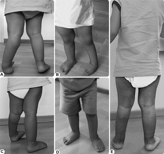
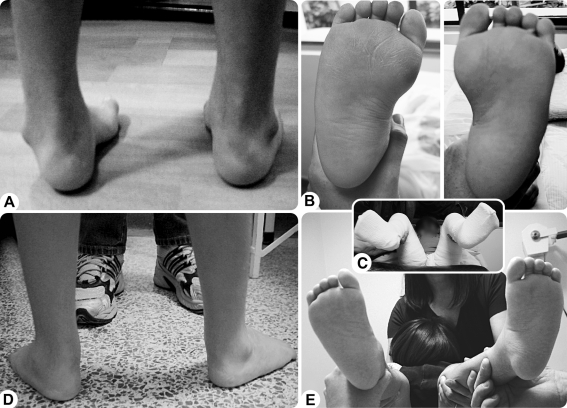
An abduction brace was used 23 hours a day for 1 to 3 months in children younger than 3 years of age and then at night only until children were 4 years old. Older children used the abduction brace at night only for 1 year or were offered anterior tibial transfer at the end of the cast phase. The angulation of the abduction in the brace was determined by the angulation obtained in the last cast when feet were considered corrected (Figs. 1, 2, 3).
Fig. 1A–E.
Photographs show a child aged 1 year 4 months with severe deformity on the left. (A) Posterior view, (B) anterior view, and (C) posterolateral view before treatment. (D) Anterior (E) and posterior view of the child’s feet after treatment.
Fig. 2A–E.
Photographs show a child aged 4 years 11 months who had previous surgery when he was 8 months old: (A) Anterior view and (B) plantar view before the surgery, (C) during surgery, and (D) posterior and (E) plantar view after treatment with eight casts.
Fig. 3A–K.
Photographs show a 5-year-old child with two previous surgeries. (A) Anterior view of the untreated feet and (B) the posterior view before treatment. (C) Anterior view of the treated feet and (D) the posterior view after treatment. (E) Plantar photograph before treatment; (F) anteroposterior radiographic view before treatment. Pretreatment photographs show a (G) Codivilla-type scar and a severe rigid cavus varus, adductus, and equinus deformity in the right foot. (H) Plantar photograph after treatment. (I) Radiographic anteroposterior and lateral views of the feet are shown; (J) the affected foot is shown; (K) the normal foot is shown.
Although the score of Pirani et al. was developed to assess virgin clubfeet and provide a quantification of the initial deformity and not designed to evaluate previously operated clubfeet [20], we chose to use the parameters of the score of Pirani et al. as a way to define the deformities in these previously operated feet. We consider the score parameters helpful to define the deformities in a clubfoot: lateral border of the foot defines adductus deformity, and reduction of calcaneocuboid joint; medial crease defines cavus deformity; talus coverage defines talonavicular reduction; and posterior crease, emptiness of the heel and reduction of equinus all relate to correction of equinus deformity.
During followup visits, we (MN, AE, CA) assessed the following parameters in all children: (1) the score of Pirani et al. (grading from 1 if needing correction to 0 if corrected for each parameter: lateral border of the foot, medial crease, talus coverage, posterior crease, emptiness of the heel, and reduction of equinus) [10, 20, 21]; (2) measurements of dorsiflexion, plantarflexion, and range of motion of the subtalar joint (using a goniometer). We also assessed muscular strength and 22 children had radiographic studies. However, neither was obtained for all patients.
In bilateral cases we considered each foot an independent observation because they did not usually have the same severity in deformity and because surgery and scar formation differed for each foot. As the score of Pirani et al. was measured initially and at final stage of correction at the same sample, the dependent variable is repeated; we therefore used a paired Student’s t-test to determine mean differences. We compared the differences in scores among the three orthopaedic surgeons performing treatment using a repeated measures analysis of variance. Using a Student’s t test we compared the group of patients that achieved a score of zero (complete correction) and the group of patients that did not achieve full correction. We correlated age with the changes (initial to final) in the score of Pirani et al. using Pearson’s correlation. Finally, we determined the relationship between number of casts (which we consider an indication of severity) and reduction of scores (expressed as percentage of reduction) using Spearman’s correlation.
Results
Of the 80 feet completing treatment 71 (89%) were plantigrade and corrected after treatment. In 11 of the original 83 feet we obtained partial corrections. Final dorsiflexion was on average 9° (range, 0°–25°) and plantar flexion was on average 31° (range, 0°–55°). Sixty-four feet (80%) had at least 30° of subtalar motion at the end of treatment. One child underwent a new posteromedial release in one foot. This child had no improvement of the varus after 10 casts, and during surgical exploration, there was fusion of the subtalar joint in varus.
All children (excluding the one that had undergone a new posteromedial release) had reductions in Pirani score. We observed a reduction (3.6 points; p < 0.001) in the mean score of Pirani et al.
We observed no correlation (p = 0.16) between age of the patients at the beginning of treatment and changes in scores.
We observed no correlation (p = 0.16) between number of casts and reduction of scores (percentage of reduction). The number of casts also did not correlate (r = 0.27; p = 0.02) with the initial score.
Patients who had full correction had lower (p = 0.002) mean initial scores than those who did not achieve full correction (3.32 versus 4.14, respectively).
We observed no difference (p = 0.799) between initial and final mean scores between the orthopaedic surgeons (MPN, CGA, AEB).
Recurrences occurred in 12 feet (14%) in nine children. In three children (five feet), recurrences could be attributed to the incorrect use of the recommended abduction brace (resolved with casts). One child stopped the treatment as a result of a torus tibial fracture not related to the abduction brace (treated with one cast); five children (six feet) had dynamic varus and adduction treated with anterior tibial transfer, three feet from those also with lateral column shortening, fasciotomy, and calcaneus tendon lengthening. In one child, deformity recurred 3 years after bilateral anterior tibial transfer and Achilles tendon lengthening. She was treated with two casts on both feet, and they became plantigrade with no dorsiflexion. One child had a planovalgus foot after correction of clubfoot with the technique requiring an insole supporting the medial longitudinal arch, but she has no pain 4 years after treatment. No child had callosities at the end of treatment, and none used special shoes. Two of the 55 children had pain at the end of treatment.
Discussion
The Ponseti technique for treating clubfoot has been popularized for idiopathic clubfoot and more recently several syndromic causes of clubfoot. Believing it might be useful for recurrent clubfoot after failed posteromedial release, we began using it for that purpose in 2001. In this study we specifically asked whether (1) correction (based on the score of Pirani et al.) could be achieved in recurrences following posteromedial releases, (2) age at treatment, the numbers of casts, or the initial score of Pirani et al. influenced the ability to correct the feet, and (3) the treatment was reproducible in three centers.
One of the major limitations of this study was the difficulty identifying a proper score for severity. We chose the score of Pirani et al. [9, 21, 22] as a primary outcome measure. This measure was originally described for assessing the severity of untreated clubfeet (“virgin clubfeet”) in infants, not for outcomes. However, we believe this score provides a surrogate for the severity of the deformity. The score is limited in assessing the hindfoot in surgically treated clubfeet, as the palpation of the calcaneus and even the posterior crease are almost always scored as 0. We were not able to accurately measure or document the stretching of the scars and the gains in suppleness of the feet although we qualitatively made this important observation in the 71 cases that were fully corrected. After presentation of some of these patients in 2007 [7, 12], one report described correction of previously operated feet. However, the number of patients in that study is small (11 children, 17 feet), the followup was inadequate to draw any definitive conclusions, and the authors did not describe how they evaluated the final correction [13]. A limitation of our study is also a relatively short followup, as further recurrences may occur beyond the minimum 24 month followup. It would be important to analyze the recurrences in this group of patients, as most of them will be occurring outside of the period when the biology of deformity is believed to be more active. Finally, we did not know the details of the posteromedial release in each foot. Whether the interosseous ligament was or was not sectioned and other details of the surgery could influence the outcomes of conservative treatment with serial castings by the Ponseti technique. If we had that information and sufficient power it would be hypothetically possible, for example, to predict the tendency of a foot to become planovalgus and then be able to stop as soon as the foot was plantigrade, not overcorrecting it with more casts.
Although the Ponseti method has not been described for operated feet, our results demonstrate that it is possible to use its principles to improve or correct relapsed or uncorrected clubfeet presenting after a previous posteromedial release. The knowledge of the biomechanics and kinematics of the subtalar joint [8, 17, 23, 24] is helpful in understanding the pathology and treatment of clubfoot, but can not fully explain how Ponseti technique overcomes the fibrosis that hypothetically involves the joints of surgically treated clubfeet. We believe that the scars and fibrotic tissues are stretched, allowing the motion and remodeling of bones and cartilage.
We observed an improvement of all feet evaluated by the score of Pirani et al., a finding observed in many different treatment centers considering idiopathic clubfeet treated by the Ponseti method (Table 1) [1, 6, 7, 9–14, 16–19, 22, 24–27].
Table 1.
Summary of the results described in the literature when applying the Ponseti method to treat clubfoot
| Author | Number of patients | Number of clubfeet | Type of clubfeet | Age at presentation | Number of casts | % initial correction | Followup | Recurrence rate | Evaluation of results |
|---|---|---|---|---|---|---|---|---|---|
| Changulani et al. [6] | 66 | 100 | Idiopathic | mean, 12 weeks | mean, 6 | 96% | 18 months | 32% | Pirani score |
| Docker et al. [9] | 50 | 77 | Idiopathic | N/A | N/A | 86% (for one of the groups) | N/A | N/A | Pirani score |
| Dyer and Davis [10] | 47 | 70 | Idiopathic | N/A | mean, 5.31 | N/A | N/A | N/A | Pirani score |
| Eberhardt et al. [11] | 29 | 41 | Idiopathic | N/A | N/A | 95,1% | 9.1 months | N/A | Pirani score |
| Garg and Dobbs [13] | 11 | 17 | Uncorrected clubfeet after PMR | mean, 4.59 years | mean, 2.81 (± TAL and ± TAT) | 100% | 27.1 months | N/A | Shoe-wear and pain |
| Gupta et al. [14] | 96 | 154 | Idiopathic and syndromic | 0–9 months | mean, 4.9 | N/A | > 6 months | 5.84% | Pirani score |
| Laaveg and Ponseti [16] | 71 | 104 | Idiopathic | 6.9 weeks | mean, 9 | 100% | mean age, 18.8 years at followup | 47% | Functional rating and radiologic measurements |
| Lavy et al. [17] | 307 | 482 | N/A | 2.8 months | N/A | 68% | N/A | N/A | N/A |
| Lourenço and Morcuende [18] | 17 | 24 | Idiopathic after walking age | 3.9 years | mean, 9 | 79% | 3.1 years | 29% | Radiologic measurements and evaluation of cosmesis and gait |
| Morcuende et al. [19] | 157 | 216 | Idiopathic | 81% younger than 6 months and 19% older than 6 months | 1–7 | 98% | N/A | 11% | N/A |
| Ponseti et al. [24] | 50 | 75 | Complex atypical clubfeet | 3 months | mean, 5 | 96% | mean, 23 months of age at last followup | 14% | ROM |
| Radler et al. [25] | 37 | 59 | Idiopathic | < 3 weeks | N/A | 93% | N/A | 3.6% | Pirani and radiologic measurements |
| Nogueira et al. (current study) | 58 | 83 | Uncorrected clubfeet after PMR | 5 years and 2 months | mean, 4 | 85.5% | 45 months | 14% | Pirani score and ROM |
N/A = not applicable or not available; ROM = range of motion.
The trend to a slightly worse score in the most severe feet could be the result of the criterion of palpation of the calcaneus that is always higher on those severe feet. However, this difference was not observed clinically in those operated patients. Contrary to the general belief that the older the patient, the more difficult the correction, we observed no relationship between outcome scores and age, or outcome score and number of casts, as findings also described by two other studies [18, 19].
We found no relationship between the initial severity and the numbers of casts. These findings are different from observations by Dyer and Davis in 70 clubfeet treated by the Ponseti technique; they reported a correlation between the number of casts needed and severity of the initial deformity [10]. These findings may be correlated by a more predictable evolution of the reduction of Pirani scores in time in the idiopathic never-operated feet. The operated feet are heterogeneous and have a more unpredictable behavior undergoing correction.
We found no difference in the final scores obtained by the three surgeons in different geographical locations. These findings suggest that the technique is reproducible in different centers.
One difficulty in applying the Ponseti treatment to older children is related with the use of long-leg casts, restricting ambulation and mobility. This can be overcome by motivating the families.
The Ponseti technique can be applied in the treatment of clubfeet recurring after posteromedial release and achieve correction in most patients. We believe these preliminary results are encouraging and may contribute to motivate other groups to follow this approach, decreasing the number of children requiring further surgery after a failed previous posteromedial release.
Acknowledgments
We thank Luis Felipe C. Neves, MD, Waldir Wilson Vilela Cipola, MD, Paulo Oliveira Machado, MD, José I. Yebra, MD, Laia S. Cequier, MD, and Marta V. Pujol, MD, for the institutional support in developing this manuscript. We also thank Harold van Bose for the English editing and reviewing the first version of this manuscript, and Valeria Baltar for the repeated statistical analysis of this study.
Footnotes
Each author certifies that he or she has no commercial associations (eg, consultancies, stock ownership, equity interest, patent/licensing arrangements, etc) that might pose a conflict of interest in connection with the submitted article.
Each author certifies that his or her institution either has waived or does not require approval for the human protocol for this investigation and that all investigations were conducted in conformity with ethical principles of research.
References
- 1.Abdelgawad AA, Lehman WB, Bosse HJ, Scher DM, Sala DA. Treatment of idiopathic clubfoot using the Ponseti method: minimum 2-year follow-up. J Pediatr Orthop B. 2007;16:98–105. doi: 10.1097/BPB.0b013e32801048bb. [DOI] [PubMed] [Google Scholar]
- 2.Aronson J, Puksarich CL. Deformity and disability from treated clubfoot. J Pediatr Orthop. 1990;10:109–112. [PubMed] [Google Scholar]
- 3.Atar D, Lehman WB, Grant AD. Complications in clubfoot surgery. Orthop Rev. 1991;20:233–239. [PubMed] [Google Scholar]
- 4.Boehm S, Limpaphayom N, Alaee F, Sinclair MF, Dobbs MB. Early results of the Ponseti method for the treatment of clubfoot in distal arthrogryposis. J Bone Joint Surg Am. 2008;90:1501–1507. doi: 10.2106/JBJS.G.00563. [DOI] [PubMed] [Google Scholar]
- 5.Bor N, Herzenberg J, Frick SL. Ponseti management of clubfoot in older infants. Clin Orthop Relat Res. 2006;444:224–228. doi: 10.1097/01.blo.0000201147.12292.6b. [DOI] [PubMed] [Google Scholar]
- 6.Changulani M, Garg NK, Rajagopal TS, Bass A, Nayagam SN, Sampath J, Bruce CE. Treatment of idiopathic clubfoot using the Ponseti method. Initial experience. J Bone Joint Surg Br. 2006;88:1385–1387. doi: 10.1302/0301-620X.88B10.17578. [DOI] [PubMed] [Google Scholar]
- 7.Cooper DM, Dietz FR. Treatment of idiopathic clubfoot: a thirty-year follow-up note. J Bone Joint Surg Am. 1995;77:1477–1489. doi: 10.2106/00004623-199510000-00002. [DOI] [PubMed] [Google Scholar]
- 8.Dobbs MB, Nunley R, Schoenecker PL. Long-term follow-up of patients with clubfeet treated with extensive soft-tissue release. J Bone Joint Surg Am. 2006;88:986–996. doi: 10.2106/JBJS.E.00114. [DOI] [PubMed] [Google Scholar]
- 9.Docker CE, Lewthwaite S, Kiely NT. Ponseti treatment in the management of clubfoot deformity—a continuing role for paediatric orthopaedic services in secondary care centres. Ann R Coll Surg Engl. 2007;89:510–512. doi: 10.1308/003588407X187739. [DOI] [PMC free article] [PubMed] [Google Scholar]
- 10.Dyer PJ, Davis N. The role of the Pirani scoring system in the management of clubfoot by the Ponseti method. J Bone Joint Surg Br. 2006;88:1082–1084. doi: 10.1302/0301-620X.88B8.17482. [DOI] [PubMed] [Google Scholar]
- 11.Eberhardt O, Schelling K, Parsch K, Wirth T. Treatment of congenital clubfoot with the Ponseti method. Z Orthop Ihre Grenzgeb. 2006;144:497–501. doi: 10.1055/s-2006-942239. [DOI] [PubMed] [Google Scholar]
- 12.Ey AMB, Yebra JI, Cequier LS, Pujol MV, Iglesia DG. Application of Ponseti method for relapses alter posteromedial release in clubfoot. J Child Orthop EPOS. 2007;1(Suppl 1):25. [Google Scholar]
- 13.Garg S, Dobbs MB. Use of the Ponseti method for recurrent clubfoot following posteromedial release. Indian J Orthop. 2008;42:68–72. doi: 10.4103/0019-5413.38584. [DOI] [PMC free article] [PubMed] [Google Scholar]
- 14.Gupta A, Singh S, Patel P, Patel J, Varshney MK. Evaluation of the utility of the Ponseti method of correction of the clubfoot deformity in a developing nation. Int Orthop. 2008;32:75–79. doi: 10.1007/s00264-006-0284-7. [DOI] [PMC free article] [PubMed] [Google Scholar]
- 15.Hatt RN, Lamphier TA. Triple hemisection: a simplified procedure for lengthening the Achilles tendon. N Engl J Med. 1947;236:166–169. doi: 10.1056/NEJM194701302360502. [DOI] [PubMed] [Google Scholar]
- 16.Laaveg SJ, Ponseti IV. Long-term results of treatment of congenital clubfoot. J Bone Joint Surg Am. 1980;62:23–31. [PubMed] [Google Scholar]
- 17.Lavy CB, Mannion SJ, Mkandawire NC, Tindall A, Steinlechner C, Chimangeni S, Chipofya E. Club foot treatment in Malawi—a public health approach. Disabil Rehabil. 2007;29:857–862. doi: 10.1080/09638280701240169. [DOI] [PubMed] [Google Scholar]
- 18.Lourenço AF, Morcuende JA. Correction of neglected idiopathic club foot by the Ponseti method. J Bone Joint Surg Br. 2007;89:378–381. doi: 10.1302/0301-620X.89B3.18313. [DOI] [PubMed] [Google Scholar]
- 19.Morcuende JA, Dolan LA, Doetz FR, Ponseti IV. Radical reduction in the rate of extensive corrective surgery for clubfoot using the Ponseti method. Pediatrics. 2004;113:376–380. doi: 10.1542/peds.113.2.376. [DOI] [PubMed] [Google Scholar]
- 20.Pirani S, Hodges D, Sekeramayi F. A reliable method of assessing the amount of deformity. SICOT/SIROT—XXII World Congress 2002 [abstract].
- 21.Pirani S, Outerbridge HK, Sawatzky B, Stothers K. A reliable method of clinically evaluating a virgin clubfoot evaluation. 1999. 21st SICOT Congress.
- 22.Ponseti IV. Treatment of congenital clubfoot. J Bone Joint Surg Am. 1992;74:448–454. [PubMed] [Google Scholar]
- 23.Ponseti IV. Congenital Clubfoot: Fundamentals of Treatment. Oxford, UK: Oxford University Press; 1996. [Google Scholar]
- 24.Ponseti IV, Zhivkov M, Davis N, Sinclair M, Dobbs M, Morcuende J. Treatment of idiopathic complex clubfoot. Clin Orthop Relat Res. 2006;451:171–176. doi: 10.1097/01.blo.0000224062.39990.48. [DOI] [PubMed] [Google Scholar]
- 25.Radler C, Suda R, Manner HM, Grill F. Early results of the Ponseti method for the treatment of idiopathic clubfeet. Z Orthop Ihre Grenzgeb. 2006;144:80–86. doi: 10.1055/s-2006-921413. [DOI] [PubMed] [Google Scholar]
- 26.Segev E, Keret D, Lokiec F, Yavor A, Wientroub S, Ezra E, Hayek S. Early experience with the Ponseti method for the treatment of congenital idiopathic clubfoot. Isr Med Assoc J. 2005;7:307–310. [PubMed] [Google Scholar]
- 27.Terrazas LG, Morcuende JA. Effect of cast removal timing in the correction of idiopathic clubfoot by the Ponseti method. Iowa Orthop J. 2007;27:24–27. [PMC free article] [PubMed] [Google Scholar]


