Abstract
Primary cardiac tumors are rare and are diverse in histology and anatomic origin. Approximately 75% are benign, and nearly 50% of these are myxomas. Herein, we report concurrent myxoma and papillary fibroelastoma, which tumors were found attached to the left atrial septum and aortic valve, respectively. Concurrent primary cardiac tumors of differing histology and origin are rare, and, to our knowledge, this is one of the few such cases reported in the medical literature.
Key words: Echocardiography; fibroma; heart neoplasms/primary/diagnosis/surgery; myxoma; neoplasms, multiple primary/diagnosis/surgery
Primary cardiac tumors are rare, with an incidence ranging from 0.0017% to 0.19% in autopsy series in unselected patients.1–3 Myxomas are the most common cardiac neoplasm, accounting for as many as 50% of all benign tumors.4 Papillary fibroelastoma, the 3rd most common cardiac neoplasm, occurs in adults and is frequently diagnosed postmortem.5 Concurrent primary cardiac tumors of differing histology and origin are rare, and few cases have been reported in the medical literature. We are reporting a case of concurrent intracardiac myxoma and papillary fibroelastoma.
Case Report
In September 2007, a 65-year-old Hispanic woman with a medical history significant for type 2 diabetes mellitus, hypertension, diabetic neuropathy, and dyslipidemia presented with exertional shortness of breath of 3 months' duration, accompanied by occasional palpitations and dizziness. The patient denied any chest pain, orthopnea, or paroxysmal nocturnal dyspnea. She denied any history of cardiac tumors, coronary artery disease, pulmonary disease, or cancer. She also denied smoking, alcohol intake, or illicit-drug use. Physical examination showed a blood pressure of 166/64 mmHg, a temperature of 98.2 °F, and a heart rate of 76 beats/min. There were no clinical features suggestive of familial myxomas (such as Carney complex or familial autosomal dominant syndrome). The cardiac examination revealed normal heart sounds and a diastolic rumbling murmur best heard at the apex. Lung examination revealed bilateral normal vesicular breathing sounds. No genetic testing was done, because the patient's history and clinical features were unremarkable. Transthoracic echocardiography (TTE) revealed a 2.2 × 3.5-cm mass in the left atrium.
Cardiac angiography performed before surgery showed normal coronary arteries with preserved left ventricular systolic function. Continued filming during the levophase showed the left atrium with a filling defect that was consistent with left atrial myxoma (Fig. 1).
Fig. 1. Cardiac angiography (during levophase) shows the left atrium with a filling defect that is consistent with left atrial myxoma.
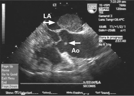
Subsequently, intraoperative transesophageal echocardiography (TEE) showed 2 masses, one in the left atrium attached to the interatrial septum in the region of the fossa ovalis, and the other attached to the right coronary cusp of the aortic valve (Figs. 2 and 3).
Fig. 2. Intraoperative transesophageal echocardiography (long-axis view) shows myxoma (arrow) in the left atrium (LA) and papillary fibroelastoma (arrow) attached to the noncoronary cusp of the aortic valve (Ao).
Fig. 3. Intraoperative transesophageal echocardiography (short-axis view) shows left atrial (LA) myxoma (arrow) in addition to papillary fibroelastoma (arrow) on the aortic valve (Ao).
Although our patient had been scheduled for a left thoracotomy to enable resection of the atrial myxoma, we made an intraoperative decision, consequent to the incidental finding of the aortic valve mass, to resect that tumor and the atrial myxoma at the same time, via a median sternotomy. The patient was placed on cardiopulmonary bypass, and an incision was made in the mid-septum and extended to the superior septum and the roof of the left atrium. The left atrial myxoma was identified and resected, together with its margin. Then a very small aortotomy revealed a mobile mass on the noncoronary aortic leaflet, which was resected for pathologic examination. The atrial myxoma was 3.1 × 3 × 1.5 cm and weighed 13 g. Hematoxylin and eosin staining of a specimen showed a myxoid matrix dominating a hypocellular tumor (Fig. 4).
Fig. 4. Hematoxylin and eosin stain (orig. ×200) shows a myxoid matrix dominating a hypocellular tumor. The tumor cells are in spindle, stellate, and epithelioid shapes. They are mixed with scattered lymphocytes and red blood cells.
An excised aortic valve mass was 0.5 × 0.1 × 0.1 cm. Hematoxylin and eosin staining showed branching papillae, and the mass was composed of central avascular collagen and variable elastic tissue, surrounded by acid mucopolysaccharide and endothelial cells (Fig. 5). The patient recovered from the procedure without sequelae. Her hospital course was uneventful, and she was discharged home. The patient was doing well at her most recent follow-up visit, 2 years after the operation.
Fig. 5. Hematoxylin and eosin stain (orig. ×40) shows papillary fibroelastoma with branching papillae, composed of central avascular collagen and variable elastic tissue, surrounded by acid mucopolysaccharide and endothelial cells.
Discussion
Myxoma can be seen in patients of any age, but most commonly between the ages of 30 and 60 years.5 These tumors are usually discovered incidentally while investigating unrelated symptoms or obtaining a baseline TTE. Most myxomas are isolated, and they are familial in less than 10% of cases. They are found mostly in the left atrium, but there have been reports of myxoma in the right atrium and in the left or right ventricles.5,6
Papillary fibroelastomas can also occur at any age but are usually seen in a population older than the age associated with myxoma. Unlike myxomas, papillary fibroelastomas are most often discovered after embolization has occurred.7–9 On the other hand, there have been reports of cases that were discovered incidentally by means of TTE or during autopsy. The occurrence of these 2 very different tumors together is rather rare. Upon review of the literature, we found 4 cases of concurrent primary cardiac tumors of differing histology and origin. Agaimy and Mandl10 reported a case in which papillary fibroelastoma of the aortic valve was found to exist concurrently with a cystic tumor (mesothelioma) of the atrioventricular nodal region. Prifti and associates11 reported a case in which 2 tumors were found on the mitral valve: a myxoma on the atrial surface and a papillary fibroelastoma on the ventricular surface. Akiyama and colleagues12 reported a case of 3 concurrent cardiac tumors: 1 left atrial myxoma and 2 papillary fibroelastomas.
Our review of other cases has placed emphasis on a crucial point: although concurrent primary cardiac tumors are rare, the possibility of additional tumors requires thorough preoperative (or intraoperative) evaluation in any patient who has a primary cardiac tumor. Our patient underwent TTE, which showed left atrial myxoma. Preoperative evaluation by TEE or by cardiac magnetic resonance imaging might have helped the cardiothoracic surgeon to identify aortic fibroelastoma and to plan the surgical approach accordingly. Although a left atrial myxoma can be approached less invasively via a right thoracotomy, the presence of concurrent tumors would require sternotomy for excision. The existence of any cardiac tumor warrants additional investigation before or during surgery.
Footnotes
Address for reprints: Fayez Shamoon, MD, Department of Cardiology, Saint Michael's Regional Medical Center, Seton Hall University, 111 Central Ave., Newark, NJ 07102
E-mail: fshamoon@aol.com
References
- 1.Straus R, Merliss S. Primary tumor of the heart. Arch Pathol 1945;39:74–8.
- 2.Heath D. Pathology of cardiac tumors. Am J Cardiol 1968;21 (3):315–27. [DOI] [PubMed]
- 3.Majano-Lainez RA. Cardiac tumors: a current clinical and pathological perspective. Crit Rev Oncog 1997;8(4):293–303. [DOI] [PubMed]
- 4.Silverman NA. Primary cardiac tumors. Ann Surg 1980;191 (2):127–38. [DOI] [PMC free article] [PubMed]
- 5.MacGowan SW, Sidhu P, Aherne T, Luke D, Wood AE, Neligan MC, McGovern E. Atrial myxoma: national incidence, diagnosis and surgical management. Ir J Med Sci 1993;162 (6):223–6. [DOI] [PubMed]
- 6.McAllister HA Jr, Hall RJ, Cooley DA. Tumors of the heart and pericardium. Curr Probl Cardiol 1999;24(2):57–116. [PubMed]
- 7.Sun JP, Asher CR, Yang XS, Cheng GG, Scalia GM, Massed AG, et al. Clinical and echocardiographic characteristics of papillary fibroelastomas: a retrospective and prospective study in 162 patients. Circulation 2001;103(22):2687–93. [DOI] [PubMed]
- 8.Howard RA, Aldea GS, Shapira OM, Kasznica JM, Davidoff R. Papillary fibroelastoma: increasing recognition of a surgical disease. Ann Thorac Surg 1999;68(5):1881–5. [DOI] [PubMed]
- 9.Gowda RM, Khan IA, Nair CK, Mehta NJ, Vasavada BC, Sacchi TJ. Cardiac papillary fibroelastoma: a comprehensive analysis of 725 cases. Am Heart J 2003;146(3):404–10. [DOI] [PubMed]
- 10.Agaimy A, Mandl L. Papillary fibroelastoma of the aortic valve coincident with a cystic tumor of the atrioventricular node [in German]. Pathologe 2000;21(3):250–4. [DOI] [PubMed]
- 11.Prifti E, Bonacchi M, Salica A. Mitral valve myxoma concomitant with papillary fibroelastoma. Ann Thorac Surg 2000; 70(1):335–6. [DOI] [PubMed]
- 12.Akiyama K, Hirota J, Tsuda Y, Ebishima H, Li C. Double primary cardiac tumors: possible association with a variety of cardiac diseases. J Cardiovasc Surg (Torino) 2006;47(1):81–2. [PubMed]


