

Abstract
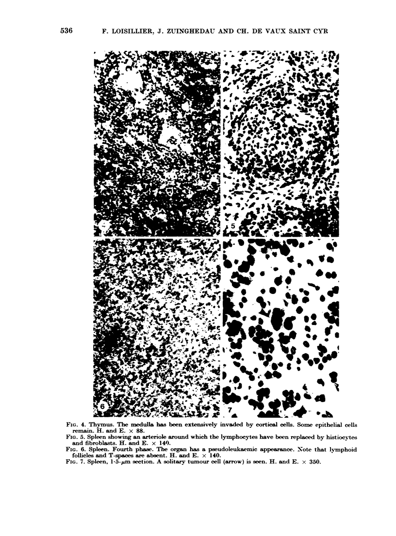
A systematic histological study of the lymphoid organs of the Syrian hamster was undertaken during development of a tumour induced with SV40 transformed cells. The essential features were: (1) A peritumoral plasma-cell reaction was present until the tumour reached a weight of 4-6 g; then it disappeared. (2) In the thymus an initial increase in mass with progressive infiltration of cortical cells into the medullary region was noted. Coincidentally with the disappearance of peritumoral plasma cells, thymus size descreased. As tumour growth proceeded, the medulla was progressively invaded by cortical and blood-borne cells which eventually made up more than 95% of the total population. (3) The spleen showed sequential changes. An early period of mild banal inflammation characterized by scattered immunoblasts in the reticulum was followed by a reduction in the number of lymphoid follicles, a loss of T lymphocytes in the periarteriolar areas and the appearance in the reticulum of young lymphocytic cells. This change coincided in time with disappearance of the peritumoral plasma cells. This change in turn gave way to a final stage characterized by progressive accumulation in the reticulum of young lymphoblastic cells which gave the spleen a pseudoleukaemic appearance and suggested a hyperimmune state. (4) The lymph nodes were the site of a mild plasmacytic stimulation in the region of the cortico-medullary junction; this persisted throughout tumour growth.
Full text
PDF


Images in this article


Selected References
These references are in PubMed. This may not be the complete list of references from this article.
- ELHASSAN A. M., STUART A. E. CHANGES IN THE LYMPHORETICULAR TISSUES OF MICE BEARING THE LANDSCHUETZ TUMOUR. Br J Cancer. 1965 Jun;19:343–352. doi: 10.1038/bjc.1965.41. [DOI] [PMC free article] [PubMed] [Google Scholar]
- Edwards A. J., Sumner M. R., Rowland G. F., Hurd C. M. Changes in lymphoreticular tissues during growth of a murine adenocarcinoma. I. Histology and weight of lymph nodes, spleen, and thymus. J Natl Cancer Inst. 1971 Aug;47(2):301–311. [PubMed] [Google Scholar]
- POPE J. H., ROWE W. P. DETECTION OF SPECIFIC ANTIGEN IN SV40-TRANSFORMED CELLS BY IMMUNOFLUORESCENCE. J Exp Med. 1964 Aug 1;120:121–128. doi: 10.1084/jem.120.2.121. [DOI] [PMC free article] [PubMed] [Google Scholar]
- Tilz G. P., de Vaux Saint Cyr C., Grabar P. New antigens in cells transformed by the SV40 virus. II. Evidence of their cytoplasmic localization in a cell line (TSV5 CL2) of hamster fibroblasts transformed by SV40. Int J Cancer. 1969 Sep 15;4(5):641–647. doi: 10.1002/ijc.2910040509. [DOI] [PubMed] [Google Scholar]
- Tournier P., Cassingena R., Wicker R., Coppey J., Suarez H. Etude du mécanisme de l'induction chez des cellules de hamster syrien transformées par le virus SV40. I. Propriétés d'une lignée cellulaire clonale. Int J Cancer. 1967 Mar 15;2(2):117–132. doi: 10.1002/ijc.2910020207. [DOI] [PubMed] [Google Scholar]
- WOODRUFF M. F., SYMES M. O. The significance of splenomegaly in tumour-bearing mice. Br J Cancer. 1962 Mar;16:120–130. doi: 10.1038/bjc.1962.12. [DOI] [PMC free article] [PubMed] [Google Scholar]
- de Vaux Saint Cyr C., Herbet A., Sobczak E., Wicker R., Grabar P. Antigènes nouveaux dans les cellules transformées par le SV40. I. Mise en évidence de trois nouveaux constituants antigéniques dans plusieurs lignées cellulaires d'origines différentes transformées par le SV40. Int J Cancer. 1969 Sep 15;4(5):616–625. doi: 10.1002/ijc.2910040506. [DOI] [PubMed] [Google Scholar]