Abstract
We have previously shown, in a two-limb position-matching task in human subjects, that exercise of elbow flexors of one arm led the forearm to be perceived as more extended, while exercise of knee extensors of one leg led the lower leg to be perceived as more flexed. These findings led us to propose that exercise disturbs position sense because subjects perceive their exercised muscles as longer than they actually are. In order to obtain further support for this hypothesis, in the first experiment reported here, elbow extensors were exercised, with the prediction that the exercised arm would be perceived as more flexed after exercise. The experiment was carried out under three load conditions, with the exercised arm resting on a support, with it supporting its own weight and with it supporting a load of 10% of its voluntary contraction strength. For each condition, the forearm was perceived as more extended, not more flexed, after exercise. This result was confirmed in a second experiment on elbow flexors. Again, under all three conditions the exercised arm was perceived as more extended. To explore the distribution of the phenomenon, in a third experiment finger flexor muscles were exercised. This had no significant effect on position sense at the elbow. In a fourth experiment, position sense at the knee was measured after knee flexors of one leg were exercised and, as for knee extensors, it led subjects to perceive their exercised leg to be more flexed at the knee than it actually was. Putting all the observations together, it is concluded that while the influences responsible for the effects of exercise may have a peripheral origin, their effect on position sense occurs centrally, perhaps at the level of the sensorimotor cortex.
Introduction
This is a study of proprioception, the ability to sense our own body's actions. The proprioceptive sense that has been singled out here is the sense of limb position. We have an acute position sense and when blindfolded, are able to match the position of one forearm by placement of the other arm with an accuracy of 2 deg or better (Allen et al. 2007).
It has been known for some time that limb position sense can be disturbed by fatigue from exercise (Skinner et al. 1986; Saxton & Donnelly, 1995; Brockett et al. 1997). In more recent experiments on position sense at the elbow joint, exercise of elbow flexor muscles of one arm produced position errors where the exercised arm was perceived as more extended than the unexercised arm (Allen et al. 2007). In related experiments, exercise of knee extensor muscles of one leg led the exercised leg to be perceived as more flexed than the unexercised leg (Givoni et al. 2007). In an attempt to include the two sets of observations under a single, unifying hypothesis we proposed that subjects perceived their exercised muscles to be longer than they actually were. We speculated that after exercise the sensory feedback from fatigued muscles might be larger for a given level of load. This would be interpreted by the brain as a longer muscle (Allen et al. 2007). If this was so, a logical consequence would be that fatigue of antagonist muscles should lead to position errors in opposite directions. If fatigue of elbow flexors of one arm produced position errors at the contralateral forearm in the direction of elbow extension, fatigue of elbow extensors should lead to errors in the direction of elbow flexion.
In the event, this hypothesis was not supported and errors from exercise of elbow extensors were in the direction of extension, as they had been after exercise of flexors. To test whether this was a non-specific effect on position sense at the elbow from exercising any arm muscle, exercise of finger flexor muscles was tested. Finally, in an attempt to broaden the implications of our findings, we sought evidence for similar patterns of errors in the leg after exercising knee flexor muscles. It had already been reported that exercise of knee extensors produced errors in the direction of knee flexion, the opposite of that seen at the elbow (Givoni et al. 2007). We therefore wanted to know whether the results at the elbow were distinct from those at the knee, or whether both conformed to the same general pattern.
Methods
A total of four experiments were carried out and they involved the testing of 34 subjects, 8 in each of the experiments on elbow flexors, elbow extensors and finger flexors and a further 10 subjects in the experiments on knee flexors. All subjects were young adults with 14 females and 20 males. Each subject gave written, informed consent before undertaking the experiments, which were approved by the Monash University Committee for Human Experimentation, and ethical aspects conformed to the Declaration of Helsinki.
In each experiment, subjects were required to attend a screening session in which a series of control position sense measurements at the elbow or knee were carried out. Subjects were asked to participate further only if they were able to achieve acceptable levels of reliability in their matching performance which was set at a standard deviation of matching errors of less than 5 deg. As a result, two subjects were excluded from the knee flexor experiment. In the finger flexor experiment, for one subject we were unable to achieve an acceptable level of fatigue after an extensive period of exercise, so they, too, were excluded.
Experiment 1: Position matching at the elbow after exercise of elbow extensors
Measurement of position sense at the elbow
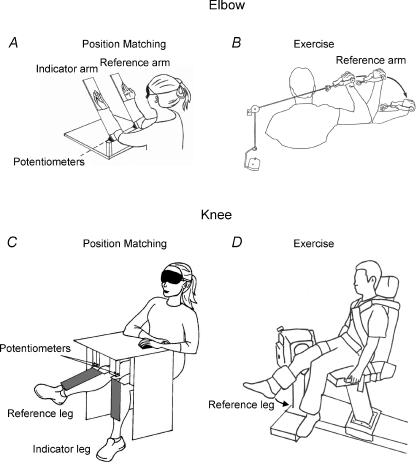
Position sense at the elbow joint in the vertical plane was measured with a custom-built piece of apparatus described previously (Allen & Proske, 2006). Subjects were blindfolded and had their forearms strapped to lightweight paddles which were hinged at a point coaxial with the elbow joint and which could be moved from the horizontal to the vertical position (Fig. 1). The upper arm rested on padded horizontal supports. Potentiometers (25 kΩ, Spectra Symbol, Salt Lake City, UT, USA) attached to the paddle hinges provided a voltage signal proportional to elbow angle. At the beginning of each experiment, potentiometer output for both paddles was checked and when calibrated gave a resolution of elbow angle of 0.2 deg. Potentiometer output was arranged so that when the forearm was horizontal, the measured angle was 0 deg, when vertical it was 90 deg.
Figure 1. Position matching at the elbow and knee.
A, position matching at the elbow. Blindfolded subjects sat at a table with their forearms strapped to lightweight paddles. The paddles were hinged at one end, the hinges aligned with the subject's elbow joint. Potentiometers attached to the paddle hinges provided a voltage signal proportional to elbow angle. B, exercise used to fatigue elbow extensors. Subjects sat in a chair with the arm to be exercised in the flexed position, resting on a horizontal support. The hand grasped a handle that was attached to a cable which ran through a pulley to a weight. Subjects were asked to slowly extend the arm to raise the weight which was adjusted to be 30% MVC. Once the arm was fully extended, the weight was lowered back to its starting position by the experimenter, ready for the next extension movement. C, position matching at the knee. Subjects were seated in a chair mounted on a steel frame. The height of the chair was adjusted so that the lateral and medial epicondyles of the knee were in line with the pivot point of the position-matching apparatus. This consisted of a pair of lightweight paddles with potentiometers at their hinge points to give a voltage output proportional to knee angle. D, exercising knee flexors with a dynamometer. Subjects were seated with the dynamometer head in line with the lateral epicondyle of the knee. A leg brace was strapped to the lower leg while hips and trunk were strapped to the chair with seatbelts. Subjects were asked to carry out concentric contractions of knee flexors in the manner of a knee curl.
In this and all other position-sense measurements at the elbow or at the knee, potentiometer output was acquired at 40 Hz using a Mac Lab running Chart software (ADInstruments, Castle Hill, NSW, Australia) on a Macintosh computer.
The test angle chosen for the position-matching trial was at approximately 45 deg to the horizontal. The actual test angle used in each trial depended on the accuracy of placement by the experimenter and the ability of the subject to maintain the arm position during the trial. Typically, angles in the range 40–50 deg were achieved.
At the beginning of each matching trial both arms were conditioned with a voluntary contraction to ensure elbow extensors were in a defined state with respect to their thixotropic property (Gregory et al. 1988). To do that, subjects placed both arms on the table in front of them and they were asked to push downwards onto the table for 2 s with an approximately half-maximal contraction. After subjects had relaxed, the experimenter moved the reference arm, the arm to be exercised, to the test angle and the subject was asked to hold it and then match its position with their other (indicator) arm. They were told to move the arm at their own pace and declare when they had achieved what they felt was a satisfactory match. The whole trial took ∼20 s. At the point at which the subject declared they had achieved a match, a marker was placed on the angle trace which was recorded continuously throughout the matching procedure. Angle values at that point were used for further analysis. Both arms always moved from an initially extended position in the direction of flexion to the test angle.
Elbow position matching was carried out under three conditions.
(1) Supported reference arm. After the conditioning contraction, subjects were asked to relax their arms and the experimenter moved the relaxed reference arm to the test angle where it was placed on a support. Subjects were then instructed to match its position with their other (indicator) arm.
To ensure that subjects remained relaxed after placement of their reference arm, they were provided with audio feedback of electromyographic (EMG) activity recorded from the surface of biceps and triceps of the reference arm and played through a loudspeaker. EMG activity was recorded using Ag–AgCl electrodes with an adhesive base and solid gel contact points (3M Health Care, London, Ontario, Canada). Electrodes were placed over the belly of each muscle. EMG was recorded at 2000 Hz and filtered at 10 Hz (high pass) and 1 kHz (low pass).
(2) Unsupported reference arm. Here the instructions were the same as in the first condition except that subjects were required to hold their reference arm at the test angle themselves (‘unsupported’) while they matched its position with the other (indicator) arm.
(3) Loaded reference arm. Here the paddle supporting the reference arm was loaded with a weight, representing approximately 10% of the maximum voluntary contraction (MVC) strength of elbow extensors. To load the extensors a counterweight was used (Walsh et al. 2006, Fig. 1). Counterweighting was achieved by attaching a rigid steel shaft directed backwards from the hinge of the reference paddle. A weight could be slid along the shaft to a position where the torque generated corresponded to a load on extensors of 10% MVC. The weight was then locked in position. When in place, the weight generated a torque in the direction of elbow flexion, thus requiring the subject to contract elbow extensors to maintain the arm at the test angle during matching.
During the matching trials, the supported and unsupported conditions were carried out first (6 trials each). The six loaded trials were carried out last because of the time it took to assemble the counterweight arrangement.
Measurement of voluntary force
To measure isometric torque at the elbow, at a given angle, the paddles were fixed in position by means of metal struts which were attached at one end to a tension transducer. The transducer consisted of four 120 Ω strain gauges (RS Components, Smithfield, NSW, Australia) in a wheatstone bridge configuration. Strain gauge output was routinely checked at the start and end of each experiment.
MVC torque was measured on three occasions: before the exercise to determine the value of the 10% load in the third condition, immediately after the exercise to determine the amount of fatigue and at the end of the position-matching trials to obtain an estimate of the amount of force recovery that had taken place during the post-exercise trials. MVC torque was measured with the paddles locked in position at the 45 deg test angle. Subjects were asked to contract their elbow extensors maximally by pushing their arms down onto the paddles while receiving verbal encouragement as well as having access to visual (computer screen display of torque) and auditory (EMG) feedback of the torque levels generated. Two, sometimes three, successive attempts at MVC measurement were made and the highest value reached was used to calculate the load for the arm.
The exercise
Subjects sat in a chair with their upper arm resting on a horizontal support with the elbow flexed. The hand grasped a handle that was attached to a cable which ran through a pulley system to a load (Fig. 1B). Subjects were asked to slowly extend the arm to raise the load which was set to be 30% MVC. Once the arm was fully extended, the experimenter lowered the weight back to its starting position and the subject was ready to carry out the next extension. Subjects did not return the weight to the starting position themselves because this would have represented an eccentric contraction for triceps and would have led to delayed muscle soreness. The number of repetitions subjects carried out to achieve the required level of fatigue (30%) depended on their fitness. We had shown previously that fatigue in the range 20–30% was necessary to achieve significant position-matching errors (Walsh et al. 2004; Givoni et al. 2007). Subjects carried out an average of 346 contractions (range, 240–420). These were done in sets of 10 repetitions with 1 min rest between sets. When subjects began to show signs of fatigue by having difficulty in raising the weight, MVC torque was remeasured. If torque had fallen by less than 30% the exercise was recommenced. This criterion was also applied to the other three experiments. If fatigue was sufficient, the second set of matching trials was begun, followed by another MVC measurement at the end of the matches to determine how much recovery from fatigue had taken place during the matches. The actual reported fatigue level for each subject was the average of the immediate post-exercise and post-matching values.
For each of the loading conditions, subjects carried out six matches before the exercise and another six afterwards, making for a total of 36 trials for the three conditions.
Experiment 2: Position matching at the elbow after exercise of elbow flexors
The position-matching procedure before and after exercise of elbow flexors was essentially the same as for elbow extensors, except that the arm was loaded, not with a cantilever arrangement, but simply by placing weights under the paddle (see Fig. 4 of Proske & Gandevia 2009). Before position matching was begun, both arms were flexion conditioned. Here, both arms were locked in position at 90 deg and subjects were asked to attempt to flex their arms using a half-maximum contraction of their elbow flexors for 2–3 s. The reference arm, the arm to be exercised, was then moved to the test angle by the experimenter and the subject was asked to match its position. MVC torque was measured by asking the subject to maximally flex their arm with the paddle locked at 45 deg (loaded matching).
To exercise elbow flexors, subjects were required to lift a weight corresponding to 30% MVC off the floor, in the process flexing their arm. After each lift the experimenter lowered the weight again to avoid eccentric flexor contractions. The number of repetitions carried out by subjects to achieve 30% fatigue was 291 (range, 200–490).
Experiment 3: Position matching at the elbow after exercise of finger flexor muscles
In this experiment the effect of finger flexor fatigue was tested on position sense at the elbow. At the start of the experiment, finger flexor MVC force was measured with a handgrip force gauge. Then subjects carried out a series of hand flexion exercises using a spring-loaded handgrip exerciser. Subjects carried out the hand flexions in sets of 10 with 10 s rest between sets followed by 1 min rest after five sets. Finger flexors were found to be more fatigue resistant than elbow muscles. To achieve a 30% fall in MVC, an average of 580 contractions were needed (range, 400–1000). Fatigue of finger flexors was also followed by a more rapid recovery of torque than for elbow muscles, although it was sufficiently slow to allow a set of measurements to be taken at the elbow while average fatigue levels remained above 20%.
Experiment 4: Position matching at the knee after exercise of knee flexors
Measurement of position sense
The apparatus for measuring position sense at the knee has been described previously (Givoni et al. 2007). Subjects were seated in an adjustable chair mounted on a steel frame. The height of the chair was adjusted so that the lateral and medial epicondyles of the knee were in line with the pivot point of each of a pair of lightweight paddles. Potentiometers at the paddle hinge points provided a voltage output proportional to knee angle (Fig. 1). The paddles rested against the front of each lower leg. Subjects wore lightweight shin guards to reduce sensations of paddle position and movement on the skin. The upper legs were held in position by knee clamps located just above the patella and the clamps were fitted once the subject was correctly seated. The initial test position for each subject was recorded and maintained throughout the experiment. Knee angle was given as degrees below the horizontal which was assigned a value of 0 deg. For the position-matching task, both legs were conditioned with an isometric contraction at 100 deg (flexion conditioning). For this, the experimenter asked the subject to contract their knee flexors (pull back both legs) against a rigid support at approximately 50% of their MVC for about 2 s. The subject was then asked to relax, while the experimenter moved one leg (reference) to the test angle of 45 deg (range, 40–50 deg). The blindfolded subject was asked to hold the reference leg, the leg to be exercised, at the test angle while they moved their other leg (indicator) to match its position. Subjects indicated that they had achieved a satisfactory match by pressing a switch. Each matching trial took ∼20 s to complete. A total of 10 matching trials was carried out before exercise and another 10 after exercise.
Measurement of voluntary force
An isokinetic dynamometer (Biodex System 3 Quickset; Biodex Medical Systems Inc., Shirley, NY, USA) was used to measure knee flexor MVC torque as well as to exercise the knee flexor muscles. To measure MVC, subjects were seated in the chair of the dynamometer (Fig. 1). The seat position was adjusted so that the pivot point of the dynamometer head was aligned with the lateral epicondyle of the knee. The leg brace was strapped to the lower leg above the ankle at 80% of the lower leg length, measured as the distance from the lateral epicondyle to the lateral malleolus. Once correctly positioned, subjects’ hips and trunk were strapped to the dynamometer chair by means of seat belts to isolate the leg and prevent upper body movement during the knee flexion contractions. Subjects were asked to generate three consecutive isometric MVCs with the knee extensors and flexors of the reference leg. To do that they were required to voluntarily flex the knee as hard as they could against the leg brace for 3 s at the test angle (45 deg), followed by ∼12 s rest. Subjects were then asked to kick their leg into extension against the brace as hard as they could to obtain an estimate of knee extensor MVC. Visual feedback of torque output, as well as verbal encouragement by the experimenter was provided during each trial.
The exercise
The exercise was also carried out on the dynamometer with subjects carrying out concentric contractions of their knee flexors in the manner of a knee curl. After each contraction the dynamometer returned to its starting position. Angular velocity was set at 60 deg s−1 and knee joint range traversed during each contraction was 10 to 90 deg of flexion, where 0 deg represented the fully extended leg. To achieve the required 30% fall in MVC torque, subjects carried out a mean of 590 contractions (range, 500–700).
Statistics
Position errors were calculated as reference limb angle minus indicator limb angle. A position-matching error of 0 deg indicated that the limbs were accurately aligned. The convention was used that a position-matching error in the direction of extension, that is, where the indicator limb was placed in a more extended position relative to the reference limb, was assigned a positive value, while an error in the direction of flexion relative to the reference limb was assigned a negative value. Angle difference values for individual trials were averaged for each subject and these were then pooled to obtain group means.
Data were analysed using Igor Pro Version 4 (Wavemetrics, Lake Oswego, OR, USA) software running on a Macintosh computer. SPSS Version 15 was used for statistical analysis. A repeated measures ANOVA with interactions was used to test for significant differences in matching errors over the two time points (pre-exercise, post-exercise) and the three loading conditions of the reference arm. For the knee flexor experiment a paired t test was used to compare errors over the two time points. A paired t test was used to test for significant changes in force over the two time points (pre-exercise, immediately post-exercise) for both the exercised and control limb. For all tests, P < 0.05 was considered statistically significant.
Results
Exercise of elbow extensors
This experiment aimed to test the hypothesis that after exercise of elbow extensors, position errors at the elbow joint were in the direction of elbow flexion, representing a longer triceps brachii muscle.
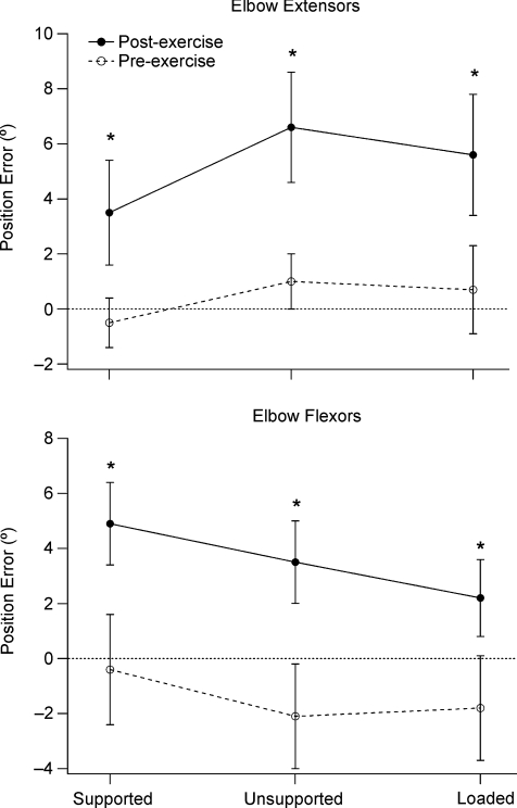
Mean force drop from exercise for the eight subjects was 28 ± 2.9%. Position errors for the group were seen to lie in the direction of extension after the exercise. To highlight the differences between the three conditions under which position sense was measured, the arm supported, unsupported and loaded, values for all three have been shown in a single plot (Fig. 2). For the supported condition the mean error during control matches of −0.5 ± 0.9 deg increased to 3.5 ± 1.9 deg after exercise. For the unsupported condition mean errors increased from 1.0 ± 1.0 to 6.6 ± 2.0 deg and for the loaded condition they increased from 0.7 ± 1.6 to 5.6 ± 2.2 deg.
Figure 2. Position errors for elbow extensors and flexors.
Upper panel: position errors before and after exercising elbow extensors, matching under different conditions. Mean position errors (±s.e.m.) for the group of subjects, before (open circles) and after (filled circles) exercise of elbow extensors of one arm, under three conditions, with the exercised reference arm lying relaxed on a support (Supported), with the subject supporting the weight of the arm themselves (Unsupported) and with the paddle holding the arm loaded with a counterweight (Loaded). Values before exercise are joined by a dashed line, values after exercise by a continuous line. Dotted line indicates zero error. Asterisks indicate significant differences between errors pre- and post-exercise. Lower panel: position errors before and after exercise of elbow flexors, matching under different conditions. Mean position errors (±s.e.m.) for the group of subjects before (open circles) and after (filled circles) exercise of elbow flexors of one arm. Matching conditions and data display as in upper panel. Asterisks indicate significant differences between errors pre- and post-exercise.
Statistical analysis using a one-way, repeated measures ANOVA showed a significant effect of exercise for all three conditions, the arm supported, unsupported or loaded (F(1,7) = 34.4; P≤ 0.05). There were no significant differences in position errors between the three load conditions (supported, unsupported and 10% MVC), or any interaction between exercise and load condition (P= 0.74).
This experiment therefore demonstrated unambiguously that exercise of elbow extensors produced errors in the direction of elbow extension. Since this was the same direction as reported previously for elbow flexors after exercise (Allen et al. 2007), the question was posed, was exercise of elbow extensors unintentionally accompanied by some fatigue of elbow flexors? The point was checked in several subjects by comparing MVC values for elbow flexors and extensors before and after exercise of the extensors. In the event, while the extensors showed the previously reported mean fall in MVC of 28%, the fall in flexor MVC was less than 5%.
Exercise of elbow flexors
Since in the previously published report on elbow flexors (Allen et al. 2007) only the unsupported condition had been tested, it was decided to repeat those observations by making measurements of position sense for all three conditions, as had been done for the elbow extensors.
For the group of eight subjects, mean MVC force drop after the exercise was 43.4 ± 7.0%. Position errors after exercise of the reference arm increased, in the direction of elbow extension, for all three conditions (Fig. 2). For the supported arm errors increased from −0.4 ± 2.0 to 4.9 ± 1.5 deg, for the unsupported arm they increased from –2.1 ± 1.9 to 3.5 ± 1.5 deg and for the loaded arm they increased from −1.8 ± 1.9 to 2.2 ± 1.4 deg.
Statistical analysis using a one-way, repeated measures ANOVA showed a significant effect of exercise on position errors for all three loading conditions (F(1,7) = 21.6; P≤ 0.05). There was no significant difference between errors for the three load conditions (supported, unsupported and 10% MVC), both before and after the exercise. There was no significant interaction between the effect of exercise on position errors and the load condition (P= 0.65).
Exercise of finger flexors
Since exercise of flexors and extensors acting at the elbow produced position errors in the same direction, it raised the question whether similar errors at the elbow could be generated by exercising any arm muscles. To test the idea, finger flexors were exercised and position-matching errors were measured at the elbow before and after exercise.
For the group of eight subjects, the exercise led to a mean force drop in finger flexors of 22.7 ± 1.9%. Since in the previous two experiments no significant differences had emerged between the three conditions, supported, unsupported and loaded, this experiment was simplified and measurements were made only with the arm supported or unsupported. After exercise of finger flexors, with the arm held supported, errors at the elbow before exercise of 0.1 ± 1.4 deg increased to 0.2 ± 2.1 deg after the exercise. This difference was not significant. For the unsupported condition, errors before exercise of 0.2 ± 1.0 deg reduced to −0.2 ± 1.6 deg. This difference, too, was not significant. It was concluded that exercising muscles not acting at the elbow joint did not alter position sense at that joint.
Exercise of knee flexors
We had previously shown that exercise of knee extensors of one leg led to errors in the direction of knee flexion (Givoni et al. 2007), the opposite from that seen at the elbow. In view of the new results at the elbow joint, we wanted to know whether exercise of knee flexors would produce errors in the same direction as for knee extensors. That prediction was tested in this experiment.
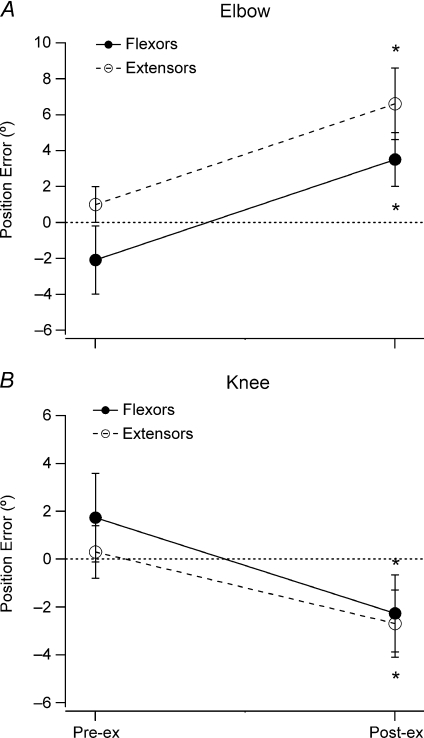
For the group of 10 subjects, exercise produced a fall in force of 31.9 ± 1.8%. In this experiment the protocol was further simplified and position matching was done using only the unsupported condition. Data for the group are shown in Fig. 3B. Small control errors in the direction of knee extension were seen before exercise (1.7 ± 1.9 deg). After exercise, the distribution of errors moved in the direction of flexion by 4 deg to −2.2 ± 1.6 deg.
Figure 3. Position matching errors at the elbow and knee before and after exercise.
A, position errors before (pre-ex) and after (post-ex) exercise of elbow flexors (filled circles, continuous line) and elbow extensors (open circles, dashed line). B, position errors before and after exercise of knee flexors (filled circles, continuous line) and knee extensors (open circles, dashed line). In both A and B matching errors are for the condition where the subject supported their exercised arm themselves. Dotted line, zero error. Values in A and for flexors in B taken from Experiments 1, 2 and 4. In B, values for extensors taken from Givoni et al. (2007). Asterisks indicate significant differences between errors pre- and post-exercise.
Statistical analysis using a paired t test (two-tailed) showed that position errors after exercise were significantly different from errors before exercise (t(9) = 3.11; P≤ 0.05). This experiment therefore demonstrated that, as for the elbow, exercising antagonists at the knee produced position-matching errors in the same direction.
Discussion
These experiments were prompted by the observation that exercise of a particular muscle group, sufficient to produce a fall in force of about 30%, leads to significant position-matching errors at the joint at which the muscle is acting. Similar observations have been made by others (Skinner et al. 1986; Saxton & Donnelly, 1995; Lattanzio et al. 1997; Fortier et al. 2009).
Interest in this subject is driven by the possibility of fatigue-related proprioceptive errors leading to injury by exercising athletes and in the elderly. Our finding that exercise of knee muscles leads to perception of a more flexed knee may mean that tiring runners will tend to overextend their knee, raising the possibility of strain injuries in knee flexors (Orchard, 2002). In the elderly, the increased frailty due to sarcopenia implies reduced proprioceptive control (Butler et al. 2008), so that any disturbance to proprioception in leg muscles from fatigue may increase the risk of falls.
In the search for an explanation for the effects of exercise on position sense, we speculated that subjects perceived the exercised muscle as longer than it actually was (Allen et al. 2007). This idea was based on the operation of an internal forward model during arm position matching (Bays & Wolpert, 2007). In a comparison between the expected afferent feedback, recalled from previous experience in a non-fatigued muscle, the larger feedback from the fatigued muscle is interpreted as the muscle being longer, that is, a more extended forearm after exercise of elbow flexors and a more flexed knee after exercise of knee extensors.
A direct test of the longer muscle hypothesis was to compare the directions of errors after exercising antagonists acting at the same joint. Exercising elbow flexors had been shown to produce errors in the direction of elbow extension (Allen et al. 2007) while exercise of elbow extensors was predicted to produce errors into flexion. The observations presented in the present study have shown that such a prediction was not supported (Fig. 2). We observed errors in the same direction from exercising each of the antagonists, both at the elbow and at the knee (Fig. 3). This led us to consider the possibility that exercise of any limb muscle can lead to a change in perceived position about a particular joint. We therefore embarked on the experiment of exercising finger flexor muscles. The negative result in that experiment served two purposes. First, it showed that exercise of muscles distant from the test joint did not lead to position errors at that joint. Whether exercise of finger flexors produces position errors at the hand remains to be shown. Second, the negative result served as a control. It could be argued that position errors reported from studies like those described here were nothing more than a gradual, spontaneous shift in matching errors over time while subjects remained strapped into the apparatus for the necessary 1–2 h. The absence of significant errors at the elbow in the finger flexor experiment made such an interpretation unlikely.
In previous experiments we had studied the effect of loading the limb on position sense at the elbow (Allen et al. 2007). The purpose had been to test whether an increase in the effort required to support the limb introduced new position errors. No new errors emerged. In that experiment elbow flexors had been loaded. When we decided to study elbow extensors, we included the loading condition to confirm that the flexor result could be extended to both antagonists as, indeed, was the case (Fig. 2).
All previous attempts at providing an explanation for the effects of exercise on limb position sense have been based on mechanisms operating at the periphery. However, animal experiments have shown that severe exercise does not disturb the normal response properties of muscle spindles (Gregory et al. 2004) or tendon organs (Gregory et al. 2002). Putting all of the observations presented here together, they have led us to the conclusion that while the factors which trigger the effects of exercise on position sense may have their origin in the periphery, they exert their influence on position-matching performance centrally, within the brain.
In framing a new hypothesis, several factors must be taken into account. First, exercising elbow muscles always produced errors in the direction of forearm extension while exercise of knee muscles produced errors in the direction of knee flexion. So the direction of the disturbance is not the same at different joints. This is unexpected and it would be interesting to know the directions of errors at other joints. Current evidence suggests that matching errors are restricted to the exercised limb; when this is acting as the reference, errors are in one direction, when it is acting as the indicator, errors lie in the opposite direction (Allen et al. 2007; Givoni et al. 2007). Finally, the available evidence suggests that exercise produces position-matching errors but not errors in movement sensation (Allen & Proske, 2006). This last observation is based on experiments using a simple manual tracking task. It deserves to be confirmed and extended.
We assign particular importance to the observation that the exercise-dependent errors in the arm were able to be expressed under all three matching conditions, with the arm held supported, unsupported or with it loaded (Fig. 2). Position errors were present even in a fully relaxed muscle, in the absence of any EMG. It is known that after exercise, in the resting subject, excitability of the region of motor cortex controlling the exercised muscle, as measured using transcranial magnetic stimulation (TMS), is seen to be reduced, as indicated by the size of the motor-evoked potential (MEP, Brasil-Neto et al. 1993). This depression lasts for at least 20 min (Sacco et al. 2000). If the fatigued muscle is made to contract, the relevant area of motor cortex is not depressed (Taylor et al. 1996) and may show an increase in excitability (Carson et al. 2002). Whether or not such excitability changes are implicated in the generation of position errors after exercise remains an open question. Our experiments have shown that position-matching errors are expressed equally well with the muscle relaxed or with it contracting. Such behaviour is not consistent with the observed excitability changes in motor cortex after exercise. It has led us to tentatively conclude that the motor cortex is not directly involved in generating the position errors.
When we experimentally disturb proprioception, for example, by vibrating a muscle, the false position signal generated by the vibration leads to perception of a longer muscle (Goodwin et al. 1972). Here, interestingly, vibration of elbow flexors leads to sensations of forearm extension while vibration of elbow extensors leads to sensations of forearm flexion. The result implies that there resides within the brain the capacity to consciously perceive the information coming from a single muscle group. It also suggests that the perceptual effects of vibration and of exercise are fundamentally different since the direction of the exercise effects is the same for flexors and extensors. The perception of a longer muscle during vibration implies that the vibration-evoked signal is being compared to some reference level, perhaps a central map of the arm, or body schema (Maravita et al. 2003). The comparison leads to a consciously perceived change in position of the arm. Are the position errors from exercise the result of an exercise-induced shift in the reference level?
Recent evidence suggests that body maps are constantly updated according to the prevailing pattern of afferent inflow. In subjects whose hand was held extended before a total nerve block, after the block had taken effect, posture of the paralysed, anaesthetised hand was perceived as slightly flexed. If the hand was held flexed before onset of the block, the final hand posture was perceived as more extended (Walsh et al. 2009). In the simplest interpretation of the observations, perceived hand posture is determined largely by activity coming from afferents in muscles stretched by that posture. As the nerve block takes effect, the stretch receptor activity falls and this fall is interpreted by the subject as a change in hand posture. The observation emphasises the lability of our body image and its dependence on immediately preceding afferent activity. It is conceivable that similar but longer lasting changes in central representation of limb position can be generated by exercise. This could be tested by experiment.
We have previously shown that the size of position errors from exercise can be correlated with the amount of fatigue (Walsh et al. 2004; Givoni et al. 2007). It may mean that processes associated with central fatigue (Meeusen et al. 2006) are implicated in the generation of the position errors. In most of our previous experiments on the effects of exercise we have used concentric exercise to fatigue the muscles. For the forearm, concentric exercise involves a shortening contraction of elbow flexors, associated with large muscle length changes and movements at the elbow joint. As a next step in our experiments we want to pose the question, is it the force being generated during the exercise and the associated fatigue that are responsible for the disturbance of position sense, or is it the accompanying movement? To answer that question, position errors will be measured after subjects have fatigued their elbow flexors by means of isometric contractions at the test angle, with no accompanying movement at the elbow joint. In a separate series the effect on position sense of repetitive movements of the passive arm will be tested.
To conclude, these experiments have shown that exercise of antagonist muscles acting at a joint produces position-matching errors in the same direction. Subjects perceive their elbow to be more extended and their knee to be more flexed. Similar errors cannot be generated by exercise of muscles remote from the test joint. The evidence points to central nervous changes, triggered by the exercise, as responsible for the errors.
References
- Allen TJ, Ansems GE, Proske U. Effects of muscle conditioning on position sense at the human forearm during loading or fatigue of elbow flexors and the role of the sense of effort. J Physiol. 2007;580:423–434. doi: 10.1113/jphysiol.2006.125161. [DOI] [PMC free article] [PubMed] [Google Scholar]
- Allen TJ, Proske U. Effect of muscle fatigue on the sense of limb position and movement. Exp Brain Res. 2006;170:30–38. doi: 10.1007/s00221-005-0174-z. [DOI] [PubMed] [Google Scholar]
- Bays PM, Wolpert DM. Computational principles of sensorimotor control that minimize uncertainty and variability. J Physiol. 2007;578:387–396. doi: 10.1113/jphysiol.2006.120121. [DOI] [PMC free article] [PubMed] [Google Scholar]
- Brasil-Neto JP, Pascual-Leone A, Valls-Sole J, Cammarota A, Cohen LG, Hallett M. Postexercise depression of motor evoked potentials: a measure of central nervous system fatigue. Exp Brain Res. 1993;93:181–184. doi: 10.1007/BF00227794. [DOI] [PubMed] [Google Scholar]
- Brockett C, Warren N, Gregory JE, Morgan DL, Proske U. A comparison of the effects of concentric versus eccentric exercise on force and position sense at the human elbow joint. Brain Res. 1997;771:251–258. doi: 10.1016/s0006-8993(97)00808-1. [DOI] [PubMed] [Google Scholar]
- Butler AA, Lord SR, Rogers MW, Fitzpatrick RC. Muscle weakness impairs the proprioceptive control of human standing. Brain Res. 2008;1242:244–251. doi: 10.1016/j.brainres.2008.03.094. [DOI] [PubMed] [Google Scholar]
- Carson RG, Riek S, Shahbazpour N. Central and peripheral mediation of human force sensation following eccentric or concentric contractions. J Physiol. 2002;539:913–925. doi: 10.1113/jphysiol.2001.013385. [DOI] [PMC free article] [PubMed] [Google Scholar]
- Fortier S, Basset FA, Billaut F, Behm D, Teasdale N. Which type of repetitive muscle contractions induces a greater acute impairment of position sense? J Electromyogr Kinesiol. 2009;20:298–304. doi: 10.1016/j.jelekin.2009.04.002. [DOI] [PubMed] [Google Scholar]
- Givoni NJ, Pham T, Allen TJ, Proske U. The effect of quadriceps muscle fatigue on position matching at the knee. J Physiol. 2007;584:111–119. doi: 10.1113/jphysiol.2007.134411. [DOI] [PMC free article] [PubMed] [Google Scholar]
- Goodwin GM, McCloskey DI, Matthews PB. The contribution of muscle afferents to kinaesthesia shown by vibration induced illusions of movement and by the effects of paralysing joint afferents. Brain. 1972;95:705–748. doi: 10.1093/brain/95.4.705. [DOI] [PubMed] [Google Scholar]
- Gregory JE, Brockett CL, Morgan DL, Whitehead NP, Proske U. Effect of eccentric muscle contractions on Golgi tendon organ responses to passive and active tension in the cat. J Physiol. 2002;538:209–218. doi: 10.1113/jphysiol.2001.012785. [DOI] [PMC free article] [PubMed] [Google Scholar]
- Gregory JE, Morgan DL, Proske U. Aftereffects in the responses of cat muscle spindles and errors of limb position sense in man. J Neurophysiol. 1988;59:1220–1230. doi: 10.1152/jn.1988.59.4.1220. [DOI] [PubMed] [Google Scholar]
- Gregory JE, Morgan DL, Proske U. Responses of muscle spindles following a series of eccentric contractions. Exp Brain Res. 2004;157:234–240. doi: 10.1007/s00221-004-1838-9. [DOI] [PubMed] [Google Scholar]
- Lattanzio PJ, Petrella RJ, Sproule JR, Fowler PJ. Effects of fatigue on knee proprioception. Clin J Sport Med. 1997;7:22–27. doi: 10.1097/00042752-199701000-00005. [DOI] [PubMed] [Google Scholar]
- Maravita A, Spence C, Driver J. Multisensory integration and the body schema: close to hand and within reach. Curr Biol. 2003;13:R531–R539. doi: 10.1016/s0960-9822(03)00449-4. [DOI] [PubMed] [Google Scholar]
- Meeusen R, Watson P, Dvorak J. The brain and fatigue: new opportunities for nutritional interventions? J Sports Sci. 2006;24:773–782. doi: 10.1080/02640410500483022. [DOI] [PubMed] [Google Scholar]
- Orchard J. Biomechanics of muscle strain injury. NZ J Sports Med. 2002;30:92–98. [Google Scholar]
- Proske U, Gandevia SC. The kinaesthetic senses. J Physiol. 2009;587:4139–4146. doi: 10.1113/jphysiol.2009.175372. [DOI] [PMC free article] [PubMed] [Google Scholar]
- Sacco P, Thickbroom GW, Byrnes ML, Mastaglia FL. Changes in corticomotor excitability after fatiguing muscle contractions. Muscle Nerve. 2000;23:1840–1846. doi: 10.1002/1097-4598(200012)23:12<1840::aid-mus7>3.0.co;2-h. [DOI] [PubMed] [Google Scholar]
- Saxton JM, Clarkson PM, James R, Miles M, Westerfer M, Clark S, Donnelly AE. Neuromuscular dysfunction following eccentric exercise. Med Sci Sports Exerc. 1995;27:1185–1193. [PubMed] [Google Scholar]
- Skinner HB, Wyatt MP, Hodgdon JA, Conard DW, Barrack RL. Effect of fatigue on joint position sense of the knee. J Orthop Res. 1986;4:112–118. doi: 10.1002/jor.1100040115. [DOI] [PubMed] [Google Scholar]
- Taylor JL, Butler JE, Allen GM, Gandevia SC. Changes in motor cortical excitability during human muscle fatigue. J Physiol. 1996;490:519–528. doi: 10.1113/jphysiol.1996.sp021163. [DOI] [PMC free article] [PubMed] [Google Scholar]
- Walsh LD, Allen TJ, Gandevia SC, Proske U. Effect of eccentric exercise on position sense at the human forearm in different postures. J Appl Physiol. 2006;100:1109–1116. doi: 10.1152/japplphysiol.01303.2005. [DOI] [PubMed] [Google Scholar]
- Walsh LD, Hesse CW, Morgan DL, Proske U. Human forearm position sense after fatigue of elbow flexor muscles. J Physiol. 2004;558:705–715. doi: 10.1113/jphysiol.2004.062703. [DOI] [PMC free article] [PubMed] [Google Scholar]
- Walsh LD, Inui N, Taylor JL, Gandevia SC. Acute changes in the body schema: development of a phantom hand. Proc Aust Neurosci Soc. 2009:P57. [Google Scholar]


