Abstract
Anomalous coronary arteries that course between the aorta and pulmonary artery are subject to compressive forces and can manifest angina, myocardial infarction and sudden death. The current report presents a young, female patient who presented with a short duration of severe, rapidly progressive angina despite optimal medical therapy. Combined computed tomography and myocardial perfusion scanning identified an anomalous dominant right coronary artery that appeared kinked at its origin between the aorta and main pulmonary artery. A robot-assisted right internal thoracic artery to right coronary artery bypass was performed, which was confirmed to be widely patent (FitzGibbon grade A) on routine intraoperative angiography. The procedure completely resolved the patient’s angina symptoms.
Keywords: Coronary artery bypass grafts, Coronary artery imaging, Coronary artery pathology, Minimally invasive surgery, Robotics
Abstract
Des artères coronaires anormales situées entre l’aorte et l’artère pulmonaire sont soumises à des forces compressives et peuvent provoquer de l’angine, un infarctus du myocarde et une mort subite. Le présent rapport présente une jeune femme qui a consulté à cause d’une grave angine de fraîche date à évolution rapide malgré une pharmacothérapie optimale. La tomodensitométrie associée à la scintigraphie myocardique de perfusion a permis de repérer une artère coronaire droite dominante anormale dont l’origine semblait coudée entre l’aorte et la principale artère pulmonaire. Un pontage coronarien de l’artère thoracique interne droite assistée par robot en a confirmé la perméabilité (score A de FitzGibbon) lors l’angiographie intraopératoire systématique. L’intervention a complètement résorbé les symptômes d’angine de la patiente.
A 37-year-old previously healthy woman presented with a six-month history of progressive exertional angina consistent with Canadian Cardiovascular Society class IVa symptoms. She denied any cardiac risk factors with the exception of her father having a myocardial infarction at 50 years of age. She had no significant medical history and her only medications were oral contraceptives. Her physical examination was unremarkable. Exercise treadmill testing suggested inferior wall ischemia with ST depression in leads II, III and aVF. A computed tomographic (CT) coronary angiography scan demonstrated an aberrant right coronary artery (RCA) that originated from the left coronary sinus adjacent to the ostium of the left main coronary artery. Imaging clearly demonstrated that the proximal portion of the RCA was ‘kinked’ (Figure 1A) due to an acute angle formed between the leftward ostial orientation, and the anteriorly and rightward-directed proximal portion, which coursed between the aorta and main pulmonary artery. Nuclear perfusion scanning confirmed the presence of inferior wall ischemia. She continued to have progressive symptoms with daily angina at rest despite optimal antianginal therapy, prompting consideration of revascularization. Because of the proximity of the RCA to the left main coronary artery, surgical options were preferred. Translocaton of the kinked RCA button was contemplated; however, coronary bypass revascularization via a minimally invasive, robotic approach was favoured because of the superior long-term outcomes of the right internal thoracic artery (RITA) to RCA anastamosis and the unique advantages of the robotic approach.
Figure 1).
A Computed tomography demonstrating ‘kinked’ anomalous origin of the right coronary artery (RCA). B Intraoperative angiogram demonstrating a widely patent right internal thoracic artery to RCA anastomosis (arrow). AO Aorta; PA Pulmonary artery
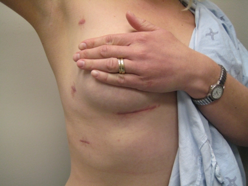
The operation was performed in a hybrid operating room where she underwent double-lumen endotracheal tube intubation and right lung collapse. The da Vinci robot was engaged at three port sites in the third, fifth and seventh intercostal spaces along the anterior axillary line, to harvest the RITA using a skeletonized technique to optimize length. The pericardium was opened longitudinally 4 cm anterior to the phrenic nerve, where the RCA was not easily visualized; therefore, the RCA was exposed through a 4 cm right inframammary crease mini-thoracotomy incision where the RITA-RCA anastomosis was constructed. Intraoperative angiography confirmed a widely patent anastomosis (FitzGibbon grade A) with excellent (Thrombolysis in Myocardial Infarction 3) flow from the graft into the dominant RCA (Figure 1B). She had an uneventful hospital course and was discharged home on the second postoperative day. At the two-month follow-up, she was well and angina free, with minimal scarring (Figure 2).
Figure 2).
Photograph of incisional scar at the two-month postoperative follow-up
DISCUSSION
Aberrant origins of the left main coronary artery from the right aortic sinus and of the RCA from the left sinus, as in our case, are the most common coronary artery anomalies and are associated with morbidity. Although poorly understood, myocardial ischemia may be the result of anomalous anatomy such as an acute angle takeoff or a proximal intermural-intervascular course of the aberrant vessel in between the aorta and pulmonary artery. The acute angle takeoff can result in a slit-like opening with a distinct possibility of collapse, and the intermural-intervascular course may predispose compression between the aorta and main pulmonary artery during exertion (1). An alternative theory of coronary hypoplasia was proposed in which an aberrant RCA arises within the aortic media, reducing its circumference and subsequent potential for growth (1). Nonetheless, it was unclear why our patient’s congenital RCA stenosis progressed so rapidly. The ‘stenosis’ at the ostium in the aberrant artery on CT often appears as a short oblique narrowed course through the aortic wall. Premature atherosclerosis may have played a role considering her father’s history; however, she did not have any other signs of atherosclerosis, nor did she have an abnormal cholesterol profile.
Failure of medical therapy, in addition to the rate of symptom progression, prompted expedited surgical intervention in our patient. Current results of percutaneous coronary intervention for aberrant origin of RCA are limited. Kansaku et al (2) concluded that coronary artery bypass grafting was the preferred treatment for aberrant RCA because of in-stent restenosis and recurrent symptoms after percutaneous coronary intervention. They postulated that frequent compression from the aorta and pulmonary trunk likely results in intimal proliferation and intrastent deformity, increasing the need for repeated revascularization (2). Theoretically, compressive forces could result in stent fracture in the long term. Various surgical techniques have been described previously, including direct reimplantation of the aberrant artery, unroofing of the intramural vessel off the aortic wall and coronary artery bypass grafting (2). While the unroofing and reimplantation technique may be theoretically preferred for younger patients, long-term patency is unknown and the potential of aortic valve distortion cannot be overlooked (2). Long-term patency of the RITA-RCA is excellent (3) and as a result, we believed it was the best choice for our patient. Because our patient was a young woman, this approach would also be amendable to a much less invasive, robot-assisted, off-pump coronary bypass, allowing her to have a quicker recovery and a 4 cm scar hidden behind a bra line. The robotic approach also provides the surgeon with superior visualization and dexterity over a thoracoscopic approach, which is important for skeletonization of the RITA graft; this is often necessary to reach the intended target. While common objections about the robotic approach include the higher capital costs, the steep learning curve for the surgical team and longer operation times, it was demonstrated that bleeding, ventilatory times, arrhythmias, hospital lengths of stay, and return to normal activity had been reduced in addition to the improved cosmesis associated with the robotic-assisted approach (4).
Finally, we were able to perform this operation without the need for a conventional coronary angiogram, reducing additional risk and expediting surgical intervention. Conventional invasive coronary angiography of an aberrant RCA can be challenging, with lengthy procedure times and suboptimal imaging. While invasive angiography could potentially describe the dynamic nature of the proximal RCA, noninvasive CT coronary angiography provided precise, high-quality images that allowed us to clearly identify the ostial RCA stenosis and confirm the large, disease-free distal RCA target preoperatively. CT coronary angiography reliably identifies areas of obstructive disease and distal targets with good sensitivity and specificity (5). Noninvasive coronary imaging modalities may play a larger role in future preoperative coronary surgery planning in selected patients.
CONCLUSION
Herein, we presented a patient with a congenital anomalous RCA correctly diagnosed using CT angiography, and treated with robot-assisted RITA-RCA bypass with symptomatic relief and excellent postoperative course.
REFERENCES
- 1.Angelini P. Coronary artery anomalies: An entity in search of an identity. Circulation. 2007;115:1296–305. doi: 10.1161/CIRCULATIONAHA.106.618082. [DOI] [PubMed] [Google Scholar]
- 2.Kansaku R, Saitoh H, Eguchi S, Maruyama Y, Ohtsuka H, Higuchi K. Advantage of vein grafts for anomalous origin of a right coronary artery. Gen Thorac Cardiovasc Surg. 2009;57:144–7. doi: 10.1007/s11748-008-0342-8. [DOI] [PubMed] [Google Scholar]
- 3.Tatoulis J, Buxton BF, Fuller JA. Patencies of 2127 arterial to coronary conduits over 15 years. Ann Thorac Surg. 2004;77:93–101. doi: 10.1016/s0003-4975(03)01331-6. [DOI] [PubMed] [Google Scholar]
- 4.Boyd WD, Kodera K, Stahl KD, Rayman R. Current status and future directions in computer-enhanced video- and robotic-assisted coronary bypass surgery. Semin Thorac Cardiovasc Surg. 2002;14:101–9. doi: 10.1053/stcs.2002.31893. [DOI] [PubMed] [Google Scholar]
- 5.Hansen M, Ginns J, Seneviratne S, et al. The value of dual-source 64-slice CT coronary angiography in the assessment of patients presenting to an acute chest pain services. Heart Lung Circ. 2010;19:213–8. doi: 10.1016/j.hlc.2010.01.004. [DOI] [PubMed] [Google Scholar]


