Abstract
Objective
We investigated the image quality of multiplanar reconstruction (MPR) using adaptive statistical iterative reconstruction (ASIR).
Methods
Inflated and fixed lungs were scanned with a garnet detector CT in high-resolution mode (HR mode) or non-high-resolution (HR) mode, and MPR images were then reconstructed. Observers compared 15 MPR images of ASIR (40%) and ASIR (80%) with those of ASIR (0%), and assessed image quality using a visual five-point scale (1, definitely inferior; 5, definitely superior), with particular emphasis on normal pulmonary structures, artefacts, noise and overall image quality.
Results
The mean overall image quality scores in HR mode were 3.67 with ASIR (40%) and 4.97 with ASIR (80%). Those in non-HR mode were 3.27 with ASIR (40%) and 3.90 with ASIR (80%). The mean artefact scores in HR mode were 3.13 with ASIR (40%) and 3.63 with ASIR (80%), but those in non-HR mode were 2.87 with ASIR (40%) and 2.53 with ASIR (80%). The mean scores of the other parameters were greater than 3, whereas those in HR mode were higher than those in non-HR mode. There were significant differences between ASIR (40%) and ASIR (80%) in overall image quality (p<0.01). Contrast medium in the injection syringe was scanned to analyse image quality; ASIR did not suppress the severe artefacts of contrast medium.
Conclusion
In general, MPR image quality with ASIR (80%) was superior to that with ASIR (40%). However, there was an increased incidence of artefacts by ASIR when CT images were obtained in non-HR mode.
Multiplanar reconstruction (MPR) of CT images plays an important role in the interpretation of the three-dimensional anatomical location or extent of disease, and is an essential technique in daily clinical practice. Multidetector row CT (MDCT) is widely used, and advances in MDCT technology have facilitated better images with thinner slice thickness and extended coverage, which has allowed more MPR images to be evaluated in greater detail. Coronal MPR images produced by MDCT supply good image quality for lung assessment and show similar image quality to axial high-resolution (HR) CT images [1,2].
To further enhance CT image quality, improvements in temporal resolution and/or spatial resolution are needed. MDCT equipped with more rapid gantry rotation or more detector arrays has already evolved for the improvement of temporal resolution. GE Healthcare (Milwaukee, WI) recently produced an MDCT unit containing a new detector composed of garnet, which has a faster response than the previous detector material. This apparatus can provide improved spatial resolution by acquiring more data.
Reconstruction algorithms are also important for improved image quality. Although the filtered back-projection algorithm has traditionally been used for image reconstruction, new reconstruction algorithms are being developed. The iterative reconstruction algorithm has already been used for image reconstruction of positron emission tomography (PET) or single photon emission CT (SPECT), resulting in improved image quality [3-6]. Iterative reconstruction was also used in early CT systems and is currently used by many manufacturers of clinical CT systems. Adaptive statistical iterative reconstruction (ASIR), recently developed for CT by GE Healthcare, is expected to improve low-contrast detectability by reducing noise when using the same radiation dose as would be used with filtered back-projection. It is also expected to reduce the radiation dose for a similar noise level compared with filtered back-projection. Until now, the quality of CT images from multiplanar reconstruction using ASIR had not been analysed. The purpose of this study was to evaluate the image quality of MPR using ASIR.
Methods and materials
The study protocol was approved by the institutional review board; the requirement for patient consent was waived. Five autopsy lungs from patients who had no history of pulmonary disease were removed from the thorax. The lungs were inflated and fixed using the method of Markarian and Dailey [7]. The lungs were distended through the main bronchus with fixative fluid that contained polyethylene glycol 400, 95% ethyl alcohol, and 40% formalin and water, in the proportions 10:5:2:3. The specimens were immersed in fixative for 2 days and the fixed lungs were then air dried.
The lungs were scanned by garnet detector CT (Discovery CT750HD, GE Healthcare) in both HR mode (2496 views) and non-HR mode (984 views). High-dose CT (400 mA tube current setting) was performed with 120 kVp, 0.984:1 pitch, 64 × 0.625 mm collimation and 0.4 s per rotation. CT images were reconstructed with 0.625 mm slice thickness, 0.625 mm interval, 20 cm field of view and the bone algorithm. Three patterns of CT images were reconstructed: without adaptive statistical iterative reconstruction (ASIR (0%)), with 40% of adaptive statistical iterative reconstruction (ASIR (40%)) and with 80% of adaptive statistical iterative reconstruction (ASIR (80%)). The level of ASIR required can be selected from 0% to 100% in steps of 10%, and represents the level of blending of ASIR and filtered back-projection techniques for image reconstruction. ASIR (0%) provides pure filtered back-projection images, ASIR (40%) provides 40% of ASIR and 60% of the filtered back-projection, and ASIR (80%) provides 80% of ASIR and 20% of the filtered back-projection. All reconstructed CT images were transferred to a workstation (Advantage Workstation 4.2, GE Healthcare), and coronal MPR images were reformatted with a section thickness of 0.6 mm and an increment of 0.6 mm.
Three MPR images from each of the five lungs were chosen from the MPR images obtained with ASIR (0%) and HR mode. The mid-MPR image of each lung with ASIR (0%) and HR mode was selected first, and two additional images about 3 cm anterior and posterior to the mid-MPR image were also selected. In total, 15 MPR images scanned with ASIR (0%) and HR mode were selected for the evaluation. The corresponding MPR images with ASIR (40%) or ASIR (80%) were also selected by two observers. Corresponding MPR images scanned with non-HR mode were also selected by the same two observers.
Two radiologists with 9 and 6 years of experience in chest CT reviewed the selected MPR images on a PACS workstation (PathSpeed, GE Healthcare). They compared MPR images of ASIR (40%) and ASIR (80%) with those of ASIR (0%) by placing the images side by side, without any prior knowledge of the image acquisition parameters. The MPR images were displayed using a window width of 1000 HU and a window level of −700 HU, and the readers graded the MPR image qualities of the following five normal structures: peripheral vessels, interlobular septa, pleura, bronchial walls and attenuations of pulmonary parenchyma. Artefacts, noise and overall image quality of each MPR image were also subjectively graded. The assessments of image quality for each item were performed independently by two readers using a visual scale (1, definitely inferior to ASIR (0%); 2, slightly inferior; 3, unchanged; 4, slightly superior; 5, definitely superior). Visual scores were summed for each item, and the mean image quality scores for the MPR images were then calculated.
Statistical analysis was performed as follows. The interobserver variation in image quality score between the two readers was evaluated using weighted κ-statistics. The interobserver agreement was classified as follows: poor, 0–0.20; fair, 0.21–0.40; moderate, 0.41–0.60; good, 0.61–0.80; and excellent, 0.81–1. The differences between the visual scores of the MPR images were analysed using the Wilcoxon signed-ranks test.
To investigate other factors that may influence the image quality of MPR, non-ionic contrast medium with an iodine concentration of 300 mg of iodine per ml (iopamidol, Iopamiron 300; Bayer HealthCare) in a 2.5 ml injection syringe was scanned with the same parameters in both HR mode and non-HR mode. MPR images with 0.6 mm slice thickness were then reconstructed with ASIR (0%), ASIR (40%) and ASIR (80%). One patient who received the same contrast medium as above was scanned with the same parameters in HR mode, and MPR images then reconstructed with ASIR (0%), ASIR (40%) and ASIR (80%). Axial and MPR images were objectively evaluated.
Results
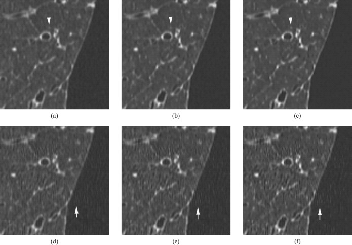
Representative MPR images are shown in Figure 1, and the MPR image quality findings are summarised in Tables 1 and 2. In HR mode, the mean scores of total image quality were 3.67 and 4.97 with ASIR (40%) and ASIR (80%), respectively. In non-HR mode, they were 2.87 and 2.53 with ASIR (40%) and ASIR (80%), respectively. The mean artefact scores in HR mode were 3.13 and 3.63 with ASIR (40%) and ASIR (80%), respectively. In non-HR mode they were 2.87 and 2.53 with ASIR (40%) and ASIR (80%), respectively. The mean scores of the other items were greater than 3, and those from HR mode were higher than those from non-HR mode. There were significant differences between ASIR (40%) and ASIR (80%) in both HR mode and non-HR mode (p<0.01).
Figure 1.
Multiplanar reconstruction (MPR) images of inflated and fixed lung in high-resolution (HR) mode (a–c) and non-HR mode (d–f). CT images were reconstructed with adaptive statistical iterative reconstruction (ASIR) (0%) (a,d), ASIR (40%) (b,e) and ASIR (80%) (c,f). Normal structures were more clearly seen using ASIR in HR mode (arrow head). Streak artefacts were more severe using ASIR in non-HR mode (arrow).
Table 1. Mean image quality scores in high-resolution mode.
| ASIR (40%) | ASIR (80%) | p-value | |
| Peripheral vessels | 3.2 | 4.0 | <0.01 |
| Interlobular septa | 3.1 | 4.1 | <0.01 |
| Pleura | 3.5 | 4.5 | <0.01 |
| Bronchial wall | 3.1 | 4.0 | <0.01 |
| Attenuation of parenchyma | 3.1 | 4.1 | <0.01 |
| Artefact | 3.1 | 3.6 | <0.01 |
| Noise | 3.9 | 5.0 | <0.01 |
| Overall image quality | 3.7 | 5.0 | <0.01 |
ASIR, adaptive statistical iterative reconstruction.
Table 2. Mean image quality scores in non-high-resolution mode.
| ASIR (40%) | ASIR (80%) | p-value | |
| Peripheral vessels | 3.0 | 3.6 | <0.01 |
| Interlobular septa | 3.1 | 4.0 | <0.01 |
| Pleura | 3.2 | 4.2 | <0.01 |
| Bronchial wall | 3.0 | 3.9 | <0.01 |
| Attenuation of parenchyma | 3.0 | 4.0 | <0.01 |
| Artefact | 2.9 | 2.5 | <0.05 |
| Noise | 3.7 | 4.6 | <0.01 |
| Overall image quality | 3.3 | 3.9 | <0.01 |
ASIR, adaptive statistical iterative reconstruction.
There was good concordance between the two observers in the assessment of interlobular septa, bronchial walls and attenuations of pulmonary parenchyma; the κ coefficient was 0.67–0.82. Moderate concordance was seen in noise and overall image quality (κ coefficient 0.41–0.47), and fair concordance was seen in peripheral vessels and pleura (κ coefficient 0.25–0.29). However, there was poor concordance in the assessment of artefacts (κ coefficient 0.07).
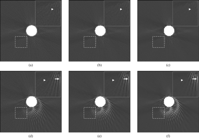
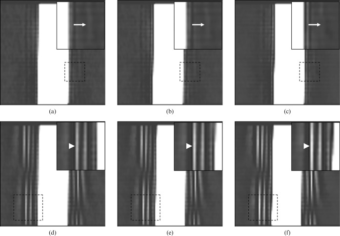
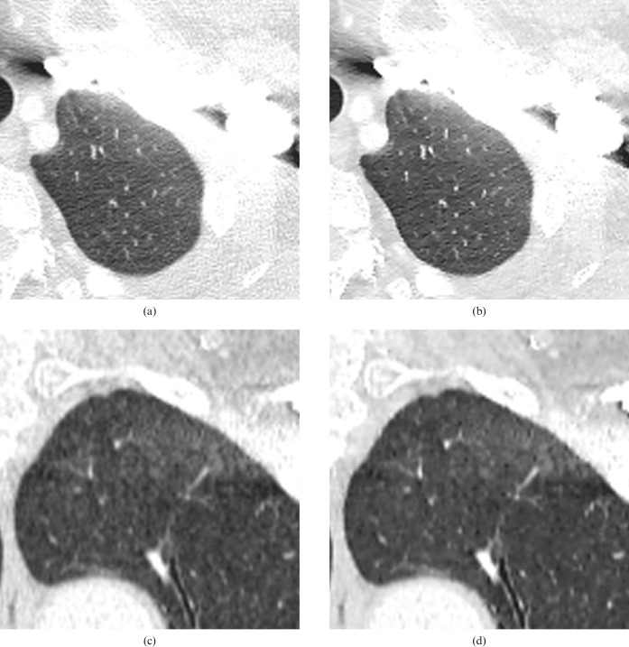
Axial images of contrast medium in the injection syringe showed that streak artefacts were prominent in non-HR mode (Figure 2). Some streak artefacts on axial images of contrast medium with ASIR (0%) were reduced on those with ASIR (80%) in non-HR mode, but some streak artefacts with ASIR (0%) were strengthened on axial images with ASIR (80%) (Figure 2d,f). The reduced streak artefacts by ASIR (80%) were mild on axial images with ASIR (0%), and the strengthened streak artefacts by ASIR (80%) were severe on axial images with ASIR (0%). Although mild artefacts were reduced and the margin of the contrast medium could be seen more clearly on the MPR images using ASIR, severe artefacts could be seen more clearly (Figure 3). The clinical data also showed that mild artefacts were reduced using ASIR, and that the normal structures, such as vessels and bronchi, could be seen more clearly using ASIR (Figure 4).
Figure 2.
Axial CT images of contrast medium in high-resolution (HR) mode (a–c) and non-HR mode (d–f). The area surrounded by a dashed line is shown, magnified, in the upper right-hand corner of the image. CT images were reconstructed with adaptive statistical iterative reconstruction (ASIR) (0%) (a,d), ASIR (40%) (b,e) and ASIR (80%) (c,f). Mild streak artefacts (arrowhead) were reduced using ASIR, but severe streak artefacts (arrow) were more prominent using ASIR, especially in non-HR mode.
Figure 3.
Multiplanar reconstruction images of contrast medium in high-resolution (HR) mode (a–c) and non-HR mode (d–f), which were obtained with adaptive statistical iterative reconstruction (ASIR) (0%) (a,d), ASIR (40%) (b,e) and ASIR (80%) (c,f). The area surrounded by a dashed line is shown, magnified, in the upper right-hand corner of the image. Mild streak artefacts (arrow) become unclear, and the margins of contrast medium become clear using ASIR in HR mode. Severe streak artefacts (arrow head) become dense and clear using ASIR in non-HR mode.
Figure 4.
Axial CT images (a,b) and multiplanar reconstruction (MPR) images of left upper lung (c,d) in high-resolution mode on enhanced CT. CT images were reconstructed with adaptive statistical iterative reconstruction (ASIR) (0%) (a,c) and ASIR (80%) (b,d). Although streak artefacts owing to contrast medium of left subclavian vein were seen in ASIR (0%) (a), mild streak artefacts were reduced in ASIR (80%) (b). Noise was reduced and the normal structures such as vessels or bronchus were more clearly seen in MPR image with ASIR (80%) (d) compared with ASIR (0%) (c).
Discussion
Currently, the filtered back-projection algorithm is the most common technique for image reconstruction of CT, and statistical iterative reconstruction has not been used in commercial CT scanners for over 30 years. Several factors contributed to the abandonment of statistical iterative reconstruction, the most important being the huge computational cost. It has been reported that the computational cost of statistical iterative reconstruction is about two to three orders of magnitude larger than that of the filtered back-projection method [8]. However, iterative reconstruction has recently become a viable option for CT images owing to increasing computational power and to speeding up of the process by the development of modern iterative reconstruction methods such as ASIR. The iterative reconstruction method now enables production of good images with enhanced image resolution, lower noise and reduced artefacts [8-10].
ASIR is usually promoted as a means to reduce the radiation dose, as a result of reduced image noise, and it has been shown to provide diagnostic quality CT images at reduced radiation doses [11]. Hara et al [12] and Prakash et al [13] used ASIR in abdominal CT and found that the radiation dose was reduced by 32–65% and 24.5–26.5%, respectively. Prakash et al [13] also reported that a radiation dose reduction of 26.8–28.8% could be obtained in chest CT using ASIR, and that ASIR improved image quality compared with conventional filtered back-projection image reconstruction [14]. Min et al [15] reported that the ASIR image reconstruction techniques enhanced in-stent assessment while decreasing image noise in a CT study of ex vivo coronary stents. Thus, ASIR can be used not only for radiation dose reduction but also improvement of image quality.
We speculated that ASIR would provide better MPR images than filtered back-projection owing to the noise reduction effect. The present study determined that ASIR does reduce the noise in MPR images of the lung, which is compatible with previous reports. It has previously been established that high-resolution CT (HRCT) using a thin slice thickness and a high spatial frequency algorithm should be applied to lung imaging to improve spatial resolution [16,17]. However, using a thin slice thickness and a high spatial frequency algorithm increases the visible image noise [17-19]. We believe that ASIR is the ideal algorithm for HRCT of the lung to reduce image noise and to obtain better image quality.
In the present study, the normal pulmonary structures, namely vessels, interlobular septa, pleura, bronchial wall and attenuation of parenchyma, were evaluated. We found that the image quality on MPR with ASIR (80%) was superior to that with ASIR (40%) in both HR mode and non-HR mode. Dot or line structures on MPR views were more clearly seen with sharp margins using ASIR, suggesting that ASIR may have an edge-enhancing effect.
We initially expected that ASIR would reduce any artefact on MPR. Interestingly, artefacts were more prominent using ASIR in non-HR mode, whereas they were reduced in HR mode (Figure 1). It is supposed that the higher sampling rates in HR mode lead to higher spatial resolution, and thus HR mode would cause less severe artefacts than non-HR mode. Our study of scanning contrast medium showed that mild artefacts were reduced using ASIR, regardless of the resolution mode (Figures 2 and 3). However, severe streak artefacts were more visible in non-HR mode than in HR mode, and were prominent when using ASIR (Figures 2d–f and 3d–f). ASIR reduces noise and allows the margin of the pulmonary structure scanned by CT to be seen more clearly. However, when using ASIR, severe streak artefacts may also be recognised as pulmonary structures, which may render severe artefacts more prominent owing to the edge-enhancement effect. In other words, ASIR reduces mild artefacts, but severe artefacts cannot be suppressed even when using ASIR. This may explain the poor concordance in the assessment of artefacts.
The present study has some limitations. First, the number of lungs was small, and fixed and inflated lungs had to be used because the necessity for two CT scan patterns (one for HR mode and one for non-HR mode) doubled the radiation dose. For assessment for clinical use, normal volunteers are required. CT images of living human lungs would have more artefacts, including respiratory or cardiac motion artefacts, which should also be investigated in the future. However, it has been reported that electrocardiographic gating improves the image quality of thin-section CT scans of the lung by reducing cardiac motion artefacts [20,21], and it is supposed that electrocardiographic gating would be useful for improving image quality even when using ASIR. The second limitation is that the MPR images were reconstructed from only three patterns: ASIR (0%), ASIR (40%) and ASIR (80%). Radiologists are not used to ASIR images that have a potentially new look [8], and axial images with ASIR (30%) or ASIR (40%) are usually used for clinical evaluation of the lung. In the present study, overall image quality of ASIR (80%) was superior to that of ASIR (40%). However, the optimal degree of ASIR for the lungs in axial images or MPR images has yet to be established. The third limitation is that only normal lung was investigated. For clinical use, the faithful representation of pulmonary disease is essential. ASIR may erase the subtle CT findings of pulmonary diseases as artefacts. Further investigation of the clinical applicability of ASIR is required.
Conclusion
Our results indicate that MPR images with ASIR (80%) are superior to those with ASIR (40%) in overall image quality, noise and visualisation of pulmonary structures. ASIR reduces slight artefacts in both HR mode and non-HR mode, although ASIR increases severe artefacts in non-HR mode. Non-HR mode has a tendency to produce more severe artefacts than HR mode under all circumstances, and thus severe artefacts are strengthened by ASIR. Therefore, although ASIR is useful for obtaining better image quality of MPR, HR mode is required to reduce streak artefacts and to obtain better MPR images.
Reference
- 1.Arakawa H, Sasaka K, Lu WM, Hirayanagi N, Nakajima Y. Comparison of axial high-resolution CT and thin-section multiplanar reformation (MPR) for diagnosis of diseases of the pulmonary parenchyma: preliminary study in 49 patients. J Thorac Imaging 2004;19:24–31 [DOI] [PubMed] [Google Scholar]
- 2.Choi EJ, Oh YW, Ham SY, Lee KY, Kang EY. Comparison between coronal reformatted images and direct coronal CT images of the swine lung specimen: assessment of image quality with 64-detector row CT. Br J Radiol 2008;81:463–7 [DOI] [PubMed] [Google Scholar]
- 3.Pretorius PH, King MA, Pan TS, de Vries DJ, Glick SJ, Byrne CL. Reducing the influence of the partial volume effect on SPECT activity quantitation with 3D modelling of spatial resolution in iterative reconstruction. Phys Med Biol 1998;43:407–20 [DOI] [PubMed] [Google Scholar]
- 4.Wells RG, King MA, Simkin PH, Judy PF, Brill AB, Gifford HC, et al. Comparing filtered backprojection and ordered-subsets expectation maximization for small-lesion detection and localization in 67Ga SPECT. J Nucl Med 2000;41:1391–9 [PubMed] [Google Scholar]
- 5.Knesaurek K, Machac J, Vallabhajosula S, Buchsbaum MS. A new iterative reconstruction technique for attenuation correction in high-resolution positron emission tomography. Eur J Nucl Med 1996;23:656–61 [DOI] [PubMed] [Google Scholar]
- 6.Riddell C, Carson RE, Carrasquillo JA, Libutti SK, Danforth DN, Whatley M, et al. Noise reduction in oncology FDG PET images by iterative reconstruction: a quantitative assessment. J Nucl Med 2001;42:1316–23 [PubMed] [Google Scholar]
- 7.Markarian B, Dailey ET. Preparation of inflated lung specimens. In: Heitzman ER. The lung: radiologic–pathologic correlations, 3rd edn. St. Louis, MO: Mosby; 1993:4–12 [Google Scholar]
- 8.Wang G, Yu H, De Man B. An outlook on x-ray CT research and development. Med Phys 2008;35:1051–64 [DOI] [PubMed] [Google Scholar]
- 9.Thibault JB, Sauer KD, Bouman CA, Hsieh J. A three-dimensional statistical approach to improved image quality for multislice helical CT. Med Phys 2007;34:4526–44 [DOI] [PubMed] [Google Scholar]
- 10.Elbakri IA, Fessler JA. Statistical image reconstruction for polyenergetic X-ray computed tomography. IEEE Trans Med Imaging 2002;21:89–99 [DOI] [PubMed] [Google Scholar]
- 11.Silva AC, Lawder HJ, Hara A, Kujak J, Pavlicek W. Innovations in CT dose reduction strategy: application of the adaptive statistical iterative reconstruction algorithm. AJR Am J Roentgenol 2010;194:191–9 [DOI] [PubMed] [Google Scholar]
- 12.Hara AK, Paden RG, Silva AC, Kujak JL, Lawder HJ, Pavlicek W. Iterative reconstruction technique for reducing body radiation dose at CT: feasibility study. AJR Am J Roentgenol 2009;193:764–71 [DOI] [PubMed] [Google Scholar]
- 13.Prakash P, Kalra MK, Kambadakone AK, Pien H, Hsieh J, Blake MA, et al. Reducing abdominal CT radiation dose with adaptive statistical iterative reconstruction technique. Invest Radiol 2010;45:202–10 [DOI] [PubMed] [Google Scholar]
- 14.Prakash P, Kalra MK, Digumarthy SR, Hsieh J, Pien H, Singh S, et al. Radiation dose reduction with chest computed tomography using adaptive statistical iterative reconstruction technique: initial experience. J Comput Assist Tomogr 2010;34:40–5 [DOI] [PubMed] [Google Scholar]
- 15.Min JK, Swaminathan RV, Vass M, Gallagher S, Weinsaft JW. High-definition multidetector computed tomography for evaluation of coronary artery stents: comparison to standard-definition 64-detector row computed tomography. J Cardiovasc Comput Tomogr 2009;3:246–51 [DOI] [PubMed] [Google Scholar]
- 16.Murata K, Khan A, Rojas KA, Herman PG. Optimization of computed tomography technique to demonstrate the fine structure of the lung. Invest Radiol 1988;23:170–5 [DOI] [PubMed] [Google Scholar]
- 17.Mayo JR, Webb WR, Gould R, Stein MG, Bass I, Gamsu G, et al. High-resolution CT of the lungs: an optimal approach. Radiology 1987;163:507–10 [DOI] [PubMed] [Google Scholar]
- 18.Zwirewich CV, Terriff B, Müller NL. High-spatial-frequency (bone) algorithm improves quality of standard CT of the thorax. AJR Am J Roentgenol 1989;153:1169–73 [DOI] [PubMed] [Google Scholar]
- 19.Mühlenbruch G, Klotz E, Wildberger JE, Koos R, Das M, Niethammer M, et al. The accuracy of 1- and 3-mm slices in coronary calcium scoring using multi-slice CT in vitro and in vivo. Eur Radiol 2007;17:321–9 [DOI] [PubMed] [Google Scholar]
- 20.Schoepf UJ, Becker CR, Bruening RD, Helmberger T, Staebler A, Leimeister P, et al. Electrocardiographically gated thin-section CT of the lung. Radiology 1999;212:649–54 [DOI] [PubMed] [Google Scholar]
- 21.Nishiura M, Johkoh T, Yamamoto S, Honda O, Kozuka T, Koyama M, et al. Electrocardiography-triggered high-resolution CT for reducing cardiac motion artefact: evaluation of the extent of ground-glass attenuation in patients with idiopathic pulmonary fibrosis. Radiat Med 2007;25:523–8 [DOI] [PubMed] [Google Scholar]


