Abstract
Purpose
Pre- and postoperative tibial posterior slope angles (PSAs) were assessed in patients who underwent cruciate-retaining total knee arthroplasty (TKA).
Material and methods
A total of 386 cruciate retaining TKA were performed in 308 patients and retrospectively reviewed. Based on the prostheses, 202 cases using NexGen® were classified as group I, 120 cases using PFC sigma® as group II, and 64 cases using Vanguard® as group III. Postoperative PSA of groups I, II, and III were compared.
Results
In groups I, II, and III, postoperative PSA was 6.0˚, 6.0˚, and 4.5˚, respectively (p < 0.001). Between preoperative measurement and final follow-up examination, mean knee score (59.7 to 97.3), function score (54.2 to 90.5), and range of motion (ROM; 126.7° to 132.2°) improved. These three values did not differ significantly among groups.
Conclusions
The 3° slope of the Vanguard® polyethylene insert caused the difference in PSAs. This design characteristic should be considered when using this implant in TKA.
Introduction
The tibial posterior slope angle (PSA) influences the flexion gap, the tension of the posterior cruciate ligament (PCL), and knee stability following total knee arthroplasty (TKA). The achievement of an appropriate PSA facilitates femoral rollback and increases the range of motion (ROM) of the knee. Several studies [6, 13, 18, 19] have reported mean PSA values in the normal knee ranging from 7–14.8°. Following cruciate-retaining TKA, proposed optimal mean PSA values have ranged from 3–5° [9] to 5–10° [10], and have included 0–3° [28] with older prosthetic designs.
The objectives of this study were to evaluate pre- and postoperative PSA in patients who underwent cruciate-retaining TKA using three current prosthesis designs, and to establish an appropriate PSA for such cases.
Subjects and methods
Subjects
This study retrospectively reviewed 386 TKAs performed in 308 patients (296 females, 12 males) using cruciate-retaining prostheses between January 2003 and March 2009. Informed consent was obtained from all patients before the review, and no patient refused to participate in the study. NexGen Legacy® prostheses (Zimmer Inc., Warsaw, IN, USA) were used in 202 knees in 157 patients (group I), PFC Sigma® systems (Johnson & Johnson Professional Inc., Raynham, MA, USA) were used in 120 knees in 96 patients (group II), and Vanguard® devices (Biomet Orthopedics Inc., Warsaw, IN, USA) were used in 64 knees in 55 patients (group III). The mean age of the patients was 66.2 years (range 42–83 years). Indications for TKA included degenerative osteoarthritis (379 knees), rheumatoid arthritis (five knees), and osteonecrosis (two knees). The three groups did not differ significantly in age, gender, or diagnosis (p = 0.372). The mean follow-up period was 2.4 years (range 1–9.0 years).
Surgical technique
All TKAs were performed by the senior author (DKB) using a midline skin incision and medial parapatellar arthrotomy. A tourniquet was used in all cases. The bone cuts were made using a measured resection technique. After the resection of the distal femur, the rotation of the femoral component was determined using the trans-epicondylar axis. The size of the femoral component was selected using the anterior-referencing method. For cases in which the anteroposterior dimension of the femoral condyles fell between size grades, the smaller femoral component was used and efforts were made to reduce the alteration of posterior femoral offset by shifting the anteroposterior cutting guide slightly backward. In cases of mediolateral overhang, the size of the femoral component was reduced by one grade. The tibial slope was usually set to 3° of the posterior slope in the sagittal plane during the resection of the proximal tibia. The extramedullary tibial cutting guide was used. Landmarks for sagittal alignment were the subcutaneous tibial crest, fibular longitudinal axis between the palpable fibular head and lateral malleolus, and the imaginary longitudinal axis of the tibia. The slide at the foot of the guide was adjusted so that the body of the guide was inclined as much as the target PSAs. After the primary resection of the proximal tibia, the tibial cut surface was trimmed and adjusted using a sharp electronic saw with confirmation of coronal and sagittal alignment. All osteophytes were removed. Any contracted medial or lateral soft tissue was evaluated carefully with palpation and then released selectively, where required, to balance the knee. The flexion and extension gaps were also assessed with a spacer block or trial component. In cases of flexion tightness or lift-off and/or disturbance of rollback during flexion, the PCL fibres were recessed and the posterior slope of the tibia was increased as necessary. In that case, the narrow electronic saw was carefully used to increase the posterior tibial slope as much as necessary.
Measurements
Pre- and postoperative true lateral views of the knee, in which the distal and posterior femoral condyles overlapped exactly, were obtained. Radiographic measurements of the PSA were taken on these lateral radiographs using a picture archiving and communication system (PACS). To reduce observational bias, two independent investigators repetitively performed all radiographic measurements. The intra- and interobserver reliabilities of all measurements were assessed using the intraclass correlation coefficient. Intra- and interobserver reliabilities were confirmed by intraclass correlation coefficient values for all measurements exceeding 0.9.
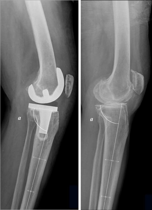
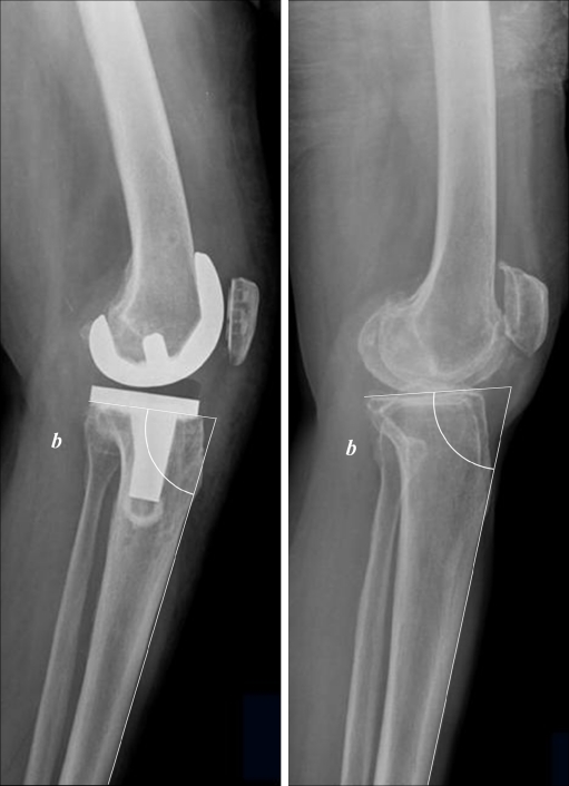
PSAs were measured with reference to the medullary canal (PSA-A) and anterior cortex (PSA-B). The reference line of the PSA-A was defined as the line connecting the centre of the medullary canal 10 cm and 20 cm distal to the tibial plateau (Fig. 1). The reference line of the PSA-B was defined as the line between 10 cm and 20 cm distal to the tibial plateau on the anterior tibial cortex (Fig. 2). Preoperative PSA was defined as the angle between the perpendicular line of reference and a line connecting the anterior and posterior borders of the medial tibial plateau. Postoperative PSA was defined as the angle between the perpendicular line of reference and the undersurface of the tibial component.
Fig. 1.
The measurement method of the proximal tibial medullary canal-referenced posterior slope (posterior slope: 90−a)
Fig. 2.
The measurement method of the proximal tibial anterior cortex-referenced posterior slope (posterior slope: 90−b)
Knee Society knee and function scores were evaluated preoperatively and at the final follow-up examination [14]. ROM was measured using a long-armed goniometer.
In addition, the increased amount of ROM after TKA with regard to the postoperative PSA-A was measured, considering the fact that preoperative ROM is the most important factor affecting postoperative ROM [24].
Statistical analyses were performed using SPSS software (vers. 12.0; SPSS Inc., Chicago, IL, USA). Pre- and postoperative PSA-A and -B values of groups I, II, and III were compared using analysis of variance (ANOVA). P values < 0.05 were considered to indicate statistical significance.
Results
Radiographic results
Preoperative PSA-As measured by investigators 1 and 2 did not differ significantly among the three groups (p = 0.972, 0.989). Mean postoperative PSA-As measured by both investigators were 6.0 ± 2.8° in group I and 6.0 ± 2.6° in group II; that in group III was 4.5 ± 2.3° (investigator 1) or 4.4 ± 2.3° (investigator 2; p < 0.001; Tables 1 and 2). Preoperative PSA-Bs measured by both investigators did not differ significantly between groups (p = 0.854, 0.876). Mean postoperative PSA-Bs were 8.1 ± 2.9° (investigator 1) or 8.2 ± 2.9° (investigator 2) in group I, 8.2 ± 2.6° in group II, and 6.5 ± 2.4° in group III (p < 0.001; Tables 1 and 2). Mean differences between PSA-A and PSA-B were 2.3 ± 1.0° (investigator 1) or 2.3 ± 1.2° (investigator 2) preoperatively and 2.1 ± 0.8° postoperatively.
Table 1.
Comparison of posterior tibial slope angle according to implant by observer 1
| Group | PSA-A | PSA-B | ||
|---|---|---|---|---|
| Preoperative | Postoperative | Preoperative | Postoperative | |
| Group I | 11.4 ± 4.8 | 6.0 ± 2.8 | 13.6 ± 4.9 | 8.1 ± 2.9 |
| Group II | 11.4 ± 4.5 | 6.0 ± 2.6 | 13.9 ± 4.6 | 8.2 ± 2.6 |
| Group III | 11.5 ± 3.3 | 4.5 ± 2.3 | 13.7 ± 3.3 | 6.5 ± 2.4 |
| P-value | 0.972 | <0.001 | 0.854 | <0.001 |
Group I cases using NexGen®, Group II cases using PFC®, Group III cases using Vanguard®, PSA-A posterior tibial slope angle considering the line passing centres of the medullary canal of proximal tibia as the reference line, PSA-B posterior tibial slope angle considering anterior cortex of proximal tibia as the reference line
Table 2.
Comparison of posterior tibial slope angle according to implant by observer 2
| Group | PSA-A | PSA-B | ||
|---|---|---|---|---|
| Preoperative | Postoperative | Preoperative | Postoperative | |
| Group I | 11.4 ± 4.8 | 6.0 ± 2.8 | 13.6 ± 4.9 | 8.2 ± 2.9 |
| Group II | 11.3 ± 4.5 | 6.0 ± 2.6 | 13.9 ± 4.6 | 8.2 ± 2.6 |
| Group III | 11.4 ± 3.3 | 4.4 ± 2.3 | 13.6 ± 3.4 | 6.5 ± 2.4 |
| P-value | 0.989 | <0.001 | 0.876 | <0.001 |
Group I cases using NexGen®, Group II cases using PFC®, Group III cases using Vanguard®, PSA-A posterior tibial slope angle considering the line passing centres of the medullary canal of proximal tibia as the reference line, PSA-B posterior tibial slope angle considering anterior cortex of proximal tibia as the reference line
Clinical results
The mean Knee Society knee score in the total sample (386 knees) was 59.7 ± 8.5 before surgery and 97.3 ± 5.1 at the final follow-up examination (p < 0.001; Table 3). These scores did not differ significantly among the three groups (p > 0.4).
Table 3.
Knee and function score according to American Knee Society scoring system
| Group | Knee score | Function score | ||
|---|---|---|---|---|
| Preoperative | Postoperative | Preoperative | Postoperative | |
| Group I | 59.7 ± 9.5 | 97.4 ± 4.9 | 54.2 ± 6.3 | 90.1 ± 7.9 |
| Group II | 59.5 ± 6.7 | 97.1 ± 6.1 | 54.0 ± 6.0 | 91.0 ± 6.8 |
| Group III | 59.7 ± 8.2 | 97.7 ± 2.4 | 54.5 ± 7.6 | 90.8 ± 3.1 |
| Total | 59.7 ± 8.5 | 97.3 ± 5.1 | 54.2 ± 6.4 | 90.5 ± 7.1 |
| P-value | 0.981 | 0.816 | 0.889 | 0.478 |
Group I cases using NexGen®, Group II cases using PFC®, Group III cases using Vanguard®
The mean flexion contracture was 5.3 ± 6.6° before surgery and 0.3 ± 2.1° at the final follow-up examination (p < 0.001). The mean further flexion was 132.0 ± 13.5° before surgery and 132.5 ± 10.6° at the final follow-up examination (p = 0.268; Table 4). Further flexion was slightly greater in group III than that in other groups, although this difference was not significant (p = 0.065; Table 4). The knees within ±3° from average postoperative PSA-A were classified as the inlier group, from which postoperative PSA-As ranged from 3° to 9° in groups I and II, and ranged from 1.5° to 7.5° in group III. Fifty-seven knees were classified as the outlier group, from which postoperative PSA-As were below 3° in groups I and II, and below 1.5° in group III. The average increased amount of ROM was 6.4 ± 16.3° in the inlier group, but 0.9 ± 12.1° in the outlier group (p = 0.023).
Table 4.
Range of motion
| Group | Flexion contracture | Further flexion | ||
|---|---|---|---|---|
| Preoperative | Postoperative | Preoperative | Postoperative | |
| Group I | 4.7 ± 7.1° | 0.1 ± 0.8° | 132.7 ± 14.0° | 133.0 ± 10.5° |
| Group II | 5.7 ± 5.7° | 0.7 ± 3.2° | 131.0 ± 13.2° | 131.6 ± 11.1° |
| Group III | 5.9 ± 7.1° | 0.2 ± 1.4° | 132.3 ± 13.0° | 133.3 ± 9.8° |
| Total | 5.3 ± 6.6° | 0.3 ± 2.1° | 132.0 ± 13.5° | 132.5 ± 10.6° |
| P-value | 0.344 | 0.060 | 0.556 | 0.480 |
Group I cases using NexGen®, Group II cases using PFC®, Group III cases using Vanguard®
Complications
Postoperative complications occurred in two knees; patellar subluxation occurred in one knee and deep infection developed in the other. The patient with patellar subluxation refused further operative treatment. The case of deep infection (acute haematogenous pattern at 2.5 years) occurred in a patient in group III and was treated successfully by two-stage revision.
Discussion
Brazier et al. [3] introduced several methods of PSA measurement using a variety of reference lines, including the anatomical axes of the tibial and fibular shafts and proximal tibia and fibula, and the anterior and posterior cortices of the proximal tibia. Among these lines, the anatomical axis and posterior cortex of the proximal tibia are most strongly correlated with the anatomical axis of the tibial shaft. The difference in PSAs based on the anatomical axis and anterior cortex of the proximal tibia ranged from 2.2 to 3.5° in Brazier’s study [3] and from 2.1 to 2.3° in our study.
Several authors [2, 8, 11] have reported variable ranges of anatomical PSA in normal knees. Dejour and Bonnin [8] reported a mean PSA of 10 ± 3° in 281 normal knees, Giffin et al. [11] reported a mean PSA of 8.8 ± 1.8° in ten cadaveric knees, and Bellemans et al. [2] found a normal PSA range of 4–9° in a cadaveric study. In our study, preoperative PSA-A ranged widely (11.4 ± 4.5°). Matsuda et al. [21] found similar PSAs in normal and varus knees. Jiang et al. [16] reported that PSA was not affected by age, gender, or arthritis. However, Chiu et al. [6] reported that the PSA of arthritic knees was 2–3° greater than that of normal knees.
Although adequate PSA plays an important role in cruciate-retaining and substituting TKA, an appropriate PSA has not been clearly determined [4, 9, 10, 22, 28]. PCL recession or PSA increase widens the flexion gap in cruciate-retaining TKA [4, 9, 28]. Whereas PCL recession reduces only anteroposterior tightness in flexion, PSA increase reduces the varus, valgus, anteroposterior, and rotational tightness in flexion. Thus, PSA increase may be more effective than PCL recession in cases with multidirectional tightness of the flexed knee [17]. In cruciate-retaining TKA, an appropriate PSA maintains the tension of the PCL properly and facilitates femoral rollback during knee flexion [17]. Proper femoral rollback increases the ROM and shifts the contact point between the femur and the tibia backward, which allows the relatively strong posterior tibial cancellous bone to support the femur [13, 17]. Walker and Garg [26] reported that knees with PSAs of 10° could flex 30° more than those with PSAs of 0° in cruciate-retaining TKA. Bellemans et al. [2] reported in a cadaveric study that 1.7° of additional flexion could be obtained by increasing the PSA as much as 1°. However, an excessive PSA may induce anteroposterior instability after TKA, which may cause anterior subluxation of the tibial component, increase the wear on the posterior portion of the polyethylene surface, and cause aseptic loosening [1, 17, 28]. Impingement of posterior femoral condyles may also occur during knee flexion due to the limitation of femoral rollback, and ROM may be limited. In contrast, an inadequate PSA or the creation of a tibial anterior slope forces the weak anterior cancellous bone to support the femoral component and increases the possibility of anterior subsidence [13, 17]. It also limits further flexion due to the tight flexion gap [17, 20, 28]. Thus, it is important to determine the proper PSA.
Recommended PSAs vary among prosthesis types due to diversity in the posterior offset of the femoral component and the slope of the polyethylene insert. The polyethylene inserts of the NexGen Legacy® and PFC Sigma® devices have slopes of 0°. The polyethylene insert of the Vanguard® system has a slope of 3°, which probably caused the difference in postoperative PSA observed in this study. The manufacturer-recommended PSAs are 7° for the NexGen Legacy®, 3–6° for the PFC Sigma®, and 2–3° for the Vanguard® device. The proper cutting angle for the PSA is still controversial, and a wide range of optimal angles has been proposed [4, 9, 10, 22, 28]. Buechel and Pappas [4] proposed an angle of 5–10° and Piazza et al. [22] proposed an angle of 6–10°. Wasielewski et al. [27] investigated polyethylene wear patterns after 29 TKAs using the Miller-Galante 1® prosthesis (Zimmer, Inc.), finding a mean PSA of 8.76° in 17 cases showing wear in the posterior portion of the polyethylene surface and a mean PSA of 5.91° in 12 cases with no posterior wear.
The posterior horn of the meniscus is thicker than the anterior horn. The biomechanical PSA determined with consideration of this structural feature differed from that measured on radiographs [7, 15]. Furthermore, most proposed optimal PSAs in cruciate-retaining TKA have been based on Caucasians. There may be ethnic differences in the recommended PSA; for example, the anatomical features of the proximal tibia may differ in Asians and Caucasians, and knee deformity is usually more pronounced in Asian patients than in Caucasian patients at the time of TKA. In this study, Knee Society knee and function scores were found to be satisfactory and mean further flexion was greater than 130° after cruciate-retaining TKA. The postoperative PSA-A measured using the medullary canal reference method was 6.0 ± 2.8°; operators should seek to achieve this angle to ensure satisfactory outcomes.
We tried to compare the increased amount of ROM after TKA with regard to the postoperative PSA-A. The inlier group of postoperative PSA-A had significantly greater ROM after TKA. Some investigators [5, 12, 13, 23, 25] also have reported different clinical outcomes, depending on PSA. However, the clinical outcome and postoperative ROM are influenced by various factors, such as the severity of preoperative deformity, preoperative PSA, alteration of the posterior femoral offset, flexion angle of the femoral component, and tension of the soft tissues (e.g., PCL). Thus, the comparative analysis of clinical outcomes according to postoperative PSA is complicated by inherent statistical bias. Furthermore, the range of postoperative PSA was narrow in this study. Thus, the comparison of ROM in relation to the postoperative PSA should be carefully interpreted. This study had another limitation through its retrospective design. The final PSA was decided by the prediction and experience of the senior author.
In summary, mean postoperative PSAs measured using the medullary canal reference method averaged 6.0°, 6.0°, and 4.5°, respectively, after cruciate-retaining TKA using the NexGen®, PFC sigma®, and Vanguard® prostheses. Patients with these PSA values experienced satisfactory pain relief and functional recovery. These postoperative PSA values may serve as a useful guideline for cruciate-retaining TKA procedures.
References
- 1.Bai B, Baez J, Testa N, Kummer FJ. Effect of posterior cut angle on tibial component loading. J Arthroplasty. 2000;15:916–920. doi: 10.1054/arth.2000.9058. [DOI] [PubMed] [Google Scholar]
- 2.Bellemans J, Robijns F, Duerinckx J, Banks S, Vandenneucker H. The influence of tibial slope on maximal flexion after total knee arthroplasty. Knee Surg Sports Traumatol Arthrosc. 2005;13:193–196. doi: 10.1007/s00167-004-0557-x. [DOI] [PubMed] [Google Scholar]
- 3.Brazier J, Migaud H, Gougeon F, Cotten A, Fontaine C, Duquennoy A. Evaluation of methods for radiographic measurement of the tibial slope. A study of 83 healthy knees. Rev Chir Orthop Reparatrice Appar Mot. 1996;82:195–200. [PubMed] [Google Scholar]
- 4.Buechel FF, Pappas MJ. New Jersey low contact stress knee replacement system. Ten-year evaluation of meniscal bearings. Orthop Clin North Am. 1989;20:147–177. [PubMed] [Google Scholar]
- 5.Catani F, Fantozzi S, Ensini A, Leardini A, Moschella D, Giannini S. Influence of tibial component posterior slope on in vivo knee kinematics in fixed-bearing total knee arthroplasty. J Orthop Res. 2006;24:581–587. doi: 10.1002/jor.20121. [DOI] [PubMed] [Google Scholar]
- 6.Chiu KY, Zhang SD, Zhang GH. Posterior slope of tibial plateau in Chinese. J Arthroplasty. 2000;15:224–227. doi: 10.1016/S0883-5403(00)90330-9. [DOI] [PubMed] [Google Scholar]
- 7.Choi CH, Kim JH, Chung HK, Choi YH. Measurement of posterior slope angle of the proximal tibia by MRI and X-ray. J Korean Orthop Assoc. 2001;36:569–573. [Google Scholar]
- 8.Dejour H, Bonnin M. Tibial translation after anterior cruciate ligament rupture. Two radiological tests compared. J Bone Joint Surg Br. 1994;76:745–749. [PubMed] [Google Scholar]
- 9.Dixon MC, Brown RR, Parsch D, Scott RD. Modular fixed-bearing total knee arthroplasty with retention of the posterior cruciate ligament. A study of patients followed for a minimum of fifteen years. J Bone Joint Surg Am. 2005;87:598–603. doi: 10.2106/JBJS.C.00591. [DOI] [PubMed] [Google Scholar]
- 10.Dorr LD, Boiardo RA (1986) Technical considerations in total knee arthroplasty. Clin Orthop Relat Res 205:5–11 [PubMed]
- 11.Giffin JR, Vogrin TM, Zantop T, Woo SL, Harner CD. Effects of increasing tibial slope on the biomechanics of the knee. Am J Sports Med. 2004;32:376–382. doi: 10.1177/0363546503258880. [DOI] [PubMed] [Google Scholar]
- 12.Higuchi H, Hatayama K, Shimizu M, Kobayashi A, Kobayashi T, Takagishi K. Relationship between joint gap difference and range of motion in total knee arthroplasty: a prospective randomised study between different platforms. Int Orthop. 2009;33:997–1000. doi: 10.1007/s00264-009-0772-7. [DOI] [PMC free article] [PubMed] [Google Scholar]
- 13.Hofmann AA, Bachus KN, Wyatt RW (1991) Effect of the tibial cut on subsidence following total knee arthroplasty. Clin Orthop Relat Res 269:63–69 [PubMed]
- 14.Insall JN, Dorr LD, Scott RD, Scott WN (1989) Rationale of the Knee Society clinical rating system. Clin Orthop Relat Res 248:13–14 [PubMed]
- 15.Jenny JY, Rapp E, Kehr P. Proximal tibial meniscal slope: a comparison with the bone slope. Rev Chir Orthop Reparatrice Appar Mot. 1997;84:435–438. [PubMed] [Google Scholar]
- 16.Jiang CC, Yip KM, Liu TK. Posterior slope angle of the medial tibial plateau. J Formos Med Assoc. 1994;93:509–512. [PubMed] [Google Scholar]
- 17.Jojima H, Whiteside LA, Ogata K (2004) Effect of tibial slope or posterior cruciate ligament release on knee kinematics. Clin Orthop Relat Res 426:194–198 [DOI] [PubMed]
- 18.Kuwano T, Urabe K, Miura H, Nagamine R, Matsuda S, Satomura M, Sasaki T, Sakai S, Honda H, Iwamoto Y. Importance of the lateral anatomic tibial slope as a guide to the tibial cut in total knee arthroplasty in Japanese patients. J Orthop Sci. 2005;10:42–47. doi: 10.1007/s00776-004-0855-7. [DOI] [PubMed] [Google Scholar]
- 19.Laskin RS, Rieger MA. The surgical technique for performing a total knee replacement arthroplasty. Orthop Clin North Am. 1989;20:31–48. [PubMed] [Google Scholar]
- 20.Massin P, Gournay A. Optimization of the posterior condylar offset, tibial slope, and condylar roll-back in total knee arthroplasty. J Arthroplasty. 2006;21:889–896. doi: 10.1016/j.arth.2005.10.019. [DOI] [PubMed] [Google Scholar]
- 21.Matsuda S, Miura H, Nagamine R, Urabe K, Ikenoue T, Okazaki K, Iwamoto Y. Posterior tibial slope in the normal and varus knee. Am J Knee Surg. 1999;12:165–168. [PubMed] [Google Scholar]
- 22.Piazza SJ, Delp SL, Stulberg SD, Stern SH. Posterior tilting of the tibial component decreases femoral rollback in posterior-substituting knee replacement: a computer simulation study. J Orthop Res. 1998;16:264–270. doi: 10.1002/jor.1100160214. [DOI] [PubMed] [Google Scholar]
- 23.Schnurr C, Csécsei G, Nessler J, Eysel P, König DP. How much tibial resection is required in total knee arthroplasty? Int Orthop. 2011;35:989–994. doi: 10.1007/s00264-010-1025-5. [DOI] [PMC free article] [PubMed] [Google Scholar]
- 24.Seon JK, Park JK, Jeong MS, Jung WB, Park KS, Yoon TR, Song EK. Correlation between preoperative and postoperative knee kinematics in total knee arthroplasty using cruciate retaining designs. Int Orthop. 2011;35:515–520. doi: 10.1007/s00264-010-1029-1. [DOI] [PMC free article] [PubMed] [Google Scholar]
- 25.Tsuji S, Tomita T, Hashimoto H, Fujii M, Yoshikawa H, Sugamoto K. Effect of posterior design changes on postoperative flexion angle in cruciate retaining mobile-bearing total knee arthroplasty. Int Orthop. 2011;35:689–695. doi: 10.1007/s00264-010-1060-2. [DOI] [PMC free article] [PubMed] [Google Scholar]
- 26.Walker PS, Garg A (1991) Range of motion in total knee arthroplasty. A computer analysis. Clin Orthop Relat Res 262:227–235 [PubMed]
- 27.Wasielewski RC, Galante JO, Leighty RM, Natarajan RN, Rosenberg AG (1994) Wear patterns on retrieved polyethylene tibial inserts and their relationship to technical considerations during total knee arthroplasty. Clin Orthop Relat Res 299:31–43 [PubMed]
- 28.Whiteside LA, Amador DD. The effect of posterior tibial slope on knee stability after Ortholoc total knee arthroplasty. J Arthroplasty. 1988;3(Suppl):S51–S57. doi: 10.1016/S0883-5403(88)80009-3. [DOI] [PubMed] [Google Scholar]


