Abstract
Endometriosis is a medical condition in women wherein endometrial cells deposited in the area outside the uterine cavity are influenced by hormonal changes, and produce symptoms depending on the site of implantation. A unique case of retroperitoneal endometriosis causing deep vein thrombosis from extrinsic compression of the right iliac vein is described. Clinical presentation with cyclical leg swelling, coincidental with menstruation and culminating with deep vein thrombosis, although very suggestive, has not been previously reported.
Keywords: DVT, Endometriosis, Stents, Thrombolysis
A 41-year-old woman with no previous pregnancies or full-term births presented to the emergency department with swelling of the right lower extremity and pelvic pain. Her symptoms began one week earlier, at which point she was seen by her primary care physician, who diagnosed deep vein thrombosis (DVT) of the right lower extremity. Anticoagulation therapy was started with enoxaparin and warfarin. The patient presented to the emergency department one week later, complaining that the pain and swelling of her right lower extremity had increased. The patient indicated that she had not taken the anticoagulation therapy that was recommended.
This patient had a four-year history of back and right leg pain associated with leg swelling – cyclical in nature and coincidental with menstruation. The only difference in the present occurrence was pain and swelling of the right leg that did not resolve after menstruation.
She described her menses as painful but regular. She had not experienced any menorrhagia, metrorrhagia or dyspareunia.
Before the present encounter, the patient used nonsteroidal anti-inflammatory drugs, muscle relaxants and opiate analgesics to control dysmenorrhea and leg pain, with moderate success.
A previous diagnostic workup for endometriosis, including a pelvic ultrasound and laparoscopy, was negative. The patient refused treatment with oral contraceptives because of her desire to get pregnant.
The patient had a medical history significant for infertility, tobacco use and intravenous drug abuse. In addition, she had a family history of colon cancer, polycystic kidney disease and liver disease.
On physical examination, the patient appeared to have moderate discomfort. The right lower extremity exhibited a reticular, erythematous skin rash, swollen from the thigh to the foot, warm to the touch and tender to palpitation. Pulses, sensation and strength were intact.
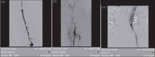
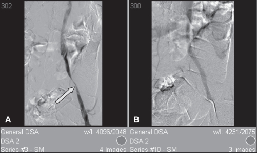
Diagnostic workup included a coagulation profile, venous duplex ultrasound of the pelvis and lower extremities, and computed tomography (CT) of the abdomen and pelvis. The venous duplex ultrasound showed DVT to the level of the right common femoral vein. The CT showed thrombosis of the right common femoral, external iliac and distal internal iliac veins. The initial pelvic CT interpretation was negative for masses. However, a review of the pelvic CT scan following a right lower extremity venogram revealed a 3 cm retroperitoneal soft tissue mass encompassing the right external iliac vein, artery and L5 nerve root. The mass was also causing some sclerosis of the right sacrum (Figure 1). The DVT extended superiorly to the level of the mass. Right transpopliteal venography showed extensive DVT involving the superficial femoral vein, common femoral vein and external iliac vein. The right common iliac vein and inferior vena cava were normal (Figure 2). DVT was managed using transcatheter thrombolysis with a tissue plasminogen activator to prevent post-thrombotic syndrome. Complete recanalization of the femoral and external iliac vein was seen after 24 h of thrombolysis. Smooth narrowing of the right external iliac vein corresponding to the soft tissue mass in the pelvis was seen following thrombolysis.
Figure 1).
Computed tomography of the sacrum. A Thrombosis of the right femoral vein (arrow). B A 3 cm soft tissue mass encircling the right external vein (arrow). C Bone window showing subtle sclerosis of the right sacrum (arrow)
Figure 2).
Transpopliteal venogram showing extensive deep vein thrombosis of the femoral and external iliac vein. The right common iliac vein and inferior vena cava are normal
Percutaneous transluminal angioplasty of the right external iliac vein with a 12 mm × 4 cm balloon catheter via the right transpopliteal approach, followed by placement of a 12 mm × 14 mm Wallstent (Boston Scientific Corporation, USA), resolved the stricture (Figure 3).
Figure 3).
Post-thrombolysis venogram showing resolution of the femoral and external iliac vein, and smooth narrowing of the external iliac vein (arrow) (A), as well as successful stenting of the external iliac vein (B)
DISCUSSION AND LITERATURE REVIEW
Endometriosis is an estrogen-dependent inflammatory process affecting 10% to 15% of fertile women, and as many as 50% of infertile women (1,2). The disease process is defined by the presence of hormone-responsive endometrial tissue outside of the uterine cavity. Localization of endometriosis can be divided into two broad anatomical categories – pelvic and extrapelvic. Pelvic lesions may be found within the ovaries, fallopian tubes or pelvic peritoneum. Extrapelvic lesions may be localized to the genitalia, perineum, urinary tract system, gastrointestinal tract, intrathoracic structures, musculoskeletal tissue, abdominal wall or nervous system (Table 1). These two anatomical subtypes differ epidemiologically, in natural history and in manner of diagnosis (3).
TABLE 1.
Sites of identified extrapelvic endometriosis and the preferred diagnostic method for their identification
| General site | Specific sites identified | Preferred diagnostic study |
|---|---|---|
| Gastrointestinal organs | Small bowel, large bowel, anal canal, Meckel’s diverticulum, liver, gallbladder, pancreas | Laparoscopy and laparotomy |
| Urinary system | Kidney, bladder, ureter, urethra | Intravenous pyelogram – ureteral; ultrasonography – bladder and renal |
| Genital tract | Vagina, rectovaginal septum, cervix, vulva, episiotomy scar | Tissue biopsy |
| Musculoskeletal tissue | Shoulder, breast, elbow, forearm, hand, pubis, groin (inguinal canal), buttocks, thigh, knee | Tissue biopsy |
| Thorax | Heart, lung parenchyma and pleura | Tissue biopsy |
| Abdominal wall | Cutaneous, rectus abdominis muscle, umbilicus, inguinal region, perineum, labia, laparoscopic trocar sites | Tissue biopsy |
| Nervous system | Cerebral cortex, spinal cord, sciatic nerve, obturator nerve | CT scan and MRI |
| Vasculature | Lymph node, thoracic aorta, femoral vein, external iliac vein | Tissue biopsy |
A number of pathological mechanisms for development and transplantation of endometriosis exist. The mechanism is dependent on the lesion site and can include retrograde menstruation, coelomic metaplasia, and vascular or lymphatic spread, as well as spontaneous and iatrogenic mechanisms (3–7).
The common clinical presentation of endometriosis is dysmenorrhea, pelvic pain and/or infertility. However, the presentation is variable depending on the site of the lesion. Pain is the most common symptom, resulting from inflammation, pressure, neuronal involvement, adhesions, psychological factors and menstrual debris (8,9). Symptoms can vary depending on the localization of extrapelvic endometriosis.
There are several case reports of extrapelvic endometriosis affecting the retroperitoneum (Table 1).
There are two case reports of endometriosis occurring around large veins. The first case reported endometriosis involving the left femoral vein (10). The patient presented with a painful groin mass that showed characteristics of endometriosis in the adventitial layer of the vein. The second case was due to endometriosis encircling the right external iliac vein causing catamenial edema of the right leg and thigh. Successful surgical resection of the endometriosis was performed. This case is different from our case in that no DVT was evident (11).
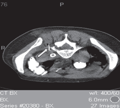
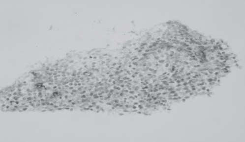
The diagnosis of extrapelvic endometriosis is difficult, usually dictated by clinical suspicion, followed by imaging studies (ultrasound, CT and magnetic resonance imaging), laparoscopy, tissue biopsy and immunohistochemistry (12). In our case, clinical presentation was suggestive of endometriosis and diagnosis was made by CT-guided tissue biopsy (Figure 4). Histology of the biopsy sample showed benign stroma of apparent endometrial type, consistent with endometriosis (Figure 5).
Figure 4).
Posterior computed tomography-guided biopsy of the retro-peritoneal paraspinal mass around the stented right external iliac vein (arrow)
Figure 5).
Right retroperitoneal paraspinal mass needle biopsy. Benign stromal tissue consisting of small spindle cells that stained positive for CD10 on immunoperoxide staining. Reproduced with permission from Dr Arthur R Gaba, Pathology and Laboratory, Henry Ford Hospital, Detroit, Michigan, USA
Historically, DVT was treated with unfractionated heparin and oral anticoagulation. The decision to treat our patient using transcatheter thrombolysis was twofold. In the short term, it was to prevent pulmonary embolization and achieve rapid reduction of pain and swelling. The long-term end point of treatment was to prevent post-thrombotic syndrome. In our experience, the best management of stenosis of the iliac veins is the placement of an endovascular stent. Self-expanding stents, such as the Wallstent or flexible nitinol stents, are preferred because they can be passively oversized to allow for lower risk of perforation while ensuring proper fixation and lower risk of migration in highly compliant veins. Our patient was to be treated with long-term anticoagulation therapy using warfarin and compression stockings.
Management of endometriosis is individualized, and driven by lesion location, presentation and patient desire for conception. The aim of conventional pharmacological management is to induce hypoestrogenism (13). Traditionally used hormone therapies include oral contraceptives, androgen derivatives (ie, danazol), progestins and gonadotropin-releasing hormone (GnRH) analogues (14–17). GnRH analogues are the most commonly used therapy of this group (18). These drugs are given to those suspected of or those who have an endometriosis diagnosis, and as adjuncts to surgery (19). The limitations of these treatment modalities are their inability to cure the disease and symptomatic recurrence after treatment (20). In situations in which conventional treatments do not produce the desired outcome, novel medical treatments are available. These include aromatase inhibitors, GnRH antagonists, mifepristone (RU 486), levonorgestrel and selective progesterone receptor modulators (21–27). Our patient was started on leuprolide injections. If this does not resolve her symptoms, laparoscopic resection of the pelvic endometrioma will be offered to her.
Through the laparoscope, not only can endometrial lesions be identified, but also, endometriotic areas can be fulgurated and adhesions lysed. With the use of a laser through the laparoscope, extensive endometriotic tissue can be vaporized and adhesions removed. Symptoms may recur following surgical resection, and the individual may require additional management (28). A surgical classification system has been proposed that suggests the proper surgical approach based on the anatomical distribution of the endometrioma (29).
REFERENCES
- 1.Cramer DW, Wilson E, Stillman RJ, et al. The relationship of endometriosis to menstrual characteristics, smoking and exercise. JAMA. 1986;255:1904–8. [PubMed] [Google Scholar]
- 2.Drake TS, Grunert GM. The unsuspected pelvic factor in the infertility investigations. Fertil Steril. 1980;34:27–31. [PubMed] [Google Scholar]
- 3.Honore GM. Extra pelvic endometriosis. Clin Obstet Gynecol. 1999;42:699–711. doi: 10.1097/00003081-199909000-00021. [DOI] [PubMed] [Google Scholar]
- 4.Markham SM, Carpenter SE, Rock JA. Extra pelvic endometriosis. Obstet Gynecol Clin North Am. 1989;16:193–4. [PubMed] [Google Scholar]
- 5.Singh KK, Lessells AM, Adam DJ, et al. Presentation of endometriosis to general surgeons: A 10-year experience. Br J Surg. 1995;82:1349–51. doi: 10.1002/bjs.1800821017. [DOI] [PubMed] [Google Scholar]
- 6.Stanley KE, Utz DC, Dockerty MB. Clinically significant endometriosis of the urinary tract. Surg Gyencol Obstet. 1965;120:492–8. [PubMed] [Google Scholar]
- 7.Williams TJ, Pratt JH. Endometriosis in 1,000 consecutive celiotomies: Incidence and management. Am J Obstet Gynecol. 1977;129:245–50. doi: 10.1016/0002-9378(77)90773-6. [DOI] [PubMed] [Google Scholar]
- 8.Droegemueller W, Herbst A, Mishell D. Comprehensive Gynecology. St Louis: Mosby; 1987. p. 1149. [Google Scholar]
- 9.Zreik TG, Olive DL. Pathophysiology: The biologic principles of disease. Am J Obstet Gynecol. 1997;24:259–68. doi: 10.1016/s0889-8545(05)70303-x. [DOI] [PubMed] [Google Scholar]
- 10.Recalde AL, Majmudar B. Endometriosis involving the femoral vein. South Med J. 1977;70:69–74. doi: 10.1097/00007611-197701000-00030. [DOI] [PubMed] [Google Scholar]
- 11.Rosengarten AM, Wong J, Gibbons S. Endometriosis causing cyclic compression of the right external iliac vein with cyclic edema of the right leg and thigh. J Obstet Gynaecol Can. 2002;24:33–5. doi: 10.1016/s1701-2163(16)30271-7. [DOI] [PubMed] [Google Scholar]
- 12.Fujishita A, Nakane PK, Koji T, et al. Expression of estrogen and progesterone receptors in endometrial and peritoneal endometriosis: An immunohistochemical and in situ hybridization study. Fertil Steril. 1997;67:856–64. doi: 10.1016/s0015-0282(97)81397-0. [DOI] [PubMed] [Google Scholar]
- 13.Olive DL. Medical therapy of endometriosis. Semin Reprod Med. 2003;21:209–22. doi: 10.1055/s-2003-41327. [DOI] [PubMed] [Google Scholar]
- 14.Olive DL, Lindheim SR, Pritt EA. New medical treatment for endometriosis. Endometriosis and infertility. Fertil Steril. 2004;81:1441–6. doi: 10.1016/j.fertnstert.2004.01.019. [DOI] [PubMed] [Google Scholar]
- 15.Olive DL, Lindheim SR, Pritts EA. New medical treatment for endometriosis. Best Pract Res Clin Obstet Gynaecol. 2004;18:319–28. doi: 10.1016/j.bpobgyn.2004.03.005. [DOI] [PubMed] [Google Scholar]
- 16.Lessey BA. Medical management of endometriosis and infertility. Fertil Steril. 2000;73:1089–96. doi: 10.1016/s0015-0282(00)00519-7. [DOI] [PubMed] [Google Scholar]
- 17.Valle RF, Sciarra JJ. Endometriosis: Treatment strategies. Ann N Y Acad Sci. 2003;997:229–39. doi: 10.1196/annals.1290.026. [DOI] [PubMed] [Google Scholar]
- 18.Rock JA, Truglia JA, Caplan RJ. Zoladex (grosgrain acetate implant) in the treatment of endometriosis: A randomized comparison with danazol. The Zoladex Endometriosis Study Group. Obstet Gynecol. 1993;82:198–205. [PubMed] [Google Scholar]
- 19.Huang H-Y. Medical treatment of endometriosis. Chang Gung Med J. 2008;31:431–40. [PubMed] [Google Scholar]
- 20.Waller KG, Shaw RW. Gonadotropin-releasing hormone analogues for the treatment of endometriosis: Long-term follow-up. Fertil Steril. 1993;59:511–5. doi: 10.1016/s0015-0282(16)55791-4. [DOI] [PubMed] [Google Scholar]
- 21.Ailawadi RK, Jobanputra S, Kataria M, Gurates B, Bulun SE. Treatment of endometriosis and chronic pelvic pain with letrozole and norethindrone acetate: A pilot study. Fertil Steril. 2004;81:290–6. doi: 10.1016/j.fertnstert.2003.09.029. [DOI] [PubMed] [Google Scholar]
- 22.Finas D, Hornung D, Diedrich K, Schultze-Mosgau A. Cetrorelix in the treatment of female infertility and endometriosis. Expert Opin Pharmacotherapy. 2006;7:2155–68. doi: 10.1517/14656566.7.15.2155. [DOI] [PubMed] [Google Scholar]
- 23.Murphy AA, Zhou MH, Malkapuram S, et al. RU486-induced growth inhibition of human endometrial cells. Fertil Steril. 2000;74:1014–9. doi: 10.1016/s0015-0282(00)01606-x. [DOI] [PubMed] [Google Scholar]
- 24.Jiang J, Wu RF, Wang ZH, et al. Effects of mifepristone on estrogen and progesterone receptors in human endometrial and endometriotic cells in vitro. Fertil Steril. 2002;77:995–1000. doi: 10.1016/s0015-0282(02)03081-9. [DOI] [PubMed] [Google Scholar]
- 25.Luukkainen T, Lahteenmaki P, Toivonen J. Levonorgestrel-releasing intrauterine device. Ann Med. 1990;22:85–90. doi: 10.3109/07853899009147248. [DOI] [PubMed] [Google Scholar]
- 26.Lockhat FB, Emembolu JO, Konje JC. The efficacy, side-effects and continuation rates in women with symptomatic endometriosis undergoing treatment with an intra-uterine administered progestogen (Levonorgestrel): A 3 year follow-up. Hum Reprod. 2005;20:789–93. doi: 10.1093/humrep/deh650. [DOI] [PubMed] [Google Scholar]
- 27.Chwalisz K, Mattia-Goldberg K, Lee M, Elger W, Edmonds A. Treatment of endometriosis with the novel selective progesterone receptor modulator (SPRM) asoprisnil. Fertil Steril. 2004;82:S83–4. [Google Scholar]
- 28.Candiani GB, Fedele L, Vercellini P, Bianchi S, DiNola G. Repetitive conservative surgery for recurrence of endometriosis. Obstet Gynecol. 1991;77:421–4. [PubMed] [Google Scholar]
- 29.Chapron C, Fauconnier A, Vieira M, et al. Anatomical distribution of deeply infiltrating endometriosis: Surgical implications and proposition for classification. Hum Reprod. 2003;18:157–61. doi: 10.1093/humrep/deg009. [DOI] [PubMed] [Google Scholar]
- 30.Dwivedi AJ, Agrawal SN, Silva YJ. Abdominal wall endometriomas. Dig Dis Sci. 2002;42:456–61. doi: 10.1023/a:1013711314870. [DOI] [PubMed] [Google Scholar]
- 31.Felson H, McGuire J, Wasserman P. Stromal endometriosis involving the heart. Am J Med. 1960;29:1072–6. doi: 10.1016/0002-9343(60)90085-1. [DOI] [PubMed] [Google Scholar]
- 32.Recalde AL, Majmudar B. Endometriosis involving the femoral vein. South Med J. 1977;70:69–74. doi: 10.1097/00007611-197701000-00030. [DOI] [PubMed] [Google Scholar]
- 33.Notzold A, Moubayed P. Endometriosis in the thoracic aorta. N Engl J Med. 1998;339:1002–3. doi: 10.1056/NEJM199810013391413. [DOI] [PubMed] [Google Scholar]
- 34.Papapietro N, Gulino G, Zobel BBB, Di Martino A, Denaro V. Cyclic sciatica related to an extra pelvic endometriosis of the sciatic nerve: New concepts in surgical therapy. J Spinal Discord Tech. 2002;15:436–9. doi: 10.1097/00024720-200210000-00016. [DOI] [PubMed] [Google Scholar]
- 35.Pellegrino VDJ, Pasternak HS, Macaulay WP. Endometrioma of the pubis: A differential in the diagnosis of hip pain. A report of two cases. Am J Bone Joint Surg. 1981;63:1333–4. [PubMed] [Google Scholar]
- 36.Strasser F, Davis R. Extra peritoneal inguinal endometriosis. Am Surg. 1977;43:421–2. [PubMed] [Google Scholar]
- 37.Elliot DL, Barker AF, Dixon LM. Catamenial hemoptysis. New methods of diagnosis and therapy. Chest. 1985;87:687–8. doi: 10.1378/chest.87.5.687. [DOI] [PubMed] [Google Scholar]


