Abstract
DNA methylation is a key epigenetic mark when occurring in the promoter and enhancer regions regulates the accessibility of the binding protein and gene transcription. DNA methylation is inheritable and can be de novo-synthesized, erased and reinstated, making it arguably one of the most dynamic upstream regulators for gene expression and the most influential pacer for development. Recent progress has demonstrated that two forms of cytosine methylation and two pathways for demethylation constitute ample complexity for an instructional program for orchestrated gene expression and development. The forum of the current discussion and review are whether there is such a program, if so what the DNA methylation program entails, and what environment can change the DNA methylation program. The translational implication of the DNA methylation program is also proposed.
Keywords: epigenetics, neural development, 5-hydroxymethylcytosine, epigenome, environmental factors, DNA demethylation
Introduction
Among the rising literature on epigenetics, besides cancer research, developmental epigenetics is the next imminent field in the epigenetic application. The cytosine of mammalian DNA can be chemically modified to influence the transcriptibility. DNA cytosine methylation, at the major groove of the α helix, together with histone modifications, presents a 3D conformation change of histone linker H1 that condenses the nucleosomal arrays (Schmid et al., 1984; Brown et al., 1995; Karymov et al., 2001; Gisselsson et al., 2005). Such conformation change hinders the accessibility or binding of bioactive proteins including transcription factors, epigenetic modification enzymes, polyribosomes, and short sequences of nucleotides. The consequence is often inactivation of transcription (Busslinger et al., 1983; Yisraeli et al., 1988; Jones and Takai, 2001). In the vertebrate genome, methylation occurs preferentially in cytosine adjacent to guanine (CpG). About 70%–80% of the CpG are methylated except those CpGs clustered as islands in the seas of the genome (Gardiner-Garden and Frommer, 1987) which prevail in the promoter region (Bird, 1986; Ramsahoye et al., 1996; Lister et al., 2009). DNA methylation distributed in promoter and enhancer regions has been shown to influence the gene transcription. Recently, the type of methylation at intragenic regions is also found to be associated with transcription (Wu et al., 2011). There are many key questions yet to be explored on how transcription is influenced. One set of major questions is whether the DNA methylations are invariantly inherited from parental cells, or are methylated de novo at each daughter cell? If methylated de novo, is there memory that allows recapitulation of the previously established methylation? It is known that mammalian tissues are heterogeneous in their methylation; such methylation heterogeneity is hypothesized to be the driving force of the orchestrated specific gene expression in the making of tissue specification (Tawa et al., 1990). How are the heterogeneity and tissue specificity of methylations among tissue and cells achieved? During the developmental stage there is a swift progression of progenitor cells into differentiation and of cellular limitation that leads to a new type of cells. How is the methylation among other epigenetic events evolved over the progression of these developmental stages? Lastly, during the establishment of methylation, would it be subject to environmental modifications?
As more methylations form and their interactions have been recently identified, this review will focus on the occurrence of DNA methylation and demethylation through neural development. Specifically, we will provide evidence that DNA methylation occurs in an orderly and programmed fashion parallel with the progression of development. A “programmed DNA methylation” which may contribute to the developmental progression is discussed. In addition, this review discusses the environmental inputs that alter the destined DNA methylation programs contributing to developmental deficits. These two lines are important since increasing evidence points to many inborn abnormalities and late onset diseases may be rooted with erroneous epigenetic coding during early development e.g. Beckwith-Wiedemann syndrome, Angelman’s syndrome, Rett syndrome, and Autistic Spectrum disorders.
Cellular DNA-methylation and -demethylation
Currently, there are two recognized forms of DNA methylation: the 5-methylcytosine (5mC) and 5-hydroxylmethylcytosine (5hmC). The 5mC is catalyzed by DNA methyl transferase (DNMT)- the DNMT3a and 3b mediate de novo methylation, while DNMT1 mediates methylation complementary to the strain which has been methylated (Hermann et al., 2004; Goll and Bestor, 2005). The 5hmC is a hydroxylated form of the 5mC mediated by the ten-11 translocation 1/2/3 (Tet1/2/3) enzyme (Kriaucionis and Heintz, 2009; Tahiliani et al., 2009; Guo et al., 2011; Ito et al., 2011). The demethylation thus far was achieved through either passive or active pathways. In passive demethylation, or replication dependent demethylation, the 5mC is passively demethylated by dilution through generations of cellular divisions in the absence of DNMT, e.g. zygotic global demethylation (Kafri et al., 1993; Inoue et al., 2011). In active demethylation, Tet1/2/3 converts the 5mC through oxidation into 5hmC. The 5hmC can be demethylated into 5-formylcytosine (5fC) and subsequently 5-carboxylcytosine (5caC) by Tet1/2/3 enzyme, or by deamination though cytidine deaminase AID (activation-induced deaminase) to 5-methyl uracil (5mU). Alternatively, the 5mC can be converted by AID/APE (activation-induced deaminase/apo-lipoprotein B mRNA editing enzyme) into thymine. The thymine, 5caC, and 5mU can be converted by TDG (Thymine-DNA glycosylase, whose primary function was thought to repair T-G mismatches) back into cystidine (Wolffe et al., 1999; Wu and Zhang, 2010; Bhutani et al., 2011). The active demethylation process prevails during development and is maintained in the adult brain e.g. in hippocampus (Guo et al., 2011) and cerebellum (unpublished observation).
The methylation and demethylation dynamics may be an important mechanism to stage the gene expression and play an active role in the regulation of development. While 5mC’s association with suppression of transcription is well accepted, the role of 5hmC is ambivalent and has not been established (Bhutani et al., 2011). The first impression that 5hmC is an intermediate form transition to demethylation, which indicates that it is transient and unstable, meets challenges. It is known that the two forms of methylation have coexisted as early as the stage of fertilization and morulation (Inoue and Zhang, 2011; Iqbal et al., 2011). Our observation of the steady coexistence of 5mC and 5hmC in the developing cells throughout neurulation and brain development argues against this notion of 5hmC as merely an intermediate product (described in next paragraph). More recent findings suggest that 5hmC is actively engaged in potential functionality departing from that of 5mC. The global analysis indicated that 5hmC was found to be bivalent in association with either genes, active or transitioning toward transcription in embryonic stem cells (Wu et al., 2011) and in developing brain (Szulwach et al., 2011). The genomic distribution of 5hmC, other than intergenic and promoter regions, is highly associated with intragenic gene body regions, particularly those with transcriptions (Wu et al., 2011). It is known that 5mC receives binding proteins MBD1/2/4 and MeCP2, but not MBD3. More recent findings indicated that MBD3 binds to 5hmC (Yildirim et al., 2011). Since the 5hmC is rather an enduring form of the DNA methylation, delineation of the function of 5hmC warrants further investigation. These observations indicate that the transition of 5mC and 5hmC are a dynamic process that may serve as a means for regulating the transition of gene transcription.
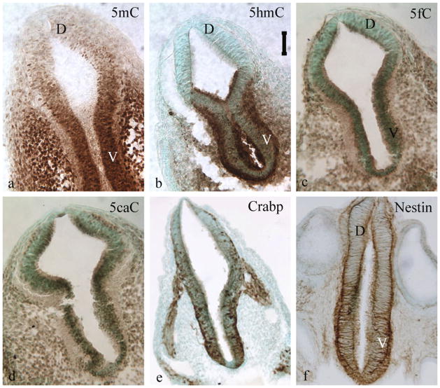
During early neural tube development, the 5mC and 5hmC appear to occur within the developing cells in tandem. Both 5mC and 5hmC exist in an observed stage of development e.g. from gestation (E) 5–17 in mice. The 5caC and 5fC are also present early on in new born cells or cells under differentiation. The co-existence of the methylation and demethylation marks indicate that a constant turn-over of 5mC to 5hmC and further demethylation is the norm in the genome of any cells at most stages of a life span (Fig. 1). The appearance of 5mC precedes 5hmC shortly in developing embryonic cells, and localize in a mosaic in chromatic regions within the nucleus correspondent to the heterochromatic and the euchromatic compartment respectively, as indicated by DAPI double staining. This arrangement indicates that the conversion of 5mC to 5hmC is associated with chromatin remodeling, which supports the observation that 5mC demethylation to 5hmC is a transition of changing transcription. The appearance of 5hmC is accompanied with the Tet1/2 e.g. in the neural tube, and heart cells. The DNA methylation binding protein appears after the arrival of 5mC and 5hmC (Zhou et al., 2011b). It lags by one day in newly emerged mouse neural tube cells during early gestation. The MBD1 and MeCP2 both appear early in the prenatal stage e.g. embryo day 10 (E10) in the neural tube by following behind the DNA methylation by a day or so in mouse development (not shown). Both increase expression over time and peak at early postnatal stage.
Figure 1.
The DNA methylation program shows spatiotemporal distribution of the immunocytochemical staining (brown diaminobenzidine) of methylation marks, 5mC (a) and 5hmC (b), and their demethylated forms, 5caC (c) and 5fC (d) in the neural tube at approximately gestation day 10 old embryo. There is a clear dorsoventral gradation in the neural tube in which DNA-methylation program (both 5mC and 5hmC, and their demethylation form 5fC and 5caC in the ventral is ahead of that of the dorsal. The progress of methylation gradations are parallel with the progression of differentiation gradation shown by immunostained cellular retinoic acid binding protein (Crabp, e) and nestin (f). D: dorsal aspect, V: ventral aspect. 5mC: 5methylcytosine, 5hmC: 5hydroxylmethylcytosine, 5fC: 5formylcytosine, 5caC: 5carboxylcytosine. Vertical scale bar = 50 μm for a–d.
The establishment of DNA methylation in the new born cell over the developmental stage can be accomplished by (a) Inheritance from parental cells, in which maintenance synthesis is followed to fill the methylation of the newly synthesized strain; or (b) De novo synthesis, in which no templates are provided, and the methylation is dependent on unknown memory in the new cells; or (c) a Combination of the two, in which part of the genomic methylations are inherited, and part are newly established. First, the DNA methylations are inherited during cell division in which DNMT1 mediates methyl transfer by interacting with proliferating cell nuclear antigen (PCNA) at the replication-fork of two unwinding stands (Schermelleh et al., 2007). Second, evidence also demonstrated that new born cells, for example many progenitor cells at the ventricular zone adjacent to the brain ventricle, are totally devoid of 5mC and 5hmC as indicated by immunocytochemical detection (Fig. 1). In this case, a de novo DNA methylation is required which is achieved by the presence of DNMT3a/3b. It is unclear whether the methylation pattern of the parent cells can be re-established entirely in these cells. Third, many of the new born cells inherited part of the parental methylation and progressively complete other methylation throughout epigenome. Apparently, de novo cytosine methylation is a major means for the re-establishment of new methylome that would most likely not be identical to that of parental cells. In any of the above cases, the outcome of the DNA methylation turn over during cell proliferation, particularly over the growth spurt during generation of new cells, new tissue, or new organs, is likely diversified which leads to a heterogeneous methylome. The subsequent transcription can be regulated differentially from parental cells and leads to new identities of the daughter cells. A compelling question is then asked, “Are these trans-cellular methylations during proliferation randomly occurring or is there some sort of order?”
DNA methylation program during neurodevelopment
It is now clear that DNA methylation is neither random nor fixed within a genome over the life span. Evidence suggests that DNA methylation, throughout development, is rather “orderly” with a clear pattern. This orderly pattern is preserved over evolution time. Such an evolving epigenetic pattern that constitutes as “memory” (Bird, 2002) may be a way for maintenance of stable cellular identities (De Carvalho et al., 2010; Deaton and Bird, 2011). On the other hand, the DNA methylation is also progressing over the embryonic and postnatal development which has been hypothesized as a driving force participating in directing life events, including shaping development and defining the stages of the life of a species, and determining cell fate for tissue specification. The DNA methylation transformation in an orderly manner during tissue or organ development is referred here as DNA methylation program (DMP).
The earliest DMP is known to occur soon after fertilization. Both 5mC and 5hmC are initially inherited from parents through the gametes. However, the parental and subsequently maternal genome is passively demethylated shortly after fertilization as cells are undergoing rapid divisions in the absence of DNMT, such that no major inherent or environmentally acquired DNA methylation history is transmitted to the offspring. Meanwhile, the global demethylation endows a totipotency to the zygotes. Soon thereafter, a less understood widespread de novo remethylation, mediated by DNMT, occurs to ensure the epigenetic regulation of genomic function (Morgan et al., 2005). Another well-established example is that during development the germ lines are also going through a global DNA demethylation and remethylation as they migrate into the gonadal ridge thus further eliminating potential carry-over of methylation memory from parents to the offspring (Brandeis et al., 1993; Kafri et al., 1993; del Mazo et al., 1994; Inoue et al., 2011). The totipotent embryonic stem (ES) cells are also going through redistribution at a genomic level, in which a distribution of 75% CpG methylation and 25% of non-CpG distribution are shifted to 99% CpG methylation (Lister et al., 2009). These drastic but unfailing processes have been conserved through evolution, which indicates that early life DNA methylation is essential for development and cellular function. The subsequent remethylation after widespread erasure is diverse during cellular proliferation. As indicated above, a methylation pattern can either be inherited, e.g. reinstated in symmetric daughter cells or partially modified, e.g. heterogeneous in asymmetrical daughter cells. Subsequently, during morulation, distinct methylation patterns characterize totipotent cells in the inner mass embryonic stem (ES) cells and trophoblasts of the external tissue (Nakanishi et al., 2012).
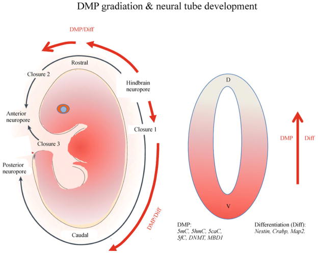
Until recently, the diverse reinstatement and modification of DNA methylation beyond morulation was not clear. The DMP progression beyond the morula stage is recently reported by Zhou et al. (2011b). This study demonstrated a neurulation-stage DMP in which 5mC, DNA methylation binding domain 1 (MBD1), and DNA methyl transferase 1 (DNMT1) exhibited distinct spatiotemporal patterns that coincided with neural differentiation in the neural tube (Zhou et al., 2011b) (Fig. 2). During neural tube development in the AP-axis, the brainstem differentiation progresses first and progresses rostrally and caudally, the DMP follow the progression of the neural axis. In the dorsoventral division, the ventralis differentiates earlier than the dorsalis, so does the DMP. Many of the neuroprogenitor cells near the ventricle bear no DNA methylation marks. The neural tubes expanded their cell number through proliferation in the absence of DNMTs. The newly arrived cells thus do not bear DNA methylation, e.g. actively proliferating ventricular zone and dorsal neural tube (Zhou et al., 2011b). They acquire DNA methylation of both 5mC and 5hmC when the proliferation ceases, begin their restriction (cell fate progression), and head for migration. Many of the differentiating neurons containing a high level of DNA methylation, will lose their 5mC and 5hmC later after arriving in target regions of the brain when transitioning into the stage of active wiring and synaptogenesis. This methylation and demethylation cycle can repeat over the progression of differentiation. The progression of DMP is cell stage-dependent. Thus, within a tissue or brain region, the late differentiating neuroprogenitor cells initiate the early DMP while the more differentiated neurons advance further in DMP. This is nicely demonstrated in the cortical layer during early development where newly arrived neurons are distributed in a top-down manner. Among the known DNA methylation marks, all do not appear at the same time. Throughout developing nervous systems and embryos in general, the 5mC appears first followed by 5hmC. It is also apparent that the DNA demethylation marks, 5caC and 5fC, have been identified early during the development e.g. at E10. This indicates that DMP includes DNA methylation and demethylation which are prevailing throughout the neural development. This likely explains the active repression and activation of different cohorts of genes during differentiation. The DNMT is similarly distributed as that of 5mC spatiotemporally in neural axial and dorsoventral patterns (Zhou et al., 2011b). The MBD1 shows a similar dorsoventral pattern to that of 5mC but appears a day late in gestation. The progression of the DMP is also demonstrated genome-wide. During neural stem cell differentiation, a signature epigenomic program is diversification of DNA methylation, in which many moderately methylated genes became hypo- and hyper-methylated (Zhou et al., 2011a). This is in consistence with the understanding that many multipotent genes were turned off, e.g. Oct 4; and many cell specific genes were turned on, e.g. MAP2 (Zhou et al., 2011a). The DMP becomes more elaborated along with developmental progression in the growing brain. The cellular 5mC and 5hmC are highly correlated with the neural progenitor cells and their progressions during differentiation throughout the brain e.g. cortical differentiation and hippocampal and dentate development. The 5hmC also demonstrated an epigenomic change in the developing hippocampus and cerebellum. When comparing early postnatal (P)7, P42, and one-year-old mice, it was found that the acquisition of 5hmC was developmentally programmed (Szulwach et al., 2011). The function of 5hmC is still unclear, but a positive correlation of 5hmC and the transition toward transcription is conjured. It was found that 5hmC is distributed rather richly in the gene body more than in promoter regions of the genes actively transcribed. Furthermore, the 5hmC levels were inversely correlated with methyl-CpG–binding protein 2 (MeCP2) in the cerebellum and hippocampus during early postnatal development (Szulwach et al., 2011). Thus the DMP includes the 5mC and 5hmC dynamics, and methylation-demethylation transition spatiotemporally during development.
Figure 2.
The progression of embryonic development has a distinct neural axial and ventrodorsal gradations. The hindbrain is developed first and the differentiation (Diff) progressed rostrally and caudally in the neural tube axis. In cross section, a ventral to dorsal progression of maturation also occurs. This maturation gradation is evident in the order of neural tube closure in both axial and ventrodorsal progression. It is also evident by many phenotypic markers e.g. nestin, Crabp, neu-N and Map2 (Zhou et al., 2011). The progression of maturation is overlapped with progression of DMP (DNA methylation program) of many DNA methylation marks as well as with histone codes. D: dorsal aspect, V: ventral aspect. Arrows indicate direction of progression of neural tube closure, DMP and differentiation.
The next question being raised is “Does developmental progression lead DMP, or reversely, does the DMP drive the developmental progression?” Apparently, DNA methylation is ensured though evolutionary hierarchy. Transient global demethylation is immediately corrected with remethylation. The disruption of DNA methylation is either lethal or leads to major developmental deficits. DNMT1, which adds methylation hemi-methylated DNA, is essential for cell viability in various mouse models; DNMT1 knockout delivers a strong blow to genome stability and cell viability (Brown and Robertson, 2007). DNMT3a/3b for de novo DNA methylation are essential for ES cells, early embryogenesis and early development, and are continuously required for late development and adult stage (Okano and Li, 2002). Pharmacologically, it was demonstrated that inhibition of DMP by treating embryos with 5-azacytidine (5-aza), a 5mC analog which inhibits DNMT for DNA methylation, retarded the embryos growth (Zhou et al., 2011b). The 5-aza also prevented migration and differentiation of neural stem cells in the culture (Singh et al., 2009). As for the function of 5hmC, knockdown (KD) of Tet1 and Tet2 in ES cells leads to defects in differentiation (Koh et al., 2011), while Tet1 knockdown also leads to defects in self renewal (Ito et al., 2010). Despite these defects in KD cells, Tet1 KD mice are viable and fertile (Dawlaty et al., 2011). This evidence puts the DMP in front of developmental progression.
Environmental modification of epigenetics during development
Many environmental factors can significantly alter the in utero environment, but how they manifest long-term developmental reprogramming is mostly unclear. Epigenetics is capable of recording certain environmental impacts, deposits these on chromatin, and serves as memory or history of the cells and organisms. The progression of the DMP in a protracted developmental period has a great opportunity to record environmental factors that may alter the trajectory of the development. The availability and quality of food that is fed during early development influences epigenetic make-up and potentially the developmental trajectory. The best exemplified cases include nutritional control of development of bees and mice. Genetically identical honeybee larvae fed with royal Jelly turn into reproductively capable queens, but when fed with common food, turn into infertile workers. Such similar fate determination in the bee population can be achieved by simply silencing the DNMT3 (Kucharski et al., 2008). In mammals, genetically identical Agouiti mice can vary in coat color and propensity for obesity and turmorigenesis depending on the expression of the A(vy) gene (Duhl et al., 1994). The A(vy) gene is regulated by methylation state of the intracisternal A particle (IAP) retrotransposon upstream; the unmethylated 5′ IAP long-terminal repeat (LTR) lead to yellow coat, while methylated 5′ IAP LTR lead to pseudoagouti (Dolinoy, 2008). The degree of DNA methylation although programmed is dynamic and dependent upon maternal nutrition and environmental exposures during early development. (Waterland and Jirtle, 2003; Dolinoy et al., 2006). The Agouti mice have been used to serve as a biosensor for environmentally induced methylation change. Bisphenol A (BPA), a chemical in many plastic drink bottles, when exposed to pregnant A(vy) mice, were found to increase yellow, obese progeny (Dolinoy et al., 2007). BPA was found to reduce DNA methylation. Supplementing the BPA treated dams with folic acid and vitamin B12 (methyl donor) was shown to counteract the reduction in DNA methylation caused by BPA. A similar test was found to validate that alcohol exposure during fetal development can affect methyl donor (Kaminen-Ahola et al., 2010). An epidemiological study revealed that nutritional status of the parents and grandparents can influence cardiovascular disease and diabetes in children (Kaati et al., 2002). Prenatal exposure to famine in “Dutch Hunger Winter” during World War II (Lumey et al., 2007) was linked to a range of developmental and adult disorders, including low birthweight, diabetes, obesity, coronary heart disease, breast cancer, and transgenerational effect (Stein et al., 2004, 2006; Kahn et al., 2009; Lumey and Stein, 2009). These abnormal developments were found to be associated with persistent epigenetic differences including less DNA methylation of the imprinted IGF2 gene (Heijmans et al., 2008). In light of epigenetics as a program that closely interact with the developmental progression, the environmental inputs effecting epigenetic progression inuterus, early postnatal development, or previous generations leading to development deficit and contributing to late onset diseases (Perera and Herbstman, 2011), is perhaps one of the most exciting underexplored territories. For example, alcohol exposure prior to or during pregnancy were found to alter DNA methylation (Liu et al., 2009; Ouko et al., 2009; Govorko et al., 2012; Otero et al., 2012) and gene expression (Green et al., 2007; Zhou et al., 2011c) of the offspring. A number of recently studied environmental factors have been demonstrated to influence epigenetics during development including nutrition (starvation, folic acid, choline), stress (maternal stress may influence programming of endocrine or immune systems in their offspring), pollutants (BPA, PAH), toxic metals or chemicals (lead, arsenic, pesticide), and substance abuse (alcohol, tobacco, etc.) are categorized in Table 1. Some of the environmental disruptors that altered DNA methylation can be inherited and cause transgenerational adult-onset diseases e.g. the pesticide vinclozolin (Anway et al., 2006). Understanding of the environmental contributors to epigenetic changes will provide clues and generate new hypotheses for mechanisms yet to be deciphered in developmental deficits and late adult on-set diseases.
Table 1.
Recent studies on environmental factors which altered epigenetics during development and their potential consequences
| Category | Environ. factors | Epigenetic changes | Phenotypes | References |
|---|---|---|---|---|
| Nutrition | Food deprivation Folate, Choline, | DNA methylation | Low birthweight, obesity, Cardiovascular disease, diabetes | (Kaati et al., 2002; Kahn et al., 2009; Stein et al., 2006; Lumey and Stein, 2009) (Food deprivation) (McKay et al., 2004; Mason and Choi, 2005) (FA) (Zeisel, 2007) (Choline) |
| Stress | Deprivation, separation | DNA methylation | Asthma Stress response, fearfulness | (Caldji et al., 1998; Meaney and Szyf, 2005; Champagne and Curley, 2009; Caldji et al., 2011) (Stress response) (Chia et al., 2011) Oxidative stress, (Wright, 2011) Stress & asthma |
| Pollutant | BPA PAH Dioxin |
Hypomethylation miRNA | Asthma, Obesity, Tumerigenesis, Breast cancer | (Kundakovic and Champagne, 2011) (BPA); (Jeffy et al., 2002; Xu et al. 2011; Tang et al., 2012) (PAH); (Wu et al., 2004) (Dioxin) |
| Toxic agent | Lead, Arsenic Pesticide (Vinclozolin), | DNA methylation, Histone | ALS, Alzheimer’s Disease. Abnormalities (prostate, kidney, immune, testis) and tumors. |
(Pilsner et al., 2009; Callaghan et al., 2011; Bakulski et al., 2012) (lead); (Kile et al., 2012, Martinez et al., 2011) (arsenic), (Anway et al., 2006) (Vinclozolin) |
| Substance of abuse | Alcohol, Tobacco | DNA methylation, Histone | Growth retardation, neurodevelopmental deficit, birthweight reduction | (Liu et al., 2009; Ouko et al., 2009; Govorko et al., 2012; Otero et al., 2012) (alcohol); (Suter et al., 2011) (Tobacco) |
Note: ALS: Amyotrophic lateral sclerosis; BPA: Bisphenol A; PAH: Policyclic aromatic hydrocarbons.
Summary
DNA methylation, 5mC and 5hmC, is an inheritable epigenetic mark which regulates the accessibility of DNA binding sites. Besides being inheritable, it can be de novo synthesized, erased and reinstated. New findings reveal that the DNA methylation process during development is not random, nor fixed, but is an orchestrated event. During development, it is spatiotemporally programmed in the growing embryo. The evolving program was observed during early development from blastocytes to neurulation, and onto brain growth. Its distribution is partially inherited and partially created de novo differentially in daughter cells, which contributes to tissue specificity. At the genomic level, it is dynamic, shifting from non-CpG to CpG, at many promoter regions to gene body as 5mC converted to 5hmC. More of such epigenetic programs are expected to be unveiled in later developmental stages and in diversified tissues. The making of the meticulous program is not yet understood. Such a program is subject to environmental inputs. Impeding the program has been found to disrupt neural differentiation and developmental progression leading to developmental deficits. Many environmental factors have been known to alter the DNA methylation program, e.g. nutrition, stress, pollutants, toxic metals/chemicals, and substance abuse. The environmental factor evoked epigenetic change opens a new hypothesis underlying developmental deficit and late onset diseases.
Figure 3.
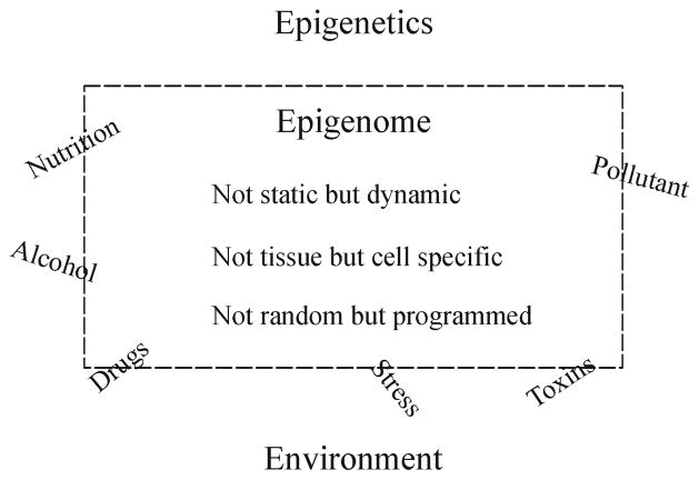
The epigenome is not static but dynamic, and not only tissue specific but are also cell-type specific; its making during development is not random but programmed in orderly manner. The epigneome is now believed permeable and subject to changes. During or after establishment of the epigenome, environment factors such as nutrition, stress, toxin, pollutant, and abusive substance can through changing epigenetics to alter the epigenome.
Acknowledgments
This article is dedicated to my mother who had profoundly influenced me for who I am. The review and the studies on epigenetic program in FCZ’s laboratory are supported by AA016698 and P50 AA07611 to FCZ. The author thanks Yuanyuan Chen for her assistance in preparation of figures and Alison Batka for the manuscript.
References
- Anway MD, Leathers C, Skinner MK. Endocrine disruptor vinclozolin induced epigenetic transgenerational adult-onset disease. Endocrinology. 2006;147(12):5515–5523. doi: 10.1210/en.2006-0640. [DOI] [PMC free article] [PubMed] [Google Scholar]
- Bakulski KM, Rozek LS, Dolinoy DC, Paulson HL, Hu H. Alzheimer’s disease and environmental exposure to lead: the epidemiologic evidence and potential role of epigenetics. Curr Alzheimer Res. 2012;9(5):563–573. doi: 10.2174/156720512800617991. [DOI] [PMC free article] [PubMed] [Google Scholar]
- Bhutani N, Burns DM, Blau HM. DNA demethylation dynamics. Cell. 2011;146(6):866–872. doi: 10.1016/j.cell.2011.08.042. [DOI] [PMC free article] [PubMed] [Google Scholar]
- Bird A. DNA methylation patterns and epigenetic memory. Genes Dev. 2002;16(1):6–21. doi: 10.1101/gad.947102. [DOI] [PubMed] [Google Scholar]
- Bird AP. CpG-rich islands and the function of DNA methylation. Nature. 1986;321(6067):209–213. doi: 10.1038/321209a0. [DOI] [PubMed] [Google Scholar]
- Brandeis M, Ariel M, Cedar H. Dynamics of DNA methylation during development. Bioessays. 1993;15(11):709–713. doi: 10.1002/bies.950151103. [DOI] [PubMed] [Google Scholar]
- Brown DC, Grace E, Sumner AT, Edmunds AT, Ellis PM. ICF syndrome (immunodeficiency, centromeric instability and facial anomalies): investigation of heterochromatin abnormalities and review of clinical outcome. Hum Genet. 1995;96(4):411–416. doi: 10.1007/BF00191798. [DOI] [PubMed] [Google Scholar]
- Brown KD, Robertson KD. DNMT1 knockout delivers a strong blow to genome stability and cell viability. Nat Genet. 2007;39(3):289–290. doi: 10.1038/ng0307-289. [DOI] [PubMed] [Google Scholar]
- Busslinger M, Hurst J, Flavell RA. DNA methylation and the regulation of globin gene expression. Cell. 1983;34(1):197–206. doi: 10.1016/0092-8674(83)90150-2. [DOI] [PubMed] [Google Scholar]
- Caldji C, Hellstrom IC, Zhang TY, Diorio J, Meaney MJ. Environmental regulation of the neural epigenome. FEBS Lett. 2011:2049–2058. doi: 10.1016/j.febslet.2011.03.032. [DOI] [PubMed] [Google Scholar]
- Caldji C, Tannenbaum B, Sharma S, Francis D, Plotsky PM, Meaney MJ. Maternal care during infancy regulates the development of neural systems mediating the expression of fearfulness in the rat. Proc Natl Acad Sci USA. 1998;95(9):5335–5340. doi: 10.1073/pnas.95.9.5335. [DOI] [PMC free article] [PubMed] [Google Scholar]
- Callaghan B, Feldman D, Gruis K, Feldman E. The association of exposure to lead, mercury, and selenium and the development of amyotrophic lateral sclerosis and the epigenetic implications. Neurodegener Dis. 2011;8(1–2):1–8. doi: 10.1159/000315405. [DOI] [PubMed] [Google Scholar]
- Champagne FA, Curley JP. Epigenetic mechanisms mediating the long-term effects of maternal care on development. Neurosci Biobehav Rev. 2009;33(4):593–600. doi: 10.1016/j.neubiorev.2007.10.009. [DOI] [PubMed] [Google Scholar]
- Chia N, Wang L, Lu X, Senut MC, Brenner C, Ruden DM. Hypothesis: environmental regulation of 5-hydroxymethylcytosine by oxidative stress. Epigenetics. 2011;6(7):853–856. doi: 10.4161/epi.6.7.16461. [DOI] [PubMed] [Google Scholar]
- Dawlaty MM, Ganz K, Powell BE, Hu YC, Markoulaki S, Cheng AW, Gao Q, Kim J, Choi SW, Page DC, Jaenisch R. Tet1 is dispensable for maintaining pluripotency and its loss is compatible with embryonic and postnatal development. Cell Stem Cell. 2011;9(2):166–175. doi: 10.1016/j.stem.2011.07.010. [DOI] [PMC free article] [PubMed] [Google Scholar]
- De Carvalho DD, You JS, Jones PA. DNA methylation and cellular reprogramming. Trends Cell Biol. 2010;20(10):609–617. doi: 10.1016/j.tcb.2010.08.003. [DOI] [PMC free article] [PubMed] [Google Scholar]
- Deaton AM, Bird A. CpG islands and the regulation of transcription. Genes Dev. 2011;25(10):1010–1022. doi: 10.1101/gad.2037511. [DOI] [PMC free article] [PubMed] [Google Scholar]
- del Mazo J, Prantera G, Torres M, Ferraro M. DNA methylation changes during mouse spermatogenesis. Chromosome Res. 1994;2(2):147–152. doi: 10.1007/BF01553493. [DOI] [PubMed] [Google Scholar]
- Dolinoy DC. The agouti mouse model: an epigenetic biosensor for nutritional and environmental alterations on the fetal epigenome. Nutr Rev. 2008;66(Suppl 1):S7–S11. doi: 10.1111/j.1753-4887.2008.00056.x. [DOI] [PMC free article] [PubMed] [Google Scholar]
- Dolinoy DC, Huang D, Jirtle RL. Maternal nutrient supplementation counteracts bisphenol A-induced DNA hypomethylation in early development. Proc Natl Acad Sci USA. 2007;104(32):13056–13061. doi: 10.1073/pnas.0703739104. [DOI] [PMC free article] [PubMed] [Google Scholar]
- Dolinoy DC, Weidman JR, Waterland RA, Jirtle RL. Maternal genistein alters coat color and protects Avy mouse offspring from obesity by modifying the fetal epigenome. Environ Health Perspect. 2006;114(4):567–572. doi: 10.1289/ehp.8700. [DOI] [PMC free article] [PubMed] [Google Scholar]
- Duhl DM, Vrieling H, Miller KA, Wolff GL, Barsh GS. Neomorphic agouti mutations in obese yellow mice. Nat Genet. 1994;8(1):59–65. doi: 10.1038/ng0994-59. [DOI] [PubMed] [Google Scholar]
- Gardiner-Garden M, Frommer M. CpG islands in vertebrate genomes. J Mol Biol. 1987;196(2):261–282. doi: 10.1016/0022-2836(87)90689-9. [DOI] [PubMed] [Google Scholar]
- Gisselsson D, Shao C, Tuck-Muller CM, Sogorovic S, Pålsson E, Smeets D, Ehrlich M. Interphase chromosomal abnormalities and mitotic missegregation of hypomethylated sequences in ICF syndrome cells. Chromosoma. 2005;114(2):118–126. doi: 10.1007/s00412-005-0343-7. [DOI] [PubMed] [Google Scholar]
- Goll MG, Bestor TH. Eukaryotic cytosine methyltransferases. Annu Rev Biochem. 2005;74(1):481–514. doi: 10.1146/annurev.biochem.74.010904.153721. [DOI] [PubMed] [Google Scholar]
- Govorko D, Bekdash RA, Zhang C, Sarkar DK. Male germline transmits fetal alcohol adverse effect on hypothalamic proopiomelanocortin gene across generations. Biol Psychiatry. 2012;72(5):378–388. doi: 10.1016/j.biopsych.2012.04.006. [DOI] [PMC free article] [PubMed] [Google Scholar]
- Green ML, Singh AV, Zhang Y, Nemeth KA, Sulik KK, Knudsen TB. Reprogramming of genetic networks during initiation of the Fetal Alcohol Syndrome. Dev Dyn. 2007;236(2):613–631. doi: 10.1002/dvdy.21048. [DOI] [PubMed] [Google Scholar]
- Guo JU, Su Y, Zhong C, Ming GL, Song H. Hydroxylation of 5-methylcytosine by TET1 promotes active DNA demethylation in the adult brain. Cell. 2011;145(3):423–434. doi: 10.1016/j.cell.2011.03.022. [DOI] [PMC free article] [PubMed] [Google Scholar]
- Heijmans BT, Tobi EW, Stein AD, Putter H, Blauw GJ, Susser ES, Slagboom PE, Lumey LH. Persistent epigenetic differences associated with prenatal exposure to famine in humans. Proc Natl Acad Sci USA. 2008;105(44):17046–17049. doi: 10.1073/pnas.0806560105. [DOI] [PMC free article] [PubMed] [Google Scholar]
- Hermann A, Gowher H, Jeltsch A. Biochemistry and biology of mammalian DNA methyltransferases. Cell Mol Life Sci. 2004;61(19–20):2571–2587. doi: 10.1007/s00018-004-4201-1. [DOI] [PMC free article] [PubMed] [Google Scholar]
- Inoue A, Shen L, Dai Q, He C, Zhang Y. Generation and replication-dependent dilution of 5fC and 5caC during mouse preimplantation development. Cell Res. 2011;21(12):1670–1676. doi: 10.1038/cr.2011.189. [DOI] [PMC free article] [PubMed] [Google Scholar]
- Inoue A, Zhang Y. Replication-dependent loss of 5-hydroxymethylcytosine in mouse preimplantation embryos. Science. 2011;334 (6053):194. doi: 10.1126/science.1212483. [DOI] [PMC free article] [PubMed] [Google Scholar]
- Iqbal K, Jin SG, Pfeifer GP, Szabó PE. Reprogramming of the paternal genome upon fertilization involves genome-wide oxidation of 5-methylcytosine. Proc Natl Acad Sci USA. 2011;108(9):3642–3647. doi: 10.1073/pnas.1014033108. [DOI] [PMC free article] [PubMed] [Google Scholar]
- Ito S, D’Alessio AC, Taranova OV, Hong K, Sowers LC, Zhang Y. Role of Tet proteins in 5mC to 5hmC conversion, ES-cell self-renewal and inner cell mass specification. Nature. 2010;466:1129–1136. doi: 10.1038/nature09303. [DOI] [PMC free article] [PubMed] [Google Scholar]
- Ito S, Shen L, Dai Q, Wu SC, Collins LB, Swenberg JA, He C, Zhang Y. Tet proteins can convert 5-methylcytosine to 5-formylcy-tosine and 5-carboxylcytosine. Science. 2011;333(6047):1300–1303. doi: 10.1126/science.1210597. [DOI] [PMC free article] [PubMed] [Google Scholar]
- Jeffy BD, Chirnomas RB, Romagnolo DF. Epigenetics of breast cancer: polycyclic aromatic hydrocarbons as risk factors. Environ Mol Mutagen. 2002;39(2–3):235–244. doi: 10.1002/em.10051. [DOI] [PubMed] [Google Scholar]
- Jones PA, Takai D. The role of DNA methylation in mammalian epigenetics. Science. 2001;293(5532):1068–1070. doi: 10.1126/science.1063852. [DOI] [PubMed] [Google Scholar]
- Kaati G, Bygren LO, Edvinsson S. Cardiovascular and diabetes mortality determined by nutrition during parents’ and grandparents’ slow growth period. Eur J Hum Genet. 2002;10(11):682–688. doi: 10.1038/sj.ejhg.5200859. [DOI] [PubMed] [Google Scholar]
- Kafri T, Gao X, Razin A. Mechanistic aspects of genome-wide demethylation in the preimplantation mouse embryo. Proc Natl Acad Sci USA. 1993;90(22):10558–10562. doi: 10.1073/pnas.90.22.10558. [DOI] [PMC free article] [PubMed] [Google Scholar]
- Kahn HS, Graff M, Stein AD, Lumey LH. A fingerprint marker from early gestation associated with diabetes in middle age: the Dutch Hunger Winter Families Study. Int J Epidemiol. 2009;38(1):101–109. doi: 10.1093/ije/dyn158. [DOI] [PMC free article] [PubMed] [Google Scholar]
- Kaminen-Ahola N, Ahola A, Maga M, Mallitt KA, Fahey P, Cox TC, Whitelaw E, Chong S. Maternal ethanol consumption alters the epigenotype and the phenotype of offspring in a mouse model. PLoS Genet. 2010;6(1):e1000811. doi: 10.1371/journal.pgen.1000811. [DOI] [PMC free article] [PubMed] [Google Scholar]
- Karymov MA, Tomschik M, Leuba SH, Caiafa P, Zlatanova J. DNA methylation-dependent chromatin fiber compaction in vivo and in vitro: requirement for linker histone. FASEB J. 2001;15(14):2631–2641. doi: 10.1096/fj.01-0345com. [DOI] [PubMed] [Google Scholar]
- Kile ML, Baccarelli A, Hoffman E, Tarantini L, Quamruzzaman Q, Rahman M, Mahiuddin G, Mostofa G, Hsueh YM, Wright RO, Christiani DC. Prenatal arsenic exposure and DNA methylation in maternal and umbilical cord blood leukocytes. Environ Health Perspect. 2012;120(7):1061–1066. doi: 10.1289/ehp.1104173. [DOI] [PMC free article] [PubMed] [Google Scholar]
- Koh KP, Yabuuchi A, Rao S, Huang Y, Cunniff K, Nardone J, Laiho A, Tahiliani M, Sommer CA, Mostoslavsky G, Lahesmaa R, Orkin SH, Rodig SJ, Daley GQ, Rao A. Tet1 and Tet2 regulate 5-hydroxymethylcytosine production and cell lineage specification in mouse embryonic stem cells. Cell Stem Cell. 2011;8(2):200–213. doi: 10.1016/j.stem.2011.01.008. [DOI] [PMC free article] [PubMed] [Google Scholar]
- Kriaucionis S, Heintz N. The nuclear DNA base 5-hydroxy-methylcytosine is present in Purkinje neurons and the brain. Science. 2009;324(5929):929–930. doi: 10.1126/science.1169786. [DOI] [PMC free article] [PubMed] [Google Scholar]
- Kucharski R, Maleszka J, Foret S, Maleszka R. Nutritional control of reproductive status in honeybees via DNA methylation. Science. 2008;319(5871):1827–1830. doi: 10.1126/science.1153069. [DOI] [PubMed] [Google Scholar]
- Kundakovic M, Champagne FA. Epigenetic perspective on the developmental effects of bisphenol A. Brain Behav Immun. 2011;25(6):1084–1093. doi: 10.1016/j.bbi.2011.02.005. [DOI] [PMC free article] [PubMed] [Google Scholar]
- Lister R, Pelizzola M, Dowen RH, Hawkins RD, Hon G, Tonti-Filippini J, Nery JR, Lee L, Ye Z, Ngo QM, Edsall L, Antosiewicz-Bourget J, Stewart R, Ruotti V, Millar AH, Thomson JA, Ren B, Ecker JR. Human DNA methylomes at base resolution show widespread epigenomic differences. Nature. 2009;462(7271):315–322. doi: 10.1038/nature08514. [DOI] [PMC free article] [PubMed] [Google Scholar]
- Liu Y, Balaraman Y, Wang G, Nephew KP, Zhou FC. Alcohol exposure alters DNA methylation profiles in mouse embryos at early neurulation. Epigenetics. 2009;4(7):500–511. doi: 10.4161/epi.4.7.9925. [DOI] [PMC free article] [PubMed] [Google Scholar]
- Lumey LH, Stein AD. Transgenerational effects of prenatal exposure to the Dutch famine. BJOG. 2009;116(6):868. doi: 10.1111/j.1471-0528.2009.02110.x. author reply 868. [DOI] [PubMed] [Google Scholar]
- Lumey LH, Stein AD, Kahn HS, van der Pal-de Bruin KM, Blauw GJ, Zybert PA, Susser ES. Cohort profile: the Dutch Hunger Winter families study. Int J Epidemiol. 2007;36(6):1196–1204. doi: 10.1093/ije/dym126. [DOI] [PubMed] [Google Scholar]
- Martínez L, Jiménez V, García-Sepúlveda C, Ceballos F, Delgado JM, Niño-Moreno P, Doniz L, Saavedra-Alanís V, Castillo CG, Santoyo ME, González-Amaro R, Jiménez-Capdeville ME. Impact of early developmental arsenic exposure on promotor CpG-island methylation of genes involved in neuronal plasticity. Neurochem Int. 2011;58(5):574–581. doi: 10.1016/j.neuint.2011.01.020. [DOI] [PubMed] [Google Scholar]
- Mason JB, Choi SW. Effects of alcohol on folate metabolism: implications for carcinogenesis. Alcohol. 2005;35(3):235–241. doi: 10.1016/j.alcohol.2005.03.012. [DOI] [PubMed] [Google Scholar]
- McKay JA, Williams EA, Mathers JC. Folate and DNA methylation during in utero development and aging. Biochem Soc Trans. 2004;32(Pt 6):1006–1007. doi: 10.1042/BST0321006. [DOI] [PubMed] [Google Scholar]
- Meaney MJ, Szyf M. Environmental programming of stress responses through DNA methylation: life at the interface between a dynamic environment and a fixed genome. Dialogues Clin Neurosci. 2005;7(2):103–123. doi: 10.31887/DCNS.2005.7.2/mmeaney. [DOI] [PMC free article] [PubMed] [Google Scholar]
- Morgan HD, Santos F, Green K, Dean W, Reik W. Epigenetic reprogramming in mammals. Hum Mol Genet. 2005;14(Spec1):R47–R58. doi: 10.1093/hmg/ddi114. [DOI] [PubMed] [Google Scholar]
- Nakanishi MO, Hayakawa K, Nakabayashi K, Hata K, Shiota K, Tanaka S. Trophoblast-specific DNA methylation occurs after the segregation of the trophectoderm and inner cell mass in the mouse periimplantation embryo. Epigenetics. 2012;7(2):173–182. doi: 10.4161/epi.7.2.18962. [DOI] [PubMed] [Google Scholar]
- Okano M, Li E. Genetic analyses of DNA methyltransferase genes in mouse model system. J Nutr. 2002;132(8 Suppl):2462S–2465S. doi: 10.1093/jn/132.8.2462S. [DOI] [PubMed] [Google Scholar]
- Otero NK, Thomas JD, Saski CA, Xia X, Kelly SJ. Choline supplementation and DNA methylation in the hippocampus and prefrontal cortex of rats exposed to alcohol during development. Alcohol Clin Exp Res. 2012 doi: 10.1111/j.1530-0277.2012.01784.x. [DOI] [PMC free article] [PubMed] [Google Scholar]
- Ouko LA, Shantikumar K, Knezovich J, Haycock P, Schnugh DJ, Ramsay M. Effect of alcohol consumption on CpG methylation in the differentially methylated regions of H19 and IG-DMR in male gametes-implications for fetal alcohol spectrum disorders. Alcohol Clin Exp Res. 2009;33(9):1615–1627. doi: 10.1111/j.1530-0277.2009.00993.x. [DOI] [PubMed] [Google Scholar]
- Perera F, Herbstman J. Prenatal environmental exposures, epigenetics, and disease. Reprod Toxicol. 2011;31(3):363–373. doi: 10.1016/j.reprotox.2010.12.055. [DOI] [PMC free article] [PubMed] [Google Scholar]
- Pilsner JR, Hu H, Ettinger A, Sánchez BN, Wright RO, Cantonwine D, Lazarus A, Lamadrid-Figueroa H, Mercado-García A, Téllez-Rojo MM, Hernández-Avila M. Influence of prenatal lead exposure on genomic methylation of cord blood DNA. Environ Health Perspect. 2009;117(9):1466–1471. doi: 10.1289/ehp.0800497. [DOI] [PMC free article] [PubMed] [Google Scholar]
- Ramsahoye BH, Davies CS, Mills KI. DNA methylation: biology and significance. Blood Rev. 1996;10(4):249–261. doi: 10.1016/s0268-960x(96)90009-0. [DOI] [PubMed] [Google Scholar]
- Schermelleh L, Haemmer A, Spada F, Rösing N, Meilinger D, Rothbauer U, Cardoso MC, Leonhardt H. Dynamics of Dnmt1 interaction with the replication machinery and its role in postreplicative maintenance of DNA methylation. Nucleic Acids Res. 2007;35(13):4301–4312. doi: 10.1093/nar/gkm432. [DOI] [PMC free article] [PubMed] [Google Scholar]
- Schmid M, Haaf T, Grunert D. 5-Azacytidine-induced under-condensations in human chromosomes. Hum Genet. 1984;67(3):257–263. doi: 10.1007/BF00291352. [DOI] [PubMed] [Google Scholar]
- Singh RP, Shiue K, Schomberg D, Zhou FC. Cellular epigenetic modifications of neural stem cell differentiation. Cell Transplant. 2009;18 (10):1197–1211. doi: 10.3727/096368909X12483162197204. [DOI] [PMC free article] [PubMed] [Google Scholar]
- Stein AD, Zybert PA, van de Bor M, Lumey LH. Intrauterine famine exposure and body proportions at birth: the Dutch Hunger Winter. Int J Epidemiol. 2004;33(4):831–836. doi: 10.1093/ije/dyh083. [DOI] [PubMed] [Google Scholar]
- Stein AD, Zybert PA, van der Pal-de Bruin K, Lumey LH. Exposure to famine during gestation, size at birth, and blood pressure at age 59 y: evidence from the Dutch Famine. Eur J Epidemiol. 2006;21 (10):759–765. doi: 10.1007/s10654-006-9065-2. [DOI] [PubMed] [Google Scholar]
- Suter M, Ma J, Harris A, Patterson L, Brown KA, Shope C, Showalter L, Abramovici A, Aagaard-Tillery KM. Maternal tobacco use modestly alters correlated epigenome-wide placental DNA methylation and gene expression. Epigenetics. 2011;6(11):1284–1294. doi: 10.4161/epi.6.11.17819. [DOI] [PMC free article] [PubMed] [Google Scholar]
- Szulwach KE, Li X, Li Y, Song CX, Wu H, Dai Q, Irier H, Upadhyay AK, Gearing M, Levey AI, Vasanthakumar A, Godley LA, Chang Q, Cheng X, He C, Jin P. 5-hmC-mediated epigenetic dynamics during postnatal neurodevelopment and aging. Nat Neurosci. 2011;14:1607–1616. doi: 10.1038/nn.2959. [DOI] [PMC free article] [PubMed] [Google Scholar]
- Tahiliani M, Koh KP, Shen Y, Pastor WA, Bandukwala H, Brudno Y, Agarwal S, Iyer LM, Liu DR, Aravind L, Rao A. Conversion of 5-methylcytosine to 5-hydroxymethylcytosine in mammalian DNA by MLL partner TET1. Science. 2009;324(5929):930–935. doi: 10.1126/science.1170116. [DOI] [PMC free article] [PubMed] [Google Scholar]
- Tang WY, Levin L, Talaska G, Cheung YY, Herbstman J, Tang D, Miller RL, Perera F, Ho SM. Maternal Exposure to Polycyclic Aromatic Hydrocarbons and 5′-CpG Methylation of Interferon-γ in Cord White Blood Cells. Environ Health Perspect. 2012;120(8):1195–1200. doi: 10.1289/ehp.1103744. [DOI] [PMC free article] [PubMed] [Google Scholar]
- Tawa R, Ono T, Kurishita A, Okada S, Hirose S. Changes of DNA methylation level during pre- and postnatal periods in mice. Differentiation. 1990;45(1):44–48. doi: 10.1111/j.1432-0436.1990.tb00455.x. [DOI] [PubMed] [Google Scholar]
- Waterland RA, Jirtle RL. Transposable elements: targets for early nutritional effects on epigenetic gene regulation. Mol Cell Biol. 2003;23(15):5293–5300. doi: 10.1128/MCB.23.15.5293-5300.2003. [DOI] [PMC free article] [PubMed] [Google Scholar]
- Wolffe AP, Jones PL, Wade PA. DNA demethylation. Proc Natl Acad Sci USA. 1999;96(11):5894–5896. doi: 10.1073/pnas.96.11.5894. [DOI] [PMC free article] [PubMed] [Google Scholar]
- Wright RJ. Epidemiology of stress and asthma: from constricting communities and fragile families to epigenetics. Immunol Allergy Clin North Am. 2011;31(1):19–39. doi: 10.1016/j.iac.2010.09.011. [DOI] [PMC free article] [PubMed] [Google Scholar]
- Wu H, D’Alessio AC, Ito S, Wang Z, Cui K, Zhao K, Sun YE, Zhang Y. Genome-wide analysis of 5-hydroxymethylcytosine distribution reveals its dual function in transcriptional regulation in mouse embryonic stem cells. Genes Dev. 2011;25(7):679–684. doi: 10.1101/gad.2036011. [DOI] [PMC free article] [PubMed] [Google Scholar]
- Wu Q, Ohsako S, Ishimura R, Suzuki JS, Tohyama C. Exposure of mouse preimplantation embryos to 2,3,7,8-tetrachlorodibenzo-p-dioxin (TCDD) alters the methylation status of imprinted genes H19 and Igf2. Biol Reprod. 2004;70(6):1790–1797. doi: 10.1095/biolreprod.103.025387. [DOI] [PubMed] [Google Scholar]
- Wu SC, Zhang Y. Active DNA demethylation: many roads lead to Rome. Nat Rev Mol Cell Biol. 2010;11(9):607–620. doi: 10.1038/nrm2950. [DOI] [PMC free article] [PubMed] [Google Scholar]
- Xu XF, Cheng F, Du LZ. Epigenetic regulation of pulmonary arterial hypertension. Hypertens Res. 2011;34(9):981–986. doi: 10.1038/hr.2011.79. [DOI] [PubMed] [Google Scholar]
- Yildirim O, Li R, Hung JH, Chen PB, Dong X, Ee LS, Weng Z, Rando OJ, Fazzio TG. Mbd3/NURD complex regulates expression of 5-hydroxymethylcytosine marked genes in embryonic stem cells. Cell. 2011;147(7):1498–1510. doi: 10.1016/j.cell.2011.11.054. [DOI] [PMC free article] [PubMed] [Google Scholar]
- Yisraeli J, Frank D, Razin A, Cedar H. Effect of in vitro DNA methylation on beta-globin gene expression. Proc Natl Acad Sci USA. 1988;85(13):4638–4642. doi: 10.1073/pnas.85.13.4638. [DOI] [PMC free article] [PubMed] [Google Scholar]
- Zeisel SH. Gene response elements, genetic polymorphisms and epigenetics influence the human dietary requirement for choline. IUBMB Life. 2007;59(6):380–387. doi: 10.1080/15216540701468954. [DOI] [PMC free article] [PubMed] [Google Scholar]
- Zhou FC, Balaraman Y, Teng M, Liu Y, Singh RP, Nephew KP. Alcohol alters DNA methylation patterns and inhibits neural stem cell differentiation. Alcohol Clin Exp Res. 2011a;35(4):735–746. doi: 10.1111/j.1530-0277.2010.01391.x. [DOI] [PMC free article] [PubMed] [Google Scholar]
- Zhou FC, Chen Y, Love A. Cellular DNA methylation program during neurulation and its alteration by alcohol exposure. Birth Defects Res A Clin Mol Teratol. 2011b;91(8):703–715. doi: 10.1002/bdra.20820. [DOI] [PubMed] [Google Scholar]
- Zhou FC, Zhao Q, Liu Y, Goodlett CR, Liang T, McClintick JN, Edenberg HJ, Li L. Alteration of gene expression by alcohol exposure at early neurulation. BMC Genomics. 2011c;12(1):124. doi: 10.1186/1471-2164-12-124. [DOI] [PMC free article] [PubMed] [Google Scholar]


