

Abstract
Macrophage migration inhibitory factor (MIF) is a pleiotropic proinflammatory cytokine produced by many mammalian tissues including skin. It is also found in many invertebrate parasites of mammals including ticks and may function to aid the parasite to evade the innate and adaptive immune responses in the host. In this study, the cDNA for a MIF gene was sequenced from Sarcoptes scabiei, the scabies mite, using RT-PCR and RACE molecular techniques. The resulting nucleotide sequence had a length of 405 base pairs and the putative amino acid sequences for the mite and tick (Dermacentor variabilis) proteins were identical. The initial steps for the project resulted in the production of expressed scabies mite cDNAs. A real time (qPCR) assay was performed with MIF from scabies mites and various tick species. Results show that mRNA encoding MIF homologues was three times more abundant in the mite samples when compared to RNA prepared from D. variabilis salivary glands and 1.3 times more abundant when compared with RNA prepared from D. variabilis midgut.
Keywords: Sarcoptes scabiei, Macrophage migration inhibitory factor, scabies, tick, Dermacentor variabilis
INTRODUCTION
Macrophage migration inhibitory factor (MIF) is a (multifunctional) pleiotropic proinflammatory cytokine that can modulate various aspects of the innate and immune responses in mammals. It is produced by many tissues including the skin where it is ubiquitously expressed in adults (Shimizu 2005, Gilliver et al. 2011). TNFα and low doses of glucocorticoids induce macrophages themselves to secrete MIF (Gilliver et al. 2011). MIF acts as a chemokine that promotes leukocyte migration including the migration of macrophages to sites of inflammation (Shimizu 2005, Santos and Morand 2009). When stimulated, macrophages themselves produce MIF that inhibits the random migration of macrophages in tissue and thus promotes inflammation (Shimizu et al 2003). MIF can induce secretion of TNFα, INFγ, IL-1β, IL-12, IL-6, IL-8 (CXCL*), MCP-1 and others from mammalian cells and expression of the cell adhesion molecules ICAM-1 and VCAM-1 (Cheng et al. 2010). It can up-regulate expression of TLR4 and thus responses to LPS (Roger et al 2001).
Increased expression of MIF is associated with many skin diseases (Gilliver et al. 2011). It has been associated with dermal endothelial cells of the skin vasculature (Gilliver et al. 2011). MIF can play both a beneficial and destructive role in skin repair (Abe et al. 2000, Ashcroft et al. 2003, Hardman et al. 2005, Zhao et al. 2005, Gilliver et al. 2008, Emmerson et al. 2009, Gilliver et al. 2011). Experiments using rodent showed that fibroblasts from skin wounds up-regulate MIF secretion that is chemotactic to keratinocytes which may facilitate wound healing (Abe et al. 2000, Zhao et al. 2005). It has been shown to promote expression of adhesion molecules and secretion of chemokines by human endothelial cells and increase leukocyte-endothelial cell interactions (Cheng et al. 2010).
MIF is also found in organisms in other phyla including arthropods, helminths and protozoa and is found in a number of invertebrate parasites of mammals including ticks, hookworms, whipworms, roundworms, filarial worms, trypanosomes and coccidians. MIF within parasites may act in many ways to evade the host’s protective innate and adaptive immune responses. Hookworms are able to thrive in the host by evading the natural immune response during tissue migration (Cho et al. 2007). This anti-inflammatory property of MIF in parasites would be an effect that is counter to the pro-inflammatory role of endogenous MIF in the host tissue. However, MIF in ticks is thought to increase inflammation at the feeding site, and thereby increase blood for the tick at the feeding site (Jaworski et al. 2009, Wasala and Jaworski 2012).
Scabies is a contagious skin disease caused by the parasitic mite, Sarcoptes scabiei that burrows in the epidermis of it mammalian hosts. This parasite infests humans, canines, rabbits, sheep, cattle, pigs and many other domestic and wild mammals. During early infestations, humans do not exhibit any skin inflammation for 4 – 8 weeks while the parasite reproduces and develops a well-established population. Scabies mites have had a long evolution with their vertebrate hosts and have developed multiple mechanisms to modulate the host defenses that would otherwise eliminate them. For instance, scabies mites are the source of substances that up-regulate production of IL-1ra by skin cells which effectively blocks the pro-inflammatory activity of IL-1 (Morgan and Arlian 2010). They are able to modulate the levels of IL-8 secreted by skin cells which inhibits the migration of neutrophils from the circulation into the skin (Arlian et al. 2003, Arlian et al. 2006, Elder et al. 2006, Mullins et al. 2009). They also influence the expression of cell adhesion molecules by endothelial cells thereby controlling the trafficking of cells into the lesion where they are located (Elder et al. 2006, Arlian et al. 2007). Mouse gene expression work suggests that they also inhibit B-cell and T-cell co-stimulatory interactions to down-regulate T-cell function (Arlian et al. 2007). Research with T-regulatory cells indicates that they down-modulate host immune response by inducing secretion of the anti-inflammatory IL-10 (Arlian et al. 2006).
Scabies mites are acarines and thus are related to ticks that have been found to have MIF present in their system. Temporally, their association with the host is different. Scabies mites are permanent obligate parasites and must constantly battle the host inflammatory and immune assault as they burrow, feed and reproduce in the host skin. In contrast, ticks need only to down regulate the protective response for short times (days) while they take a blood meal and reproduction and development occur off the host or when not feeding. Ticks with MIF have been given anti-MIF compounds that reduce inflammation at the feeding site (Jaworski et al., 2009). The purpose of this study was to determine if Sarcoptes scabiei expressed genes for the macrophage migration inhibitory factor that may function as an additional mechanism that contributes to their ability to overcoming the host’s protective response to them. The objective of the present study was to produce scabies mite cDNAs and screen these for a MIF homolog in the scabies mite.
MATERIALS AND METHODS
Samples
Twenty-five mg of Sarcoptes scabiei were collected while alive, washed briefly sequentially with phosphate buffered saline with 0.05% Tween® 20, water, and 70% ethanol. The 25 mg of scabies mites were then placed in TRIzol® Reagent solution (Ambion) and frozen at −80°C.
Dermacentor variabilis tissues were dissected from unfed females. Briefly tissues were dissected in cold PBS buffer and transferred directly into TRIzol® Reagent solution (Ambion). Tissues were retained at −80°C.
Animals used for rearing scabies mites and ticks were reared and cared for according to protocols approved by the Institutional Animal Care and Use Committees at Oklahoma State University and Wright State University.
RNA isolation
Scabies mite total RNA was extracted from the mites using the TRIzol® Reagent (Ambion) manufacturer’s RNA protocol. The final RNA pellet was reconstituted with nuclease-free water (Ambion, Canada) and stored at −80°C. Template solutions were aliquoted into two concentrations of 50 ng/ml and 250 ng/ml. The total RNA in the samples was quantified using the ND-1000 nanodrop spectrophotometer (Fisher). Samples with an A260/280 ratio below 1.7 were not used. Total RNA extraction from tick tissues followed the same protocol.
Primers and amplification conditions
DvMIF Exp1 and DvMIF Exp2 primers (Table 1 and Figure 1) were used to amplify scabies MIF. Amplification conditions were 94°C for 30 sec, 35 cycles of 94°C for 5 sec, then 55°C for 2 min, then stored at 4°C. Other portions of the scabies MIF cDNA were amplified with other primers as indication on figure 1. Gene specific primers used were SsRvse3′RACE (GSP2) and SsFwd5′RACE (GSP1, Table 1). SsRACE amplification conditions were 94°C for 30 sec, 35 cycles of 94°C for 5 sec, 68°C for 2 min, then stored at 4°C. Human 18S rRNA amplification conditions were 94°C for 30 sec, 35 cycles of 94°C for 5 sec, 60°C for 2 min, then stored at 4°C.
Table 1.
Primers synthesized for RACE and RT-PCR assays.
| Gene | Forward primer | Reverse primer |
|---|---|---|
| Human 18S RNA | 5′-TTCGAACGTCTGCCCTATCAA-3′ | 5′-GATGTGGTAGCCGTTTCTCAGG-3′ |
| MIF | 5′-AAGCCGCTTTCGTATGTTGTGG-3′ | 5′-TTCCTTGATGCCCAGGGTCTTT-3′ |
| DvMIF | 5′CTCCTTTGGAGAGAGGCAGCCAAT GCTG-3′ | 5′TGTTGTGGTGCACATCAGTCCTGGCCAAT-3′ |
| IsMIF | 5′TGACGAGCCTGTGCCTCGCAAACCTGTA-3′ | 5′TCCTTGATGCCCAGGGTCTTTCTCAATGTGCTC-3′ |
| DvMIF Exp1 | 5′-TGTGTGCTTCTTCTGTTGCGAGT-3′ | 5′-AATCCGAGATACGCAGACTTCTCTCC-3′ |
| DvMIF Exp2 | 5′CATATGTGTGTGCTTCTTCTGTTGCGAGT-3′ | 5′TTCGAAAATCCGAGATACGCAGACTTCTCTCC-3′ |
| SsRACE | 5′-GGCCATTGTGTATTTGGAGCCCTG-3′ | 5′-TTCCTTGATGCCCAGGGTCTTTGC-3′ |
Figure 1.

Sarcoptes scabiei MIF nucleotide sequences and deduced amino acid sequence. Genbank accession number JQ894503.
For reverse transcriptase polymerase chain reaction (RT-PCR) and reverse transcriptase – relative quantitative-PCR (RT-qPCR), MIF primers (Table 1) were generated from the Tick MIF sequence (Jaworski et al, 2001) and yielded a180 bp product. All primers were designed using the Integrated DNA Technologies website. One primer set was evaluated for use as internal controls for gene expression in the RT-PCR assays (Table 1). Human 18S ribosomal RNA primer set (Table 1) was used as an internal control to normalize gene expression for MIF quantification (Bowen et al, 2010). The product resulting from this primer set was sequenced to verify scabies 18S rRNA.
cDNA library construction
For first strand cDNA synthesis 50 ng of scabies RNA was used. After the first strand cDNA was purified, a poly A+ tail was added to the 3′ end of the sequence. For the second strand cDNA synthesis 10 μl of the first strand reaction was used. Adaptor ligation was performed on the dsDNA from the second strand synthesis using the Marathon cDNA Amplification kit® (California, USA) protocol. The adaptor ligation reaction was incubated at 16°C overnight. After the incubation, the reaction was heated at 70°C for 5 min to inactivate the ligase. Undiluted scabies cDNA was stored at −20°C for future use. Two templates of scabies adaptor ligated cDNA were diluted to concentrations of 1/25 and 1/50 adaptor-ligated ds-cDNA in Tricine-EDTA Buffer® for use in RACE reactions. The diluted ds-cDNA was heated at 94°C for 2 min to denature the ds-cDNA and produce an adaptor primer library of scabies mite cDNAs. Our purpose in generating this adaptor primer library was not to establish transcriptomic data for scabies mites, but to qualitatively assess the presence of expressed a MIF gene in the mites.
Sequencing
DNA bands were excised from 1.5% agarose/EtBr gels and purified with the Gene Clean II kit® (California, USA). Gel electrophoresis was conducted at 50 V for ~ 30 min using 17-μl product per well in a 0.1% EtBr in 1.5% agarose gel. Sequencing was carried out at the OSU Protein and Recombinant DNA Core Facility using the Applied Biosystems BigDye® terminator cycle sequencing kit version 1.1 and analyzed with an Applied Biosystems Model 3730 DNA Analyzer®. The resulting sequence was analyzed against possible other sequences using the Basic Local Alignment Search Tool nucleotide collection (BLASTn) on the National Center for Biotechnology Information (NCBI) website.
Two-dimensional gel electrophoresis and immunoblotting
An extract was prepared by grinding 90 mg of scabies mites, washed and collected as above in 20 volumes of endotoxin-free water (Lonza, Walkersville, MD). Following overnight extraction at 4°C, the homogenate was centrifuged at 12,000 ×g and the supernatant was collected. The pellet was resuspended in a second 20 volumes of water, homogenized, extracted and the supernatant collected as before. The supernatants were pooled and sterile filtered into a sterile vial.
Two-dimensional gel electrophoresis was performed using the reagents and protocol provided with the ReadyPrep 2-D Starter Kit from Bio-Rad (Hercules, CA). Prior to analysis, a 200 μL aliquot of the scabies extract was treated with the Bio-Rad ReadyPrep 2-D Cleanup Kit according to the manufacturer’s instructions and the resulting pellet was resuspended in ReadyPrep Rehydration/Sample Buffer. This sample was loaded onto a ReadyStrip pH 4–7 IPG strip using passive rehydration and isoelectric focusing was performed as prescribed. Proteins on the strip were reduced and alkylated and the strip was loaded onto a 10–20% Bio-Rad Criterion polyacrylamide electrophoresis gel. Electrophoresis was performed for 60 min at a constant 200 V.
Following electrophoresis, proteins were transferred for 90 min at 50 V to a ProBlott PVDF membrane (Applied Biosystems, Foster City, CA) in 10 mM CAPS, pH 11 with 10% methanol. Blocking and incubation of the blot was carried out in Dulbecco’s phosphate-buffered saline, 0.2% Tween 20, 5% nonfat milk and 3% goat serum. The blot was probed with rabbit antiserum to MIF (AB1 and AB2, each 1/100), followed by alkaline phosphatase-conjugated goat anti-rabbit Ig (Fisher) and developed with AP-Blue Membrane Substrate (Sigma). These antisera were previously characterized and shown to recognize purified tick MIF (expressed, cloned and purified) by immunoblotting (Bowen et al., 2009, Jaworski et al., 2001).
Reverse transcriptase-PCR and reverse transcriptase-relative quantitative PCR
A MJ Research Inc.® PTC-100 Reverse Transcriptase Polymerase Chain Reaction (RT-PCR) thermocycler was used to amplify scabies MIF. A modified Ambion AgPath-ID™ One-Step RT-PCR Kit (Ambion, Canada) protocol was used. MIF amplification conditions were 50°C for 4 min, 95°C for 15 min, 35 cycles of 94°C for 1 min (denature cycle), 58°C for 1 min (annealing cycle), 72°C for 1 min (extension cycle), followed by 72°C for 10 min then stored at 4°C. Three 25μl samples of scabies RNA and one negative control were used. Gel electrophoresis was performed at 50 V for 30 minutes using 14-μl product per well in a 0.1% ethidium bromide (EtBr) in 1.5% agarose gel to confirm presence of an amplicon.
Reverse transcriptase-relative quantitative-PCR (RT-qPCR) was completed using a modified Ambion AgPath- ID™ One-Step RT-PCR Kit (Ambion, Canada). FastStart Universal SYBR Green Master with Rox (Roche Diagnostics, Indiana, USA) replaced the AgPath 2x RT-PCR Buffer. Primers (as described in Table 1) were diluted to 10 μM concentrations. AgPath-ID protocols were used for appropriate reagent volumes for a 25 μl final volume. 10 ng of template was used for each reaction. Samples were mixed in 96-well plates and then placed into an Applied Biosystems 7500 Real Time PCR system. Amplification conditions were 50°C for 4 min, 95°C for 15 min, 40 cycles of 95°C for 15 sec and 55°C for 1 min. Microsoft Excel was used for Ct values and MIF abundance levels were normalized against human 18S ribosomal RNA control (Bowen et al, 2010). Three replicates were averaged and the averages were then used to calculate the Ct value for each sample (Bowen et al, 2010).
RESULTS
The nucleotide sequence for Sarcoptes scabiei MIF was determined and the putative amino acid sequence was deduced (Figure 1, Genbank accession number JQ894503). The putative amino acid sequence traversed the complete open reading frame (ORF) of 405 nucleotides (Figure 1). The nucleotide sequence for Sarcoptes scabiei MIF homologue had 98% identity to Dermacentor variabilis MIF homologue (Figure 2). An alignment of the amino acid sequences showed a high identity between the three tick species and the scabies mite MIF homologues (Figure 3). In fact, the putative amino acid sequences for S. scabiei and D. variabilis were identical. The putative amino acid sequence from the scabies mite MIF homologous gene contained a specific peptide sequence, formerly found only in ticks (Figure 3). Utilizing antibody to this specific peptide, the presence of a tick MIF homologue protein in scabies mite extract was confirmed by immunoblotting (Figure 4). Two-dimensional gels were blotted and probed with rabbit antiserum to tick MIF peptide to demonstrate specific antibody binding to a spot with apparent molecular weight of 11.0 kDa and a pI 6.6 (Figure 4).
Figure 2.

Alignment of MIF nucleotide sequences from Dermacentor variabilis (Dv, Genbank accession number JQ026322), Amblyomma americanum (Aa, Genbank accession number AF126688), Haemaphysalis longicornis (Hl, Genbank accession number AB255601), and Sarcoptes scabiei (Ss, Genbank accession number JQ894503).
Figure 3.
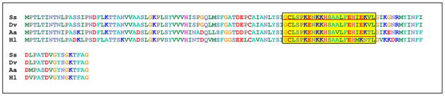
Amino acid sequence alignment of putative MIF genes between Dermacentor variabilis (Dv), Amblyomma americanum (Aa), Haemaphysalis longicornis (Hl), and Sarcoptes scabiei (Ss). Boxed region shows the specific peptide to which specific tick MIF antibodies were produced.
Figure 4.

Identification of Sarcoptes scabiei Macrophage migration inhibitory factor (MIF) by specific tick anti-MIF peptide antibodies. Protein extracts from Sarcoptes scabiei separated by two-dimensional gel electrophoresis and transferred to PVDF membranes as described in the methods. Panel A. Immunoblot of 2D gel probed with peroxidase-labeled goat anti-rabbit Ig to located rabbit antibody in the mites preparation (red spots). Panel B. Immunoblot of 2D gel probed with anti-MIF peptide antibodies. Panel C. Immunoblot of 2D gel probed with serum from scabies infested rabbits. MIF is indicated with arrow in Panels B and C.
To gain some information about the relative abundance of scabies MIF homologue compared to tick MIF homologue, a relative quantitative real time PCR assay (qPCR) was done. Scabies MIF homologous gene was over three times that of D. variabilis salivary gland MIF homologue (Figure 5). D. variabilis salivary gland RNA was set at 1.0 as the reference sample to show comparisons between it and D. variabilis midgut mRNA samples and the mRNA tissue samples from S. scabiei. RNA encoding MIF homologues in D. variabilis midgut samples was 1.3 times more abundant than that of MIF homologues in the D. variabilis salivary gland mRNA; and the mRNA encoding scabies mite MIF homologue was over three fold higher than the reference sample.
Figure 5.

MIF gene real time relative quantitative PCR assay (qPCR) using RNA purified from tissues of Sarcoptes scabiei (SsMIF), and salivary glands (DvSGMIF) and midgut epithelium (DvMGMIF)from unfed female Dermacentor variabilis. Fold differences relative to MIF in DvSGMIF are shown on the figure.
DISCUSSION
This study identified a MIF gene homologue from the scabies mite. Surprisingly, while the nucleotide sequence differed slightly, the putative amino acid sequence was exactly the same as the putative amino acid sequence from D. variabilis ticks. We ruled out the possibility of contamination of human MIF or MIF homologues from symbiotic organisms. The scabies mite amino acid sequence contained a specific peptide that previously had only been found in tick MIF homologues (Jaworski et al. 2001; Wasala and Jaworski 2012). In a phylogenetic analysis of ticks, nematodes, and vertebrate MIF homologues, the tick MIF homologues were closely related and clustered with nematode MIFs, while the human and other vertebrate MIF homologous genes were distinctly separated (Jaworski et al, 2001; Wasala and Jaworski 2012). Previously the genomic data for the tick MIF gene was characterized from Amblyomma americanum ticks (Jaworski et al, 2001). The genomic structure for the tick MIF gene was different from that of MIF genes in other organisms. The organization of the tick gene revealed three exons separated by wide intronic regions. Pastrana et al., 1998 compared the gene structure between Brugia malayi (a parasitic nematode), human, mouse, and Caenorhabditis elegans (a nonparasitic nematode) MIF genes. All of these organisms, including ticks had a similar length first exon. Subsequent spacing between the exons and introns differed greatly between ticks and the other organisms. Also, the B. malayi and C. elegans MIF homoogues consisted of a different number of exons than three. We suggest that scabies mite MIF encoding gene is likely to have the same gene structure as A. americanum.
MIF gene homologues have been found in several tick species. For A. americanum (Jaworski et al., 2001), a MIF protein was expressed and purified and tested in vitro for its biological properties compared to human recombinant MIF. This protein was equivalent to human recombinant MIF in its ability to inhibit random macrophage migration and it had ten times less dopachrome tautomerase activity than the human MIF. Through the results of both in vitro and in vivo studies, it is postulated that MIF’s function is to induce inflammation at the tick feeding site and thereby increase the blood available for ingestion (Jaworski et al. 2001, 2009; Wasala and Jaworski 2012). Ticks feed by creating an incision through the epidermis and dermis that ruptures the blood vessels and then suck blood from the hemorrhage pool as it accumulates in the lesion in the dermis. In contrast, scabies mites burrow into the avascular strateum corneum of the epidermis. They feed on the plasma that seeps into the burrow near their mouthparts (Arlian et al. 1988). They do not feed on blood and do not normally rupture or penetrate blood vessels as do ticks. Scabies mites seem to have evolved a two-fold strategy for survival in the host skin: (1) they down-modulate key aspects of the host innate and adaptive immune responses aimed at eliminating them, and (2) they promote host responses that enhance the accumulation of plasma in the vicinity of their mouthparts to facilitate feeding. Therefore, the desirability of MIF for scabies mites feeding is similar to that for ticks even though ticks and mites utilize different components of blood.
MIF is generally regarded as a pro-inflammatory molecule. Vasodilation to increase vascular permeability to enhance food (plasma) availability is advantageous to the scabies mite. We previously found that scabies mites induce secretion of VEGF by skin keratinocytes and fibroblasts (Arlian et al. 2003). VEGF induces increased vascularization and plasma leakage in the vicinity of the burrowing mite providing them with a source of nutrients. Thus, our findings that these mites also up-regulate a MIF-like gene suggests that this may be a redundant mechanism evolved to insure that they have a food and fluid source in the otherwise dry strateum corneum where they live.
The presence of a gene for MIF in scabies mites is of particular interest. In humans first infested with scabies mites, there is a delay before there is an inflammatory reaction that is manifested by skin clinical symptoms. Our histological studies describing inflammatory cell infiltration into the scabietic lesion of infested hosts show that early infiltrates in the dermis and at the epidermal/dermal junction are composed of many neutrophils and plasma cells and few macrophages (Arlian et al. 1994, Arlian et al. 1996, Stemmer et al. 1996, Arlian et al. 1997). During a primary scabies infestation, very few macrophages were observed in cellular infiltrates in the vicinity of burrowing mites (Arlian et al. 1994, Arlian et al. 1996, Stemmer et al. 1996, Arlian et al. 1997). If MIF is present in mite salivary secretions, perhaps it is secreted into the host during burrowing and inhibits the ability of macrophages to migrate to the location of a mite during the time when there are few skin signs. This would offer the mite a substantial survival advantage by allowing a mite population to develop. Thus, scabies mite MIF could explain the absence of macrophages that we have noted in our earlier studies.
In this study, we also utilized our established MIF qPCR assay to compare the relative abundance of scabies mites MIF homologue to that of tick MIF homologue. From these studies, we found that scabies MIF homologue was abundant at a higher level than that of D. variabilis tick MIF homologue. Previously, Bowen et al. (2010) and Wasala et al. (2012) established that MIF encoding gene homologues in A. americanum and D. variabilis, respectively, were most abundant in mRNA from the midgut cells of feeding ticks, and that MIF protein pools were present in the midgut before attachment to a host. The ticks used in our experiment were unfed, while the scabies mites were feeding before being removed from the host. This intake of host nutrients could lead to a higher abundance level of MIF in the scabies mite versus the MIF of unfed ticks. These studies also indicate that MIF is not a house-keeping gene in scabies mites.
MIF alone, or along with other immune-modulating molecules, is a potential target for vaccine development to reduce the transmission, prevalence, and impact of scabies in susceptible populations. Jaworski et al. (2009) showed that by using a tick MIF peptide to immunize rabbit hosts, A. americanum ticks did not attach as readily to the immunized hosts and the feeding period was lengthened due to an unproductive feeding lesion. Some scenarios for using anti-mite MIF or peptide would be to increase macrophage activity and inflammation in the host thereby potentially decreasing mite feeding and reducing mite fecundity and survival.
Macrophage migration factor found in scabies mites
amino acid sequence identical to tick MIF
MIF abundance in scabies mites was higher than tick tissue MIFs
Acknowledgments
The authors acknowledge the helpful review by Jack Dillwith and Justin Talley. This paper was approved for publication by the Director of the Oklahoma Agricultural Experiment Station and supported under project OKL02623. In addition, we would like to express our appreciation to the United States Navy Medical Service Corps In-service Procurement Program for providing support for NMC. The project described was supported in part by Award Number R01AI01725 from the National Institute of Allergy and Infectious Diseases. The content is solely the responsibility of the authors and does not necessarily represent the official views of the National Institute of Allergy and Infectious Diseases of the National Institutes of Health.
Footnotes
Publisher's Disclaimer: This is a PDF file of an unedited manuscript that has been accepted for publication. As a service to our customers we are providing this early version of the manuscript. The manuscript will undergo copyediting, typesetting, and review of the resulting proof before it is published in its final citable form. Please note that during the production process errors may be discovered which could affect the content, and all legal disclaimers that apply to the journal pertain.
LITERATURE CITED
- Abe R, Shimizu T, Ohkawara A, Nishihira J. Enhancement of macrophage migration inhibitory factor (MIF) expression in injured epidermis and cultured fibroblasts. Biochim Biophys Acta. 2000;1500:1–9. doi: 10.1016/s0925-4439(99)00080-0. [DOI] [PubMed] [Google Scholar]
- Arlian LG, Fall N, Morgan MS. In vivo evidence that Sarcoptes scabiei (Acari: Sarcoptidae) is the source of molecules that modulate splenic gene expression. J Med Entomol. 2007;44:1054–1063. doi: 10.1603/0022-2585(2007)44[1054:ivetss]2.0.co;2. [DOI] [PubMed] [Google Scholar]
- Arlian LG, Morgan MS, Neal JS. Modulation of cytokine expression in human keratinocytes and fibroblasts by extracts of scabies mites. Am J Trop Med Hyg. 2003;69:652–656. [PubMed] [Google Scholar]
- Arlian LG, Morgan MS, Paul CC. Evidence that scabies mites (Acari: Sarcoptidae) influence production of interleukin-10 and the function of T-regulatory cells (Tr1) in humans. J Med Entomol. 2006;43:283–287. doi: 10.1603/0022-2585(2006)043[0283:etsmas]2.0.co;2. [DOI] [PubMed] [Google Scholar]
- Arlian LG, Morgan MS, Rapp CM, Vyszenski-Moher DL. The development of protective immunity in canine scabies. Vet Parasitol. 1996;62:133–142. doi: 10.1016/0304-4017(95)00854-3. [DOI] [PubMed] [Google Scholar]
- Arlian LG, Rapp CM, Stemmer BL, Morgan MS, Moore PF. Characterization of lymphocyte subtypes in scabietic skin lesions of naive and sensitized dogs. Vet Parasitol. 1997;68:347–358. doi: 10.1016/s0304-4017(96)01093-x. [DOI] [PubMed] [Google Scholar]
- Arlian LG, Rapp CM, Vyszenski-Moher DL, Morgan MS. Sarcoptes scabiei: histopathological changes associated with acquisition and expression of host immunity to scabies. Exp Parasitol. 1994;78:51–63. doi: 10.1006/expr.1994.1005. [DOI] [PubMed] [Google Scholar]
- Arlian LG, Runyan RA, Vyszenski-Moher DL. Water balance and nutrient procurement of Sarcoptes scabiei var. canis (Acari: Sarcoptidae) J Med Entomol. 1988;25:64–68. doi: 10.1093/jmedent/25.1.64. [DOI] [PubMed] [Google Scholar]
- Ashcroft GS, Mills SJ, Lei K, Gibbons L, Jeong MJ, Taniguchi M, Burow M, Horan MA, Wahl SM, Nakayama T. Estrogen modulates cutaneous wound healing by downregulating macrophage migration inhibitory factor. J Clin Invest. 2003;111:1309–1318. doi: 10.1172/JCI16288. [DOI] [PMC free article] [PubMed] [Google Scholar]
- Bowen CJ, Jaworski DC, Wasala NB, Coons LB. Macrophage migration inhibitory factor expression and protein localization in Amblyomma americanum (Ixodidae) Experimental and Applied Acarology. 2010;50:343–352. doi: 10.1007/s10493-009-9324-5. [DOI] [PubMed] [Google Scholar]
- Bucala Richard, Morand EF, Leech Michelle. Macrophage migration inhibitory factor: An emerging therapeutic target in Rheumatoid arthritis. Arthritis and Rheumatism. 2003;48(2):291–299. doi: 10.1002/art.10728. [DOI] [PubMed] [Google Scholar]
- Cheng Q, McKeown SJ, Santos L, Santiago FS, Khachigian LM, Morand EF, Hickey MJ. Macrophage migration inhibitory factor increases leukocyte-endothelial interactions in human endothelial cells via promotion of expression of adhesion molecules. J Immunol. 2010;185:1238–1247. doi: 10.4049/jimmunol.0904104. [DOI] [PubMed] [Google Scholar]
- Cho Y, Jones BF, Vermeire JJ, Leng L, DiFedele L, Harrison LM, Xiong H, Kwong YA, Chen Y, Bucala R, Lolis E, Cappello M. Structural and functional characterization of a secreted hookworm Macrophage Migration Inhibitory Factor (MIF) that interacts with the human MIF receptor CD74. Journal of Biological Chemistry. 2007;282(32):23447–23456. doi: 10.1074/jbc.M702950200. [DOI] [PMC free article] [PubMed] [Google Scholar]
- Elder BL, Arlian LG, Morgan MS. Sarcoptes scabiei (Acari: Sarcoptidae) mite extract modulates expression of cytokines and adhesion molecules by human dermal microvascular endothelial cells. J Med Entomol. 2006;43:910–915. doi: 10.1603/0022-2585(2006)43[910:ssasme]2.0.co;2. [DOI] [PMC free article] [PubMed] [Google Scholar]
- Emmerson E, Campbell L, Ashcroft GS, Hardman MJ. Unique and synergistic roles for 17beta-estradiol and macrophage migration inhibitory factor during cutaneous wound closure are cell type specific. Endocrinology. 2009;150:2749–2757. doi: 10.1210/en.2008-1569. [DOI] [PubMed] [Google Scholar]
- Estes SA, Kummel B, Arlian L. Experimental canine scabies in humans. Journal of the American Academy of Dermatology. 1983;9:397–401. doi: 10.1016/s0190-9622(83)70148-9. [DOI] [PubMed] [Google Scholar]
- Gilliver SC, Emmerson E, Bernhagen J, Hardman MJ. MIF: a key player in cutaneous biology and wound healing. Exp Dermatol. 2011;20:1–6. doi: 10.1111/j.1600-0625.2010.01194.x. [DOI] [PubMed] [Google Scholar]
- Gilliver SC, Ruckshanthi JP, Hardman MJ, Nakayama T, Ashcroft GS. Sex dimorphism in wound healing: the roles of sex steroids and macrophage migration inhibitory factor. Endocrinology. 2008;149:5747–5757. doi: 10.1210/en.2008-0355. [DOI] [PubMed] [Google Scholar]
- Hardman MJ, Waite A, Zeef L, Burow M, Nakayama T, Ashcroft GS. Macrophage migration inhibitory factor: a central regulator of wound healing. Am J Pathol. 2005;167:1561–1574. doi: 10.1016/S0002-9440(10)61241-2. [DOI] [PMC free article] [PubMed] [Google Scholar]
- Jaworski DC, Jasinskas A, Metz CN, Buccala R, Barbour AG. Identification and characterization of a homologue of the pro-inflammatory cytokine Macrophage Migration Inhibitory Factor in the tick, Amblyomma americanum. Insect Molecular Biology. 2001;10(4):323–331. doi: 10.1046/j.0962-1075.2001.00271.x. [DOI] [PubMed] [Google Scholar]
- Jaworski DC, Bowen CJ, Wasala NB. Amblyomma americanum (L): Tick macrophage migration inhibitory factor peptide immunization lengthens lone star tick feeding intervals in vivo. Experimental Parasitology. 2009;121:384–387. doi: 10.1016/j.exppara.2008.12.003. [DOI] [PubMed] [Google Scholar]
- Kuhn C, Lucius R, Matthes HF, Meusel G, Reich B, Kalinna BH. Characterisation of recombinant immunoreactive antigens of the scab mite Sarcoptes scabiei. Veterinary Parasitology. 2008;153:329–337. doi: 10.1016/j.vetpar.2008.02.007. [DOI] [PubMed] [Google Scholar]
- Morgan MS, Arlian LG. Response of human skin equivalents to Sarcoptes scabiei. J Med Entomol. 2010;47:877–883. doi: 10.1603/me10012. [DOI] [PMC free article] [PubMed] [Google Scholar]
- Mullins JS, Arlian LG, Morgan MS. Extracts of Sarcoptes scabiei De Geer downmodulate secretion of IL-8 by skin keratinocytes and fibroblasts and of GM-CSF by fibroblasts in the presence of proinflammatory cytokines. J Med Entomol. 2009;46:845–851. doi: 10.1603/033.046.0415. [DOI] [PMC free article] [PubMed] [Google Scholar]
- Pastrana Diana V, Raghavan Nithy A, Fitzgerald Peter, Eisinger Stephen W, Metz Christine, Bucala Richard, Schleimer Robert P, Bickel Carol, Scott Alan L. Filarial nematode parasites secrete a homologue of the human cytokine Macrophage migration inhibitory factor. Infection and Immunity. 1998;66(12):5955–5963. doi: 10.1128/iai.66.12.5955-5963.1998. [DOI] [PMC free article] [PubMed] [Google Scholar]
- Roger T, David J, Glauser MP, Calandra T. MIF regulates innate immune responses through modulation of Toll-like receptor 4. Nature. 2001;414:920–924. doi: 10.1038/414920a. [DOI] [PubMed] [Google Scholar]
- Routh HB, Mirensky YM, Parish LC, Witkowski JA. Ectoparasites as sexually transmitted diseases. Seminars in Dermatology. 1994;13(4):243–247. [PubMed] [Google Scholar]
- Sambrook J, Russell DW. Rapid amplification of 5′ cDNA ends. Molecular Cloning: A Laboratory Manual. 2001;8(9):8.54–8.60. [Google Scholar]
- Santos LL, Morand EF. Macrophage migration inhibitory factor: a key cytokine in RA, SLE and atherosclerosis. Clin Chim Acta. 2009;399:1–7. doi: 10.1016/j.cca.2008.09.014. [DOI] [PubMed] [Google Scholar]
- Shimizu T. Role of macrophage migration inhibitory factor (MIF) in the skin. J Dermatol Sci. 2005;37:65–73. doi: 10.1016/j.jdermsci.2004.08.007. [DOI] [PubMed] [Google Scholar]
- Shimizu T, Abe R, Nishihira J, Shibaki A, Watanabe H, Nakayama T, Taniguchi M, Ishibashi T, Shimizu H. Impaired contact hypersensitivity in macrophage migration inhibitory factor-deficient mice. European Journal of Immunology. 2003;33:1478–1487. doi: 10.1002/eji.200323751. [DOI] [PubMed] [Google Scholar]
- Steinhoff M, Meinhardt A, Steinhoff A, Gemsa D, Bucala R, Bacher M. Evidence for a role of macrophage migration inhibitory factor in psoriatic skin disease. British Journal of Dermatology. 1999;141:1061–1066. doi: 10.1046/j.1365-2133.1999.03206.x. [DOI] [PubMed] [Google Scholar]
- Stemmer BL, Arlian LG, Morgan MS, Rapp CM, Moore PF. Characterization of antigen presenting cells and T-cells in progressing scabietic skin lesions. Vet Parasitol. 1996;67:247–258. doi: 10.1016/s0304-4017(96)01038-2. [DOI] [PubMed] [Google Scholar]
- Walton SF, Currie BJ. Problems in diagnosing scabies, a global disease in human and animal populations. Clinical Microbiology Reviews. 2007;20(2):268–279. doi: 10.1128/CMR.00042-06. [DOI] [PMC free article] [PubMed] [Google Scholar]
- Wasala NB, Jaworski DC. Dermacentor variabilis: Characterization and modeling of macrophage migration inhibitory factor with phylogenetic comparisons to other ticks, insects and parasitic nematodes. Exp Parasitol. 2012;130:232–238. doi: 10.1016/j.exppara.2011.12.010. [DOI] [PubMed] [Google Scholar]
- Wasala NB, Bowen CJ, Jaworski DC. Expression and regulation of macrophage migration inhibitory factor (MIF) in feeding American dog ticks (Dermacentor variabilis Say) Exp Appl Acarol. 2012;57:179–187. doi: 10.1007/s10493-012-9550-0. [DOI] [PubMed] [Google Scholar]
- Zhao Y, Shimizu T, Nishihira J, Koyama Y, Kushibiki T, Honda A, Watanabe H, Abe R, Tabata Y, Shimizu H. Tissue regeneration using macrophage migration inhibitory factor-impregnated gelatin microbeads in cutaneous wounds. Am J Pathol. 2005;167:1519–1529. doi: 10.1016/S0002-9440(10)61238-2. [DOI] [PMC free article] [PubMed] [Google Scholar]