

Abstract
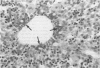
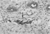
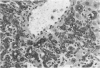
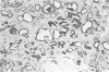
A polyclonal anti-cytokeratin antibody has been used to examine the expression of this intermediate filament both during normal development in the rat and in a variety of pathological states in the rat and mouse. Bile duct proliferation induced by the administration of alpha-naphthylisothiocyanate (ANIT) as well as the oval cell proliferation induced by 3'-methyl-4-dimethylaminoazobenzene (3-MeDAB) have been used to examine the expression of the rodent cytokeratins in the proliferating cells regarded as being of bile duct origin. Examples of cholangiofibrosis and cholangiocarcinomas were also examined for evidence of cytokeratin expression using this antibody, as well as proliferations of a morphological intermediate type between epithelial and mesenchymal. In all cases we have been able to demonstrate continuity of phenotypic expression of the cytokeratins recognized by this antibody in cells which are recognized as bile duct in origin, even where their morphological appearance does not resemble an epithelial cell type. Because this antibody can be used on formalin-fixed, paraffin-processed tissues, after trypsin treatment, it is proposed that it can be used routinely in the toxicological evaluation (even retrospectively) of bile duct related proliferations and tumours.
Full text
PDF


Images in this article


Selected References
These references are in PubMed. This may not be the complete list of references from this article.
- GOLDFARB S., SINGER E. J., POPPER H. Experimental cholangitis due to alpha-naphthyl-isothiocyanate (ANIT). Am J Pathol. 1962 Jun;40:685–698. [PMC free article] [PubMed] [Google Scholar]
- Hayner N. T., Braun L., Yaswen P., Brooks M., Fausto N. Isozyme profiles of oval cells, parenchymal cells, and biliary cells isolated by centrifugal elutriation from normal and preneoplastic livers. Cancer Res. 1984 Jan;44(1):332–338. [PubMed] [Google Scholar]
- Imoto M., Nishimura D., Fukuda Y., Sugiyama K., Kumada T., Nakano S. Immunohistochemical detection of alpha-fetoprotein, carcinoembryonic antigen, and ferritin in formalin-paraffin sections from hepatocellular carcinoma. Am J Gastroenterol. 1985 Nov;80(11):902–906. [PubMed] [Google Scholar]
- Johnson N. F., Edwards R. E., Munday D. E., Rowe N., Wagner J. C. Pluripotential nature of mesotheliomata induced by inhalation of erionite in rats. Br J Exp Pathol. 1984 Jun;65(3):377–388. [PMC free article] [PubMed] [Google Scholar]
- PRICE J. M., HARMAN J. W., MILLER E. C., MILLER J. A. Progressive microscopic alterations in the livers of rats fed the hepatic carcinogens 3'-methyl-4-dimethylaminoazobenzene and 4'-fluoro-4-dimethylaminoazobenzene. Cancer Res. 1952 Mar;12(3):192–200. [PubMed] [Google Scholar]
- Thung S. N., Gerber M. A., Sarno E., Popper H. Distribution of five antigens in hepatocellular carcinoma. Lab Invest. 1979 Aug;41(2):101–105. [PubMed] [Google Scholar]
- Van Eyken P., Sciot R., Desmet V. Intrahepatic bile duct development in the rat: a cytokeratin-immunohistochemical study. Lab Invest. 1988 Jul;59(1):52–59. [PubMed] [Google Scholar]