

Dear Editors,
A 40‐year‐old man with type 2 diabetes mellitus of 2 years duration was admitted as a result of a non‐healing plantar ulcer, 2 cm in diameter, located at the distal first metatarsal (Figure 1). One year ago, he had been hospitalised because of a severely infected ulcer at the same location, which required plantar and dorsal incisions for surgical drainage. Following 6 weeks of treatment comprising antibiotherapy, offloading and wound care, the wound was closed with a fasciocutaneous flap. Two months after discharge, however, a plantar ulcer reoccurred at the surgical site and persisted to date. Physical examination showed palpable foot pulses, severe peripheral sensory neuropathy and significant restriction of the range of motion in the metatarsophalangeal joint. An X‐ray showed synostosis between the metatarsal heads and proximal phalanges of the first and second toes (Figure 2).
Figure 1.
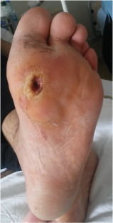
Chronic non‐healing plantar ulcer located at the distal first metatarsal.
Figure 2.
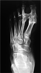
An X‐ray showing synostosis between the metatarsal heads and proximal phalanges of the first and second toes.
Iatrogenic synostosis usually occurs as a result of trauma or soft‐tissue damage, haemorrhage or subperiosteal dissection across the interosseous membrane during surgery. Once occurred, the treatment of choice is conservative. Some patients, however, may require resection of synostosis to improve motion and to prevent complications.
Bony synostosis may lead to structural foot deformities and complications, such as hallux rigidus, limited joint mobility and high plantar pressure, which are strongly associated with ulceration of the great toe and the first metatarsal 1, 2, 3. A careful surgical technique, particularly avoiding dissection of the interosseous membrane, may help prevent synostosis following surgical interventions of the diabetic foot.
Ali Memis, MD1, Omer Ersen, MD2, Ali Kemal Sivrioglu, MD3, Mesut Mutluoglu, MD1 & Hakan Ay1, MD
1Department of Undersea and Hyperbaric Oxygen, Gulhane Haydarpasa Training Hospital, Istanbul, Turkey
drmutluoglu@gmail.com
2Department of Orthopaedics, Maresal Cakmak Military Hospital, Erzurum, Turkey
3Department of Radiology, Kasimpasa Military Hospital, Istanbul, Turkey
References
- 1. Lavery LA, Armstrong DG, Vela SA, Quebedeaux TL, Fleischli JG. Practical criteria for screening patients at high risk for diabetic foot ulceration. Arch Intern Med 1998;158:157–62. [DOI] [PubMed] [Google Scholar]
- 2. Lavery LA, Peters EJG, Armstrong DG. What are the most effective interventions in preventing diabetic foot ulcers? Int Wound J 2008;5:425–33. [DOI] [PMC free article] [PubMed] [Google Scholar]
- 3. Lavery LA. Effectiveness and safety of elective surgical procedures to improve wound healing and reduce re‐ulceration in diabetic patients with foot ulcers. Diabetes Metab Res Rev 2012;28(Suppl 1):60–3. [DOI] [PubMed] [Google Scholar]