

Abstract
Two male and two female pet rabbits of about 1 year of age, naturally infested with Notoedres cuniculi were studied in the present investigation. Clinical examination revealed presence of crustaceous lesions on the head, ear pinnae, both the limbs and external genitalia. Microscopic examination of skin scrapings confirmed presence of Notoedres mange infestation. Biochemical parameters revealed altered values of total serum protein, albumin, globulin, and activities of aspartate amino transferase, alanine amino transferase and alkaline phosphatase respectively. The affected rabbits were treated with two injections of Ivermectin (400 μg/kg, subcutaneously, at interval of 15 days) along with supportive therapy. This treatment showed appreciable improvement in clinical signs and restoration in biochemical parameters.
Keywords: Rabbits, Notoedres cuniculi, Ivermectin, Biochemical parameters
Introduction
Mange is a commonly encountered in rabbit production, where considerable scourge has been attributed by Notoedres cuniculi (Aulakh et al. 2003). Notoedric mange is present in a large rabbit colony and would act a serious menace unless adequate preventive measures are undertaken to control it (Osborne 1947). Present communication reports clinico-biochemical and therapeutic studies on Notoedric mange in pet rabbits.
Case history and clinical examination
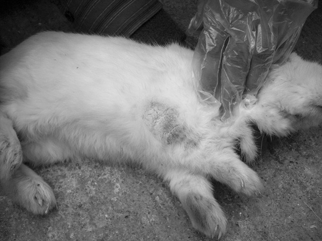
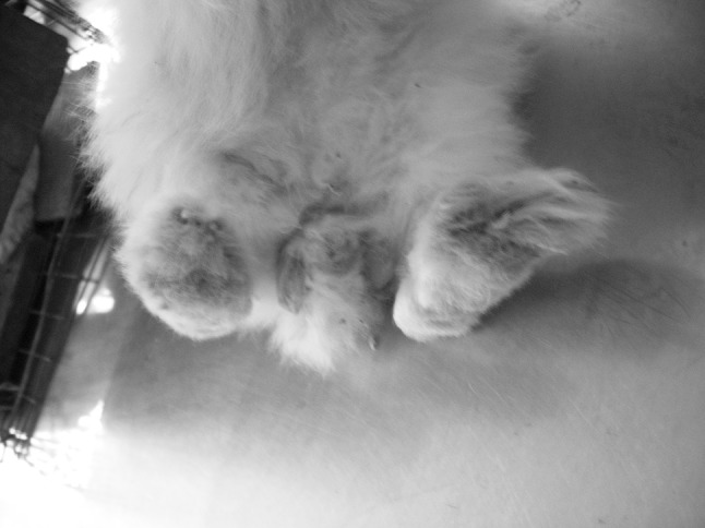
Two male and two female pet rabbits of about 1 years of age were presented with crustaceous lesions on the head, ears, fore limbs, hind limbs and the external genitalia. Anthology of history revealed that the owner housed the rabbit in a moist, dirty and damp environment, and maintained exclusively on homemade food. Owner first noticed scaling inside both ear pinnae (2 weeks prior to presentation) and intermittent scratching with front paws. Physical examination revealed presence of crustaceous lesions on the head and ear pinnae (Fig. 1) with brownish discharge, both the limbs (Fig. 2) and external genitalia (Fig. 3).
Fig. 1.

Crustaceous lesions on the head and ear pinnae
Fig. 2.
Crustaceous lesion on the limbs
Fig. 3.
Crustaceous lesion on the external genitalia
Laboratory examination
Collection of skin scrapings from affected parts of the body and its microscopic evaluation was performed as per standard procedures outlined by Soulsby (1982). The microscopic examination revealed Notoedres mange infestation on the basis of morphology having concentric thumbprint-like rings on dorsal surface (Fig. 4) and absence of dorsal scales with distinct dorsal anus. About 2 mL blood sample was collected and harvested for serum from each rabbits for estimation of biochemical parameters viz total serum protein, albumin, globulin, and aspartate amino transferase (AST), alanine amino transferase (ALT) and alkaline phosphatase (ALP) on semi-auto-analyzer (Microlab-300, Merck) using commercial diagnostic kits manufactured by Merck. The results of biochemical parameters were statistically analyzed for comparison before and after treatment as per methods suggested by Snedecor and Cochran (1994).
Fig. 4.
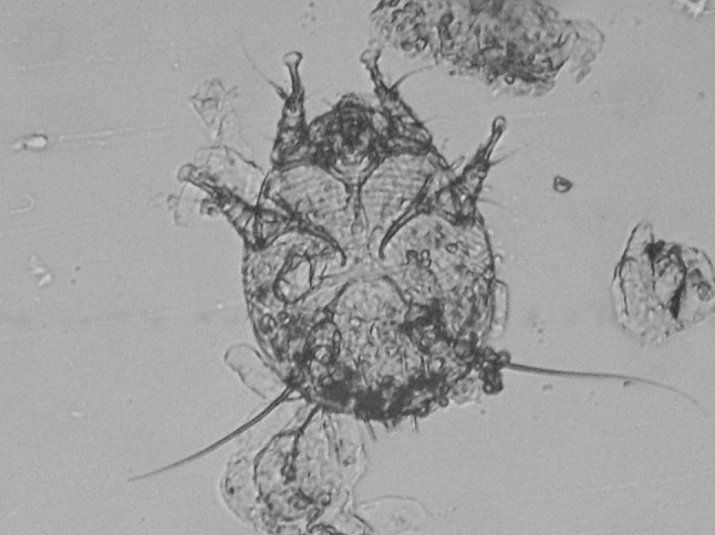
Notoedres cuniculi mange having concentric rings on dorsal surface
Therapeutic management
Following confirmation of parasitic infestation and evaluation of biochemical profiles, affected rabbits were treated with two injections of Ivermectin (400 μg/kg, S/C.) at fortnight interval. The supportive therapy of antihistaminics (chlorpheniramine maleate @ 1 mg/kg body wt), multivitamins (injection of vitamin B1+B6+B12 @ 0.5 mL I/M) and topical application of 0.5 % Povioine iodine solution for initial 5 days.
Results and discussion
The physical examination of the affected rabbis showed presence of crustaceous lesions on the head, ear pinnae, both the limbs and external genitalia. These findings are in agreement with findings of Osborne (1947) and Darzi et al. (2007), who reported similar clinical representation in rabbits affected by Notoedric mange. The affected rabbits were treated with two injections of Ivermectin (at fortnight interval) with 5 days supportive therapy. This treatment showed gradual clinical improvement in all rabbits after 1 week. The crustaceous lesions of affected parts were gradually reduced and disappeared with appreciable improvement in skin coat on 15th (Fig. 5) and 30th day (Fig. 6) respectively as compared to before treatment (Fig. 7). On the 30th day, no mite or their developmental stages were demonstrated on the scrapings examinations from the affected lesions.
Fig. 5.

Improvement in skin coat on 15th day post treatment
Fig. 6.

Improvement in skin coat on 30th day post treatment
Fig. 7.

Skin coat before treatment
Thus in the present investigation, two doses of Ivermectin along with supportive therapy showed complete recovery. These findings were in agreement with findings of Kurade et al. (1996) and Acar et al. (2007), where they successfully treated clinical cases of rabbit mange with Ivermectin and supportive therapy.
The results of alterations in biochemical profile before and after treatment (on 30th day after first injection of Ivermection) have been depicted in Table 1. The significant (p ≥ 0.05) increase in concentration of albumin (4.18 ± 0.09 g/dL), along with nonsignificant (p ≤ 0.05) increase in concentration of total protein (6.69 ± 0.08 g/dL) and decrease in concentration of globulin (2.50 ± 0.14 g/dL) was recorded after treatment (on 30th day). The enzymes showed significant (p ≥ 0.05) decrease in concentration of AST and ALT (24.31 ± 1.51 IU/L, 26.96 ± 3.01 IU/L); along with nonsignificant (p ≤ 0.05) decrease in concentration of ALP (60.54 ± 18.60 IU/L) after treatment, respectively. Thus biochemical profile showed restoration in altered values of all the parameters after treatment.
Table 1.
Showing (Mean ± SE) changes in biochemical parameters
| S. No. | Parameters | Before treatment | After treatment (on 30th day) | Calculated t value | Table t value at 5 % level |
|---|---|---|---|---|---|
| 1 | Total protein (g/dL) | 6.58 ± 0.17 | 6.69 ± 0.08 | 0.69 | 3.18 |
| 2 | Albumin (g/dL) | 3.86 ± 0.11 | 4.18 ± 0.09 | 7.70* | 3.18 |
| 3 | Globulin (g/dL) | 2.73 ± 0.22 | 2.50 ± 0.14 | 1.31 | 3.18 |
| 4 | AST (IU/L) | 63.06 ± 6.94 | 24.31 ± 1.51 | 5.07* | 3.18 |
| 5 | ALT (IU/L) | 50.68 ± 2.21 | 26.96 ± 3.01 | 6.99* | 3.18 |
| 6 | ALP (IU/L) | 80.58 ± 13.16 | 60.54 ± 18.60 | 0.90 | 3.18 |
* Significant at 5 %
Acknowledgments
Authors are thankful to the Associate Dean, COVAS, Parbhani, for providing necessary facilities to conduct this work.
References
- Acar A, Kurtdede A, Ural K, Cingi CC, Karakurum MC, Yagci BB, Sari B. An ectopic case of Psoroptes cuniculi infestation in a pet rabbit. Turk J Vet Anim Sci. 2007;31(6):423–425. [Google Scholar]
- Aulakh GS, Singh S, Singla LD, Singla N. Pathology and therapy of natural notoedric acariosis in rabbits. J Vet Parasitol. 2003;17:127–129. [Google Scholar]
- Darzi MM, Mir MS, Shahardar RA, Pandit BA. Clinicopathological, histochemical and therapeutic studies on concurrent sarcoptic and notoedric acariosis in rabbits (Oryctolagus cuniculus) Vet Arhiv. 2007;77:167–175. [Google Scholar]
- Kurade NP, Bhat TK, Jithendran KP. Effect of Ivermectin against ear mange mite (Psoroptes cuniculi) in naturally infested rabbits. World Rabbit Sci. 1996;4(1):25–27. [Google Scholar]
- Osborne HG. Mange in rabbits. Can J Comp Med. 1947;11(5):144–145. [PMC free article] [PubMed] [Google Scholar]
- Snedecor GW, Cochran WG. Statistical Methods. 6. Ames: IOWA State University Press; 1994. [Google Scholar]
- Soulsby EJL. Helminths, arthropods and protozoa of domestic animals. 7. London: ELBS and Bailliere Tindall; 1982. pp. 763–778. [Google Scholar]