

Abstract
Oblique crown-root fracture in the cervical third of the root is a common event following trauma to the anterior region of the mouth. As a result, sound tooth structure coronal to the attachment apparatus may not be available for restorative needs. Invasion of biological width by fracture line presents a clinical challenge in restorative planning. Placing a restoration margin on sound tooth structure within the dentogingival biological width might result in violation of biological width and should be considered a restorative failure. Maintaining a healthy periodontal attachment apparatus is crucial for long term prognosis and esthetics of the restored tooth. Surgical crown lengthening, surgical extrusion or orthodontic extrusions are the few alternative modalities to expose the fracture line. This case presentation demonstrates a predictable solution in overcoming an oblique crown-root fracture caused by trauma during a road accident. Orthodontic extrusion was used to elevate the fractured tooth from within the alveolar socket to allow the placement of crown margins on sound tooth structure without harming the biologic width. Combining fiberotomy with the extrusion procedure in this case eliminated the need for the surgical procedure. This allowed proper fabrication of post and core and the placement of the crown on sound tooth structure, fulfilling the biological and mechanical principles including obligatory ferrule effect.
Keywords: Circumferential supracrestal fibrotomy, Oblique crown root fracture, Orthodontic extrusion
Introduction
Crown-root fracture is defined as fracture involving enamel, dentin, and root cementum. Maxillary incisors are the most common teeth involved in dental trauma and most of the times, the crowns are damaged. According to the system adopted by the World Health Organization in its application of International Classification of Diseases to Dentistry and Stomatology (N.502.54), crown-root fractures have been classified as complicated and uncomplicated.1
A crown-root fracture (Type B according to dean’s classification) extends below the cemento-enamel junction and invades the biologic width.2 Oblique crown-root fractures that extend below both the gingival margin and the alveolar bone are difficult to restore. Efforts must be made to avoid the tooth loss and restore the form and function, with respect to the gingival portion of the attachment apparatus in order to prevent soft tissue deformities, which usually results from the violation of biologic width while restoring the defect.3 The subgingival location of the fracture makes the bonding procedures and a tight seal restoration extremely difficult or impossible. The objective in the treatment of such fractures must be aimed at exposing the fracture margins supragingivally and hence that all the procedures can be managed with moisture control.4
There are several options for the treatment of tooth fracture involving the biologic width which include (a) extraction, (b) surgical crown lengthening (c) surgical extrusion and (d) orthodontic extrusion.5
Extraction seems to be the easiest choice, yet it involves mutilation of adjacent dental tissues typically that occurs during subsequent prosthetic rehabilitation, or the patient may require a more complex implant therapy.
Attempts to expose the fracture line by alveolar recontouring alone may compromise the functional root length. In addition, poor esthetics may result from any attempt to recontour the labial tissues with simple or complex periodontal techniques. By using the intra-alveolar transplantation method, bone support around the root is lost compared with the orthodontic extrusion.6-9 Orthodontic extrusion or forced eruption was proposed by Heithersay for the treatment of horizontal root fractures. Orthodontic extrusion is a conservative procedure that allows retention of a tooth without loss of bone or periodontal support.10
This case report describes a multidisciplinary approach involving periodontal and restorative considerations for the management of complicated oblique crown-root fracture assisted by orthodontic extrusion.
Case Report
This was a case of a 24-year-old male patient reported to the Department of Conservative Dentistry and Endodontics with a fractured tooth in the upper left anterior region, 7 days after trauma from an accident.
Clinical examination did not reveal any soft tissue injury. Clinical and radiographic examination revealed an oblique crown-root fracture of the maxillary left lateral incisor. The fracture line was located 1.5 mm supragingivally on the buccal aspect (Figure 1a) and at the level of the alveolar crest on the palatal aspect. On the proximal aspect, the fracture line was located 1 mm subgingivally (Figure 1b and c). There was no damage to the adjacent teeth.
Figure 1.
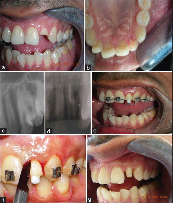
(a) Pre-operative labial view, (b) pre-operative palatal view (c) pre-operative radiograph (d) sectional obturation (e) sectional orthodontic appliance (f) circumferential supracrestal fibrotomy (g) crown preparation done.
Based on the clinical and radiographic findings, a diagnosis of oblique complicated crown-root fracture, dean’s Type B (plane of fracture angled cervically in a facial-to-lingual direction when viewed proximally) was made.
A definitive treatment plan was made, which involved endodontic therapy of the residual tooth, followed by rapid orthodontic extrusion to move the fracture line 3 mm above the alveolar crest and a prosthetic rehabilitation of the tooth.
Management
Local anesthesia was administered. Endodontic access cavity was done on the palatal surface using a no. 2 round bur and EX 24 bur (non-end cutting tapered fissure; Mani, Tochigi, Japan). Pulp extirpation was performed using a barbed broach (denstply-malliefer, Ballaigues, Switzerland) and K-files (Mani Inc., Tochigi, Japan). The canal was thoroughly debrided with a copious irrigation of sodium hypochlorite (2.5%) and saline (0.9%). Coronal flaring of the root canal was done using Gates-Glidden drills no 1-3 (Mani Inc., Tochigi, Japan). The working length was determined using apex locator (Propex, DENTSPLY-Malleifer, Ballaigues, Switzerland) and confirmed radiographically. Cleaning and shaping of the root canal system was completed using a step-back technique (apical enlargement was done up to ISO no 40). Canals were copiously irrigated with sodium hypochlorite and saline. The canal was dried with sterile paper points and calcium hydroxide (Ultracal XS, Ultradent, South Jordan, UT) was placed in the root canal and the access cavity was temporized with Cavit G (3M ESPE, Germany). The patient was recalled after 1 week for obturation.
After a week, the tooth was asymptomatic and a sectional obturation using thermoplastic obturation technique (E&Q plus Meta Biomed Co ltd-Korea) and AHplus as a sealer was done (Figure 1d). A temporary screw post was cemented using zinc phosphate cement to aid in extrusion. A sectional Begg’s appliance using teeth from maxillary left canine to right canine as anchorage units was used (Figure 1e). The orthodontic appliances were activated weekly, with a force of approximately 50 g for 3 weeks. A 3.5 mm extrusion was achieved at the end of the activation period. The weekly activations were accompanied with fiberotomy performed using a scalpel and a 15C blade (Swann-Morton) jointly with root planning from the top of the alveolar bone crest using curettes (Gracey 5/6, Hu-Freidy) (Figure 1f). The incisions were made under papillary anesthesia, using the smallest possible quantity of anesthetic. Following the active period of extrusion, the teeth were stabilized for 8 weeks, using the same fixed orthodontic appliance, to prevent relapse. Following the retentive phase, the provisional post cemented for the extrusion was removed. A fiber post (Luscent-Anchor; Dentatus) of appropriate size was then cemented using dual cure resin cement (Panavia F2, Kurarray). Core build up was done using light cure composite (Z100, 3M ESPE, Germany) (Figure 1g). Finally the tooth was restored with an esthetic and functional porcelain crown (Figure 2a and b).
Figure 2.
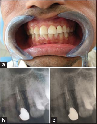
(a) Porcelain-fused-to-metal crown cemented (b) post-operative radiograph (c) 2 years follow-up radiograph.
The patient was examined every 3 months during the follow-up period of 24 months (Figure 2c). The tooth did not show any signs of root resorption during the treatment and follow-up periods.
Discussion
Tooth fracture involving cervical third of the root constitutes an insult to the biologic width. A predictable esthetic restoration is not limited to the restored tooth but has to include the gingival unit. Violation of biologic width while placement of the restorative margins should be considered as a potential restorative failure because it may lead to irreversible damage in the form of gingival inflammation, alveolar crest resorption and recession.3 There should be 2-3 mm of biologic width on all teeth to protect the teeth from progression of infection from the gingival sulcus into periodontium and must be reestablished before esthetic and functional recovery.2 In this case, mere placement of a restoration was not possible because the fracture line involved the biologic width. In such a case, two main factors must be addressed:
The fracture margin access and
The possibility of performing a tight seal restoration.10
Surgical exposure of sound tooth structure is fraught with compromise especially in the anterior segment where esthetics is a consideration. Gingival and osseous surgery cannot be limited to the involved tooth and must be extended to the adjacent teeth in order to blend the gingival and osseous contours. The ultimate result is a sacrifice of supporting bone on several teeth, root sensitivity and esthetic deformities in the form of long clinical crowns and open embrasures. Furthermore surgery shortens the root and increases the crown-root ratio.11
The use of orthodontic extrusion also referred to as forced eruption, has been suggested as an alternative to periodontal crown lengthening. Orthodontic extrusion is considered to be the easiest tooth movement with a good prognosis and a low risk of relapse.5,7,12-14 Forced eruption is usually limited to one, two or three maxillary anterior teeth or premolars with as much as 5 mm of extrusion possible.15 Before starting with forced eruption, a few factors should be considered. The most important is crown−to−root ratio, which shall remain at least 1:1.16 Root extrusion, when compared to surgical crown elongation maintains more ideal proportions: it decreases root length with remaining crown length unchanged.6-8,14 Other factors like root morphology, tooth localization, its periodontal status and the aesthetics needs are also of great importance.17 When concerning aesthetics, the clinician should also remember that a root diameter decreases at the end of the extrusion. Proper contouring of final restoration becomes a very important issue.
The extrusion rate used in this case was similar to that recommended by other authors. After 21 days of controlled extrusion, 3 mm of the root was exposed with a speed of 1 mm per week.7,8,18 Forces in the range of 30-60 g has been advocated to extrude the tooth.8,18-21 In the present case, 50 g of force was used for extrusion of the tooth. A retentive phase of 8-12 weeks, followed by activation is recommended to stabilize the extruded tooth in its new position. In the present case, 8 weeks of stabilization was done with the help of the orthodontic appliance itself.
Whenever a tooth is extruded the alveolar bone and gingival attachment descends with the extruded tooth. Ingber reported the need for surgical crown lengthening after the retention phase to re-establish the biologic width through the removal of gingival and bone tissues that follow the tooth in its coronal trajectory, thus permitting appropriate prosthetic therapy.7
The coronal migration of periodontal tissues in dental extrusion is induced by the tension provoked by the gingival fibers and by the periodontal ligament.22 Supracrestal fiberotomy, which involves resection of the supra-alveolar fibers stretched by the extruded tooth to eliminate the tensile stress on the alveolar crestal bone and to prevent the tendency of attachment apparatus to follow the extruded tooth is a well-documented procedure.23,24 Combining fiberotomy during the process of extrusion makes the entire treatment more conservative and non-surgical, and offers an esthetically excellent, long lasting soft tissue contours.
Finally, the use of fiber post gives good esthetic results and increases the retention and distributes the stresses evenly along the root.
Conclusion
Functional and esthetic needs should be balanced with the demands of healthy periodontium. Maintaining a healthy periodontal attachment apparatus is crucial for a positive long-term prognosis. Orthodontic extrusion combined with fiberotomy presents the most suitable and predictable treatment modality for the management of oblique crown-root fractures that invade the biologic width.
Footnotes
Conflicts of Interest: None
Source of Support: Nil
References
- 1.Andreason JO, Andreason FM. Traumatic Injuries of Teeth. 2nd ed. Copenhagen: Saunders Publications, Munksgaard; 1988. [Google Scholar]
- 2.Trushkowsky RD. Esthetic, biologic and restorative considerations in coronal segment reattachment for a fractured tooth: A clinical report. J Prosthet Dent. 1998;79(2):115–9. doi: 10.1016/s0022-3913(98)70202-1. [DOI] [PubMed] [Google Scholar]
- 3.Padbury A, Jr, Eber R, Wang HL. Interactions between the gingiva and the margin of restorations. J Clin Periodontol. 2003;30(5):379–85. doi: 10.1034/j.1600-051x.2003.01277.x. [DOI] [PubMed] [Google Scholar]
- 4.Olsburgh S, Jacoby T, Krejci I. Crown fractures in the permanent dentition: Pulpal and restorative considerations. Dent Traumatol. 2002;18(3):103–15. doi: 10.1034/j.1600-9657.2002.00004.x. [DOI] [PubMed] [Google Scholar]
- 5.Fournier A. Orthodontic management of subgingivally fractured teeth. J Clin Orthod. 1981;15(7):502–3. [PubMed] [Google Scholar]
- 6.Heithersay GS. Combined endodontic-orthodontic treatment of transverse root fractures in the region of the alveolar crest. Oral Surg Oral Med Oral Pathol. 1973;36(3):404–15. doi: 10.1016/0030-4220(73)90220-x. [DOI] [PubMed] [Google Scholar]
- 7.Ingber JS. Forced eruption: Part II. A method of treating nonrestorable teeth – Periodontal and restorative considerations. J Periodontol. 1976;47(4):203–16. doi: 10.1902/jop.1976.47.4.203. [DOI] [PubMed] [Google Scholar]
- 8.Oesterle LJ, Wood LW. Raising the root. A look at orthodontic extrusion. J Am Dent Assoc. 1991;122(7):193–8. doi: 10.14219/jada.archive.1991.0229. [DOI] [PubMed] [Google Scholar]
- 9.Baker IM. Esthetic extrusion of a nonrestorable tooth. J Clin Orthod. 1990;24(5):323–5. [PubMed] [Google Scholar]
- 10.Villat C, Machtou P, Naulin-Ifi C. Multidisciplinary approach to the immediate esthetic repair and long-term treatment of an oblique crown-root fracture. Dent Traumatol. 2004;20(1):56–60. doi: 10.1111/j.1600-4469.2004.00206.x. [DOI] [PubMed] [Google Scholar]
- 11.Koyuturk AE, Malkoc S. Orthodontic extrusion of subgingivally fractured incisor before restoration. A case report: 3-years follow-up. Dent Traumatol. 2005;21(3):174–8. doi: 10.1111/j.1600-9657.2005.00291.x. [DOI] [PubMed] [Google Scholar]
- 12.Silness J. Periodontal conditions in patients treated with dental bridges. 3. The relationship between the location of the crown margin and the periodontal condition. J Periodontal Res. 1970;5(3):225–9. doi: 10.1111/j.1600-0765.1970.tb00721.x. [DOI] [PubMed] [Google Scholar]
- 13.Ivey DW, Calhoun RL, Kemp WB, Dorfman HS, Wheless JE. Orthodontic extrusion: Its use in restorative dentistry. J Prosthet Dent. 1980;43(4):401–7. doi: 10.1016/0022-3913(80)90209-7. [DOI] [PubMed] [Google Scholar]
- 14.Hovland EJ. Horizontal root fractures. Treatment and repair. Dent Clin North Am. 1992;36(2):509–25. [PubMed] [Google Scholar]
- 15.Wang WG, Wang WN. Forced eruption: An alternative to extraction or periodontal surgery. J Clin Orthod. 1992;26(3):146–9. [PubMed] [Google Scholar]
- 16.Simon JH. Root extrusion. Rationale and techniques. Dent Clin North Am. 1984;28(4):909–21. [PubMed] [Google Scholar]
- 17.Johnson GK, Sivers JE. Forced eruption in crown-lengthening procedures. J Prosthet Dent. 1986;56(4):424–7. doi: 10.1016/0022-3913(86)90381-1. [DOI] [PubMed] [Google Scholar]
- 18.Biggerstaff RH, Sinks JH, Carazola JL. Orthodontic extrusion and biologic width realignment procedures: Methods for reclaiming nonrestorable teeth. J Am Dent Assoc. 1986;112(3):345–8. doi: 10.1016/s0002-8177(86)23014-7. [DOI] [PubMed] [Google Scholar]
- 19.Bondemark L, Kurol J, Hallonsten AL, Andreasen JO. Attractive magnets for orthodontic extrusion of crown-root fractured teeth. Am J Orthod Dentofacial Orthop. 1997;112(2):187–93. doi: 10.1016/s0889-5406(97)70245-2. [DOI] [PubMed] [Google Scholar]
- 20.Kocadereli I, Taşman F, Güner SB. Combined endodontic-orthodontic and prosthodontic treatment of fractured teeth. Case report. Aust Dent J. 1998;43(1):28–31. doi: 10.1111/j.1834-7819.1998.tb00148.x. [DOI] [PubMed] [Google Scholar]
- 21.Emerich-Poplatek K, Sawicki L, Bodal M, Adamowicz-Klepalska B. Forced eruption after crown/root fracture with a simple and aesthetic method using the fractured crown. Dent Traumatol. 2005;21(3):165–9. doi: 10.1111/j.1600-9657.2005.00287.x. [DOI] [PubMed] [Google Scholar]
- 22.Edwards JG. A surgical procedure to eliminate rotational relapse. Am J Orthod. 1970;57(1):35–46. doi: 10.1016/0002-9416(70)90203-4. [DOI] [PubMed] [Google Scholar]
- 23.Pontoriero R, Celenza F, Jr, Ricci G, Carnevale G. Rapid extrusion with fiber resection: A combined orthodontic-periodontic treatment modality. Int J Periodontics Restorative Dent. 1987;7(5):30–43. [PubMed] [Google Scholar]
- 24.Carvalho CV, Bauer FP, Romito GA, Pannuti CM, De Micheli G. Orthodontic extrusion with or without circumferential supracrestal fiberotomy and root planing. Int J Periodontics Restorative Dent. 2006;26(1):87–93. [PubMed] [Google Scholar]